Review
. 2021 Mar 22;6(6):e146006.
doi: 10.1172/jci.insight.146006.

Therapeutic manipulation of innate lymphoid cells

Review

Therapeutic manipulation of innate lymphoid cells

Laura M Cobb et al. JCI Insight. .

Abstract

Since their relatively recent discovery, innate lymphoid cells (ILCs) have been shown to be tissue-resident lymphocytes that are critical mediators of tissue homeostasis, regeneration, and pathogen response. However, ILC dysregulation contributes to a diverse spectrum of human diseases, spanning virtually every organ system. ILCs rapidly respond to environmental cues by altering their own phenotype and function as well as influencing the behavior of other local tissue-resident cells. With a growing understanding of ILC biology, investigators continue to elucidate mechanisms that expand our ability to phenotype, isolate, target, and expand ILCs ex vivo. With mounting preclinical data and clinical correlates, the role of ILCs in both disease pathogenesis and resolution is evident, justifying ILC manipulation for clinical benefit. This Review will highlight areas of ongoing translational research and critical questions for future study that will enable us to harness the full therapeutic potential of these captivating cells.


Conflict of interest statement

Conflict of interest: MRV has filed for patent protection around the methodology described in this manuscript for the generation of the described ILC subpopulations using UCB-derived hematopoietic stem cells: US-9862928-B2, “Generation of natural killer cells and lymphoid tissue inducer-like (LTi-like) NK-22 cells.”

Figures

Figure 1
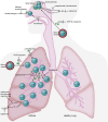
Figure 1. Targeting ILCs in diseases of the airway.
Activated ILC2s accumulate in the lung in asthma and promote disease via 2 mechanisms: (a) direct activation by TSLP (thymic stromal lymphopoietin), IL-33, and IL-25 in response to allergens, viruses, and environmental stress and (b) IgE-dependent antigen responses, which induce mast cell secretion of lipid mediators, such as PGD2 (prostaglandin D2) and CysLTs (cysteinyl leukotrienes), to attract and activate ILC2s. ILC2s also promote type 2 responses in allergic rhinitis (AR) and chronic rhinosinusitis with nasal polyposis (CRSwNP), where they accumulate in polyps. Seasonal increases in ILC2s in AR are suppressed by allergen immunotherapy. Montelukast (a CysLT1 antagonist), approved for asthma and AR, inhibits ILC2 activation. β2 Adrenergic receptor (β2AR) agonists negatively regulate ILC2s in asthma by inhibiting their proliferation and effector function. In both asthma with or without AR and CRSwNP, systemic steroids decrease ILC2 cytokine production and promote ILC2 apoptosis. Dupilumab inhibits IL-4Rα, the shared receptor of IL-4 and IL-13, and is approved in asthma and CRSwNP. Anti–IL-5 mAbs (mepolizumab, benralizumab, reslizumab) are approved in asthma and are being tested in CRSwNP. CRTH2 (chemoattractant receptor-homologous molecule expressed on Th2 cells) antagonists to block PGD2-mediated ILC2 trafficking as well as antibodies against TSLP and IL-33 to inhibit ILC2 activation are in testing. Illustrated by Rachel Davidowitz.
Figure 2
Figure 2. Targeting ILCs in obesity and type 2 diabetes mellitus.
In healthy white adipose (A), ILC2s promote an antiinflammatory type 2 environment via eosinophil recruitment and induction of alternatively activated macrophages (AAMs). Specialized beige adipocytes interspersed in WAT can utilize the mitochondrial uncoupling protein 1 (UCP1) for thermogenesis, increasing caloric expenditure and protecting from obesity. “Beiging” is promoted by ILC2 production of methionine-enkephalin (MetEnk) peptides. Thus, expansion of ILC2s via IL-25, IL-33, or ILC2 adoptive transfer promotes weight loss and improved glucose tolerance. Additionally, engagement of the glucocorticoid-induced TNF receptor (GITR) on ILC2s via a GITR agonist also promotes beiging. ILC2s are decreased in obese adipose (B), while ILC1s are increased. IL-12–dependent IFN-γ production is sustained by adipose ILC1s and induces classically activated macrophages (CAMs) to secrete IL-6 and TNF-α. This promotes a proinflammatory type 1 environment, which fosters development of glucose intolerance and leads to type 2 diabetes mellitus. Therefore, therapeutically activating the IL-33/GITR/ILC2-beiging pathway signifies a novel method for treating obesity and T2DM. Illustrated by Rachel Davidowitz.
Figure 3
Figure 3. Targeting ILCs in Crohn’s disease.
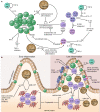
(A) In Crohn’s disease (CD), ILC1s accumulate in the intestine, while NCR ILC3s are reduced. Myeloid cells, including DCs, produce IL-12, stimulating ILC1s to produce IFN-γ and TNF-α. IL-12 also induces transdifferentiation of NCR and NCR+ ILC3s into ILC1s. IL-23 promotes differentiation of ILC1s into IL-22–producing NCR+ ILC3s. Regulatory ILCs (ILCregs) downregulate ILC1s and ILC3s via IL-10 secretion to suppress production of cytokines other than IL-22. ILCregs also produce TGF-β, which promotes their own expansion and survival. Thalidomide and lenalidomide modulate ILC transdifferentiation by selectively degrading Aiolos (ILC1 specific) and Ikaros (ILC nonspecific), increasing ILC3-associated Helios. Sphingosine-1 phosphate receptor 1 (S1PR1) antagonists downregulate ILC3 NCR expression. TNF-α inhibitors directly block TNF-α produced by ILC1s. Ustekinumab and JAK inhibitors target IL-12 and IL-23, while IL-23–specific mAbs target IL-23 and an anti–IL-22 Fc mAb targets IL-22. An anti-NKG2D mAb might modulate ILC1s and NCR+ ILC3s, which express NKG2D in mice. (B) In healthy intestine, ILC3s reside largely within lymphoid aggregate cryptopatches (CPs). Early in inflammation (left), IL-23 secretion by myeloid cells is GM-CSF dependent, and GM-CSF production by ILC3s is sustained by IL-23. Later in inflammation (right), GM-CSF recruits additional myeloid cells and mobilizes ILC3s from CPs into adjacent intestinal mucosa. In response to IL-23, ILC3s produce IL-17 and IFN-γ, promoting intestinal inflammation. IL-23 can be blocked by anti–IL-23 mAbs, ustekinumab, and JAK inhibitors. α4β7 Integrin blockade with vedolizumab might inhibit ILC migration from CPs into intestinal mucosa. A FimH antagonist reduces bacterial adherence to the gut, decreasing innate immune activation and improving intestinal permeability. S1PR1 antagonists prevent ILC1/3 maturation and migration and modify ILC functions. Illustrated by Rachel Davidowitz.

References

    1. Artis D, Spits H. The biology of innate lymphoid cells. Nature. 2015;517(7534):293–301. doi: 10.1038/nature14189. - DOI - PubMed
    1. Eberl G, et al. The brave new world of innate lymphoid cells. Nat Immunol. 2015;16(1):1–5. doi: 10.1038/ni.3059. - DOI - PubMed
    1. Vivier E, et al. Innate lymphoid cells: 10 years on. Cell. 2018;174(5):1054–1066. doi: 10.1016/j.cell.2018.07.017. - DOI - PubMed
    1. An Z, et al. Pleiotropic role and bidirectional immunomodulation of innate lymphoid cells in cancer. Front Immunol. 2019;10:3111. - PMC - PubMed
    1. Mattiola I, Diefenbach A. Innate lymphoid cells and cancer at border surfaces with the environment. Semin Immunol. 2019;41:101278. - PubMed

Publication types