Review
. 2021 Apr 5;220(4):e202012095.
doi: 10.1083/jcb.202012095.

The cell biology of Parkinson's disease

Affiliations
Review

The cell biology of Parkinson's disease

Nikhil Panicker et al. J Cell Biol. .

Abstract

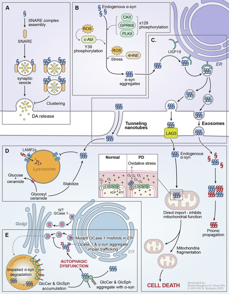
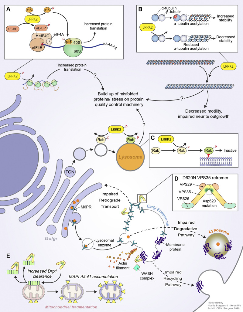
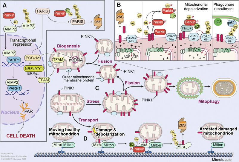
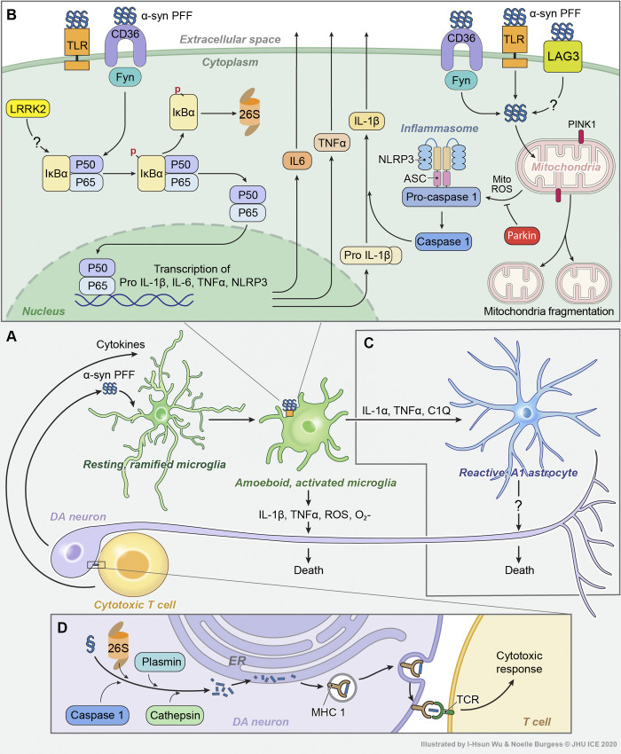
Parkinson's disease (PD) is a progressive neurodegenerative disorder resulting from the death of dopamine neurons in the substantia nigra pars compacta. Our understanding of PD biology has been enriched by the identification of genes involved in its rare, inheritable forms, termed PARK genes. These genes encode proteins including α-syn, LRRK2, VPS35, parkin, PINK1, and DJ1, which can cause monogenetic PD when mutated. Investigating the cellular functions of these proteins has been instrumental in identifying signaling pathways that mediate pathology in PD and neuroprotective mechanisms active during homeostatic and pathological conditions. It is now evident that many PD-associated proteins perform multiple functions in PD-associated signaling pathways in neurons. Furthermore, several PARK proteins contribute to non-cell-autonomous mechanisms of neuron death, such as neuroinflammation. A comprehensive understanding of cell-autonomous and non-cell-autonomous pathways involved in PD is essential for developing therapeutics that may slow or halt its progression.


Figures

Figure 1.
Figure 1.
α-Syn and GBA1 signaling in PD. (A) Monomeric α-syn acts as a chaperone for SNARE proteins, promoting synaptic transmission/DA release. Certain aggregated species of α-syn can reduce DA release by inhibiting synaptic vesicle clustering. (B) Progressive accumulation of α-syn aggregates is a fundamental characteristic of PD progression. Numerous posttranslational modifications have been reported to modulate α-syn aggregation, including pS129 (unclear effect) via multiple kinases, pY39 (pro-aggregation) by c-Abl, and 4-hydroxy-2-nonenal (4HNE) and other modifications mediated by oxidative stress. (C) Aggregated α-syn is incorporated into the ER-transport machinery by USP19 and can propagate from cell to cell via tunneling nanotubes or exosomes, or by direct LAG3-mediated uptake into efferent neurons. (D) Endocytosed α-syn fibrils can seed the templated aggregation of endogenous α-syn and drive prion-like propagation. α-Syn aggregates can also be directly imported into mitochondria, eliciting mitochondrial fragmentation and death. The function of lysosomes, where GCase1 localizes to and metabolizes glycolipids, is also impaired by the presence of α-syn aggregates. (E) GBA1-associated PD is thought to be closely linked to dysregulation of α-syn proteostasis. Likewise, sporadic PD is thought to drive impairments in GCase1 activity. In patients carrying mutant GBA1 alleles, coding mutations in the GCase1 protein may lead to misfolding in the ER, leading to direct coaggregation with α-syn or indirectly causing α-syn aggregation by impairing autophagy. In sporadic PD, loss of GCase1 function may be driven by coaggregation with α-syn, which then serves as a positive feedback loop to accelerate further α-syn aggregation. In addition, accumulation of GCase1 substrates such as GlcCer and glucosylsphingosine (GlcSph) is sufficient to trigger α-syn aggregation.
Figure 2.
Figure 2.
LRRK2 and VPS35 signaling in PD. (A) LRRK2 may facilitate misfolded protein buildup via increasing global protein translation. It does so by phosphorylating (depicted by a red P) 4E-BP and the 40S ribosomal subunit S15. (B) LRRK2 can associate with β-tubulin via its Roc domain, physically impeding α-tubulin acetylation (depicted by a red A), which may lead to decreased microtubule stability and impaired neurite outgrowth. (C) LRRK2 can phosphorylate multiple RAB proteins within their switch II domains, inactivating them and promoting their membrane targeting. Increased LRRK2 activity may compromise vesicular sorting machinery in neurons, leading to the accumulation of misfolded proteins. (D) The VPS 35 retromer forms a dimer of trimers comprising VPS26/29/35, with the D620N mutation in VPS35 disrupting an acidic residue present at the region critical for retromer complex dimerization, potentially impairing proper assembly of the retromer dimer of trimers. The D620N mutation has been associated with impaired retrograde transport of Golgi-endosome cargo receptors, leading to a deficiency of receptors at the Golgi and impairing forward transport of lysosomal enzymes. D620N VPS35 also exhibits deficiencies in endosome-plasma membrane recycling of surface membrane proteins due to impaired association with the WASH complex. (E) VPS35 is also involved in regulation of mitochondrial dynamics. The D620N mutation can lead to mitochondrial fragmentation through MAPL/Mul1 accumulation or increased Drp1 clearance.
Figure 3.
Figure 3.
Parkin and PINK1 signaling in PD. (A) Loss of parkin E3 ligase activity via PD-associated mutations or PTM results in the accumulation of its substrates. Accrual of the amino-acyl tRNA cofactor AIMP2 results in poly(ADP-ribose) polymerase-1 (PARP1)–dependent neuron death. Buildup of PARIS/ZNF746, a transcription inhibitor, prevents mitochondrial biogenesis by repressing the expression of PGC-1α, the master mitochondrial biogenesis regulator. (B) PINK1 is a serine/threonine kinase that phosphorylates (depicted by a red PD) both ubiquitin and parkin at S65, allowing parkin to be recruited to damaged mitochondria. This is followed by the parkin-mediated polyubiquitination of OMM proteins, culminating in the clearance of damaged mitochondria via mitophagy. (C) PINK1/parkin signaling maintains a balance between mitochondrial fission and fusion. (D) Finally, PINK1/parkin inhibit mitochondrial transport by phosphorylating and ubiquitinating the protein Miro, causing mitochondrial detachment from the microtubule. Ub, ubiquitin.
Figure 4.
Figure 4.
Non-autonomous signaling pathways in PD. (A) Hyperactivated microglia, reactive A1 astrocytes, and cytotoxic T cells can independently or cooperatively act to mediate DA neuron death in PD. (B) α-Syn oligomers or aggregates released from dysfunctional DA neurons can activate the NF-κB pathway in microglia. LRRK2 may play a role in this process. This results in a pro-inflammatory cytokine response. Endocytosed α-syn oligomers or PFFs can also mediate mitochondrial dysfunction, resulting in the production of mitochondria-derived reactive oxygen species (mitoROS), which activates the NLRP3 inflammasome, resulting in IL-1β processing and secretion. Microglia-released pro-inflammatory cytokines and ROSs can directly elicit DA neuron death. Parkin/PINK1 signaling dampens inflammasome activation by preventing mitoROS release. (C) Activated microglia can produce IL-1α, TNF-α, and C1Q to elicit the formation of reactive A1 astrocytes, which directly kill neurons via an unknown mechanism. (D) Aberrant auto-processing of α-syn in DA neurons can result in the generation of α-syn antigenic epitopes, which, when presented by MHC class I molecules, can drive autoimmune cytotoxic T cell responses that result in DA neuron death.

Similar articles

Cited by

References

    1. Abounit, S., Bousset L., Loria F., Zhu S., de Chaumont F., Pieri L., Olivo-Marin J.C., Melki R., and Zurzolo C.. 2016. Tunneling nanotubes spread fibrillar α-synuclein by intercellular trafficking of lysosomes. EMBO J. 35:2120–2138. 10.15252/embj.201593411 - DOI - PMC - PubMed
    1. Alegre-Abarrategui, J., Christian H., Lufino M.M., Mutihac R., Venda L.L., Ansorge O., and Wade-Martins R.. 2009. LRRK2 regulates autophagic activity and localizes to specific membrane microdomains in a novel human genomic reporter cellular model. Hum. Mol. Genet. 18:4022–4034. 10.1093/hmg/ddp346 - DOI - PMC - PubMed
    1. Alim, M.A., Hossain M.S., Arima K., Takeda K., Izumiyama Y., Nakamura M., Kaji H., Shinoda T., Hisanaga S., and Ueda K.. 2002. Tubulin seeds alpha-synuclein fibril formation. J. Biol. Chem. 277:2112–2117. 10.1074/jbc.M102981200 - DOI - PubMed
    1. Aloisi, F. 2001. Immune function of microglia. Glia. 36:165–179. 10.1002/glia.1106 - DOI - PubMed
    1. Amo, T., Saiki S., Sawayama T., Sato S., and Hattori N.. 2014. Detailed analysis of mitochondrial respiratory chain defects caused by loss of PINK1. Neurosci. Lett. 580:37–40. 10.1016/j.neulet.2014.07.045 - DOI - PubMed

Publication types

MeSH terms