Re-Evaluation of Rat Corneal Damage by Short-Wavelength UV Revealed Extremely Less Hazardous Property of Far-UV-C†
- PMID: 33749837
- PMCID: PMC8251618
- DOI: 10.1111/php.13419
Re-Evaluation of Rat Corneal Damage by Short-Wavelength UV Revealed Extremely Less Hazardous Property of Far-UV-C†
Abstract
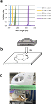
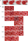
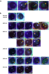
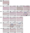
Corneal damage-induced various wavelength UV (311, 254, 235, 222 and 207 nm) was evaluated in rats. For 207 and 222-UV-C, the threshold radiant exposure was between 10 000 and 15 000 mJ cm-2 at 207 nm and between 3500 and 5000 mJ cm-2 at 222 nm. Penetrate depth to the cornea indicated by cyclobutene pyrimidine dimer (CPD) localization immediately after irradiation was dependent on the wavelength. 311 and 254 nm UV penetrate to corneal endothelium, 235 nm UVC to the intermediate part of corneal stroma, 222 and 207 nm UVC only to the most outer layer of corneal epithelium. CPD observed in corneal epithelium irradiated by 222 nm UVC disappeared until 12 h after. The minimum dose to induce corneal damage of short-wavelength UV-C was considerably higher than the threshold limit value (TLV® ) promulgated by American Conference of Governmental Industrial Hygienists (ACGIH). The property that explains why UV-C radiation at 207 and 222 nm is extremely less hazardous than longer UV wavelengths is the fact that this radiation only penetrates to the outermost layer of the corneal epithelium. These cells typically peel off within 24 h during the physiological turnover cycle. Hence, short-wavelength UV-C might be less hazardous to the cornea than previously considered until today.
© 2021 The Authors. Photochemistry and Photobiology published by Wiley Periodicals LLC on behalf of American Society for Photobiology.
Figures


Similar articles
-
Wavelength-dependent ultraviolet induction of cyclobutane pyrimidine dimers in the human cornea.Photochem Photobiol Sci. 2013 Aug;12(8):1310-8. doi: 10.1039/c3pp25408a. Photochem Photobiol Sci. 2013. PMID: 23364620
-
Safety Evaluation of Far-UV-C Irradiation to Epithelial Basal Cells in the Corneal Limbus.Photochem Photobiol. 2023 Jul-Aug;99(4):1142-1148. doi: 10.1111/php.13750. Epub 2022 Dec 14. Photochem Photobiol. 2023. PMID: 36437576
-
Evaluation of acute corneal damage induced by 222-nm and 254-nm ultraviolet light in Sprague-Dawley rats.Free Radic Res. 2019 Jun;53(6):611-617. doi: 10.1080/10715762.2019.1603378. Epub 2019 May 27. Free Radic Res. 2019. PMID: 30947566
-
Ultraviolet-induced photochemical damage in ocular tissues.Health Phys. 1989 May;56(5):671-82. doi: 10.1097/00004032-198905000-00012. Health Phys. 1989. PMID: 2651362 Review.
-
Mechanistic considerations on the wavelength-dependent variations of UVR genotoxicity and mutagenesis in skin: the discrimination of UVA-signature from UV-signature mutation.Photochem Photobiol Sci. 2018 Dec 5;17(12):1861-1871. doi: 10.1039/c7pp00360a. Photochem Photobiol Sci. 2018. PMID: 29850669 Review.
Cited by
-
Wavelength-dependent DNA Photodamage in a 3-D human Skin Model over the Far-UVC and Germicidal UVC Wavelength Ranges from 215 to 255 nm.Photochem Photobiol. 2022 Sep;98(5):1167-1171. doi: 10.1111/php.13602. Epub 2022 Feb 18. Photochem Photobiol. 2022. PMID: 35104367 Free PMC article.
-
222 nm far-UVC light markedly reduces the level of infectious airborne virus in an occupied room.Sci Rep. 2024 Mar 20;14(1):6722. doi: 10.1038/s41598-024-57441-z. Sci Rep. 2024. PMID: 38509265 Free PMC article.
-
Effects of 222 nm Germicidal Ultraviolet Light on Aerosol and VOC Formation from Limonene.ACS EST Air. 2024 May 4;1(7):725-733. doi: 10.1021/acsestair.4c00065. eCollection 2024 Jul 12. ACS EST Air. 2024. PMID: 39021671 Free PMC article.
-
Far-Ultraviolet Light at 222 nm Affects Membrane Integrity in Monolayered DLD1 Colon Cancer Cells.Int J Mol Sci. 2024 Jun 27;25(13):7051. doi: 10.3390/ijms25137051. Int J Mol Sci. 2024. PMID: 39000160 Free PMC article.
-
Bactericidal effect of far ultraviolet-C irradiation at 222 nm against bacterial peritonitis.PLoS One. 2024 Nov 12;19(11):e0311552. doi: 10.1371/journal.pone.0311552. eCollection 2024. PLoS One. 2024. PMID: 39531431 Free PMC article.
References
-
- Diffey, B. L. (2002) Human exposure to solar ultraviolet radiation. J. Cosmet. Dermatol. 1, 124–130. - PubMed
-
- Buonanno, M. , Stanislauskas M., Ponnaiya B., Bigelow A. W., Randers‐Pehrson G., Xu Y., Shuryak I., Smilenov L., Owens D. M. and Brenner D. J. (2016) 207‐nm UV light‐A promising tool for safe low‐cost reduction of surgical site infections. II: In‐vivo safety studies. PLoS One 11, e0138418. - PMC - PubMed
Publication types
MeSH terms
Substances
LinkOut - more resources
Full Text Sources
Other Literature Sources
Medical

