. 2021 Sep;73(9):1683-1693.
doi: 10.1002/art.41727. Epub 2021 Aug 11.

Eosinophil ETosis-Mediated Release of Galectin-10 in Eosinophilic Granulomatosis With Polyangiitis

Affiliations

Eosinophil ETosis-Mediated Release of Galectin-10 in Eosinophilic Granulomatosis With Polyangiitis

Mineyo Fukuchi et al. Arthritis Rheumatol. 2021 Sep.

Abstract

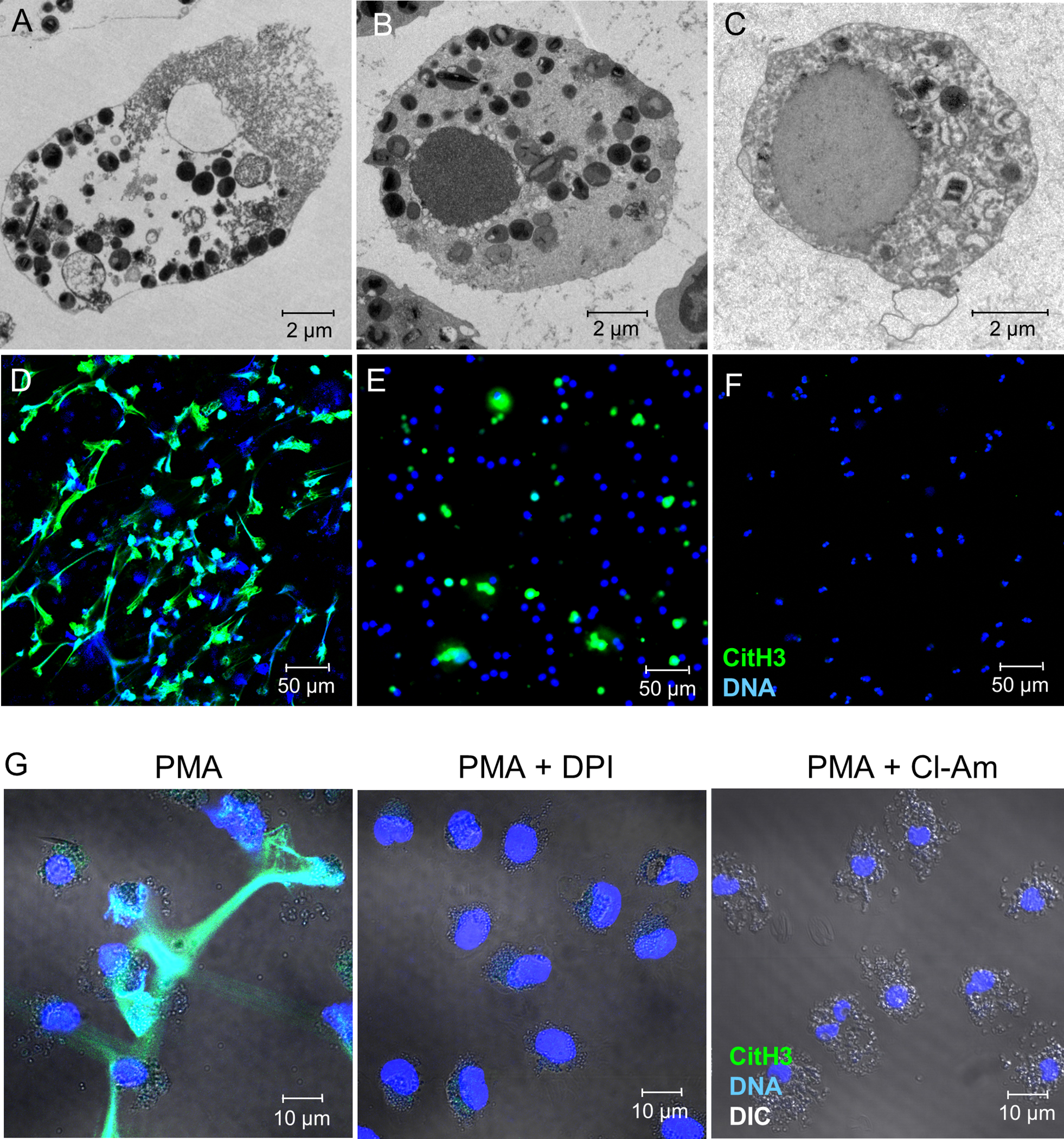
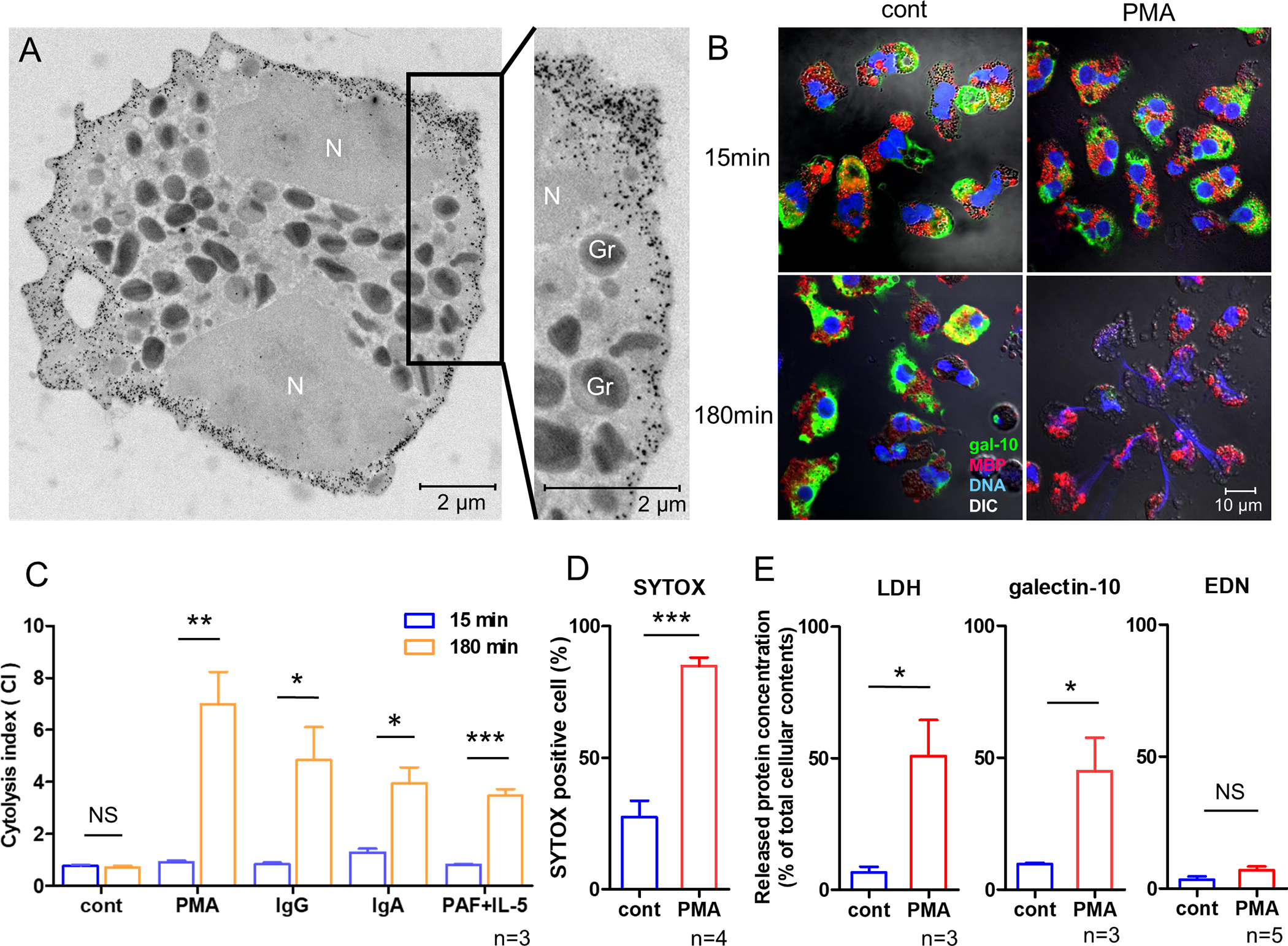
Objective: Eosinophils are tissue-dwelling immune cells. Accumulating evidence indicates that a type of cell death termed ETosis is an important cell fate involved in the pathophysiology of inflammatory diseases. Although the critical role of eosinophils in eosinophilic granulomatosis with polyangiitis (EGPA; formerly Churg-Strauss syndrome) is well established, the presence of eosinophil ETosis (EETosis) is poorly understood. We undertook this study to better understand the characteristics of EETosis.

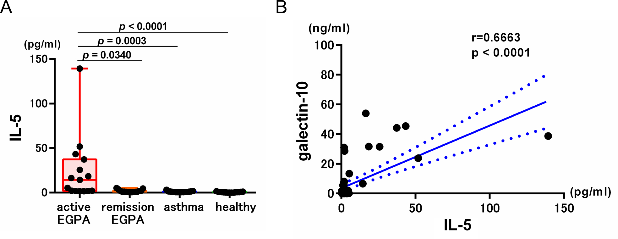
Methods: In vitro studies using blood-derived eosinophils were conducted to characterize EETosis. The occurrence of EETosis in tissues from patients with EGPA was studied by immunostaining and electron microscopy. Serum concentrations of eosinophil-derived proteins in healthy controls, patients with asthma, and EGPA patients with active disease or with disease in remission (n = 15 per group) were examined.

Results: EETosis was reliant on reactive oxygen species and peptidylarginine deiminase type 4-dependent histone citrullination, resulting in the cytolytic release of net-like eosinophil extracellular traps, free galectin-10, and membrane-bound intact granules. The signature of EETosis, including loss of cytoplasmic galectin-10 and deposition of granules, was observed in eosinophils infiltrating various tissues from EGPA patients. Serum eosinophil granule proteins and galectin-10 levels were increased in EGPA and positively correlated with disease activity as assessed by the Birmingham Vasculitis Activity Score (r = 0.8531, P < 0.0001 for galectin-10). When normalized to blood eosinophil counts, this correlation remained for galectin-10 (r = 0.7168, P < 0.0001) but not for granule proteins. Galectin-10 levels in active EGPA positively correlated with serum interleukin-5 levels.

Conclusion: Eosinophils infiltrating diseased tissues in EGPA undergo EETosis. Considering the exclusive expression and large pool of cytoplasmic galectin-10 in eosinophils, elevated serum galectin-10 levels in patients with EGPA might reflect the systemic occurrence of cytolytic EETosis.


Figures

Figure 1.
Figure 1.. Citrullinated histone H3-loaded EETs are released during EETosis.
Using purified human eosinophils, EETosis was induced by stimulation with PMA for 180 min (A, D), apoptosis was induced by treatment with anti-Fas antibody for 48 h (B, E), and necrosis was induced by heat treatment for 7 min followed by incubation at 37°C for 60 min (C, F). Cells were assessed by transmission electron microscopy (A–C) and immunofluorescence staining of citrullinated histone H3 (CitH3; green) and DNA (blue) (D–F) was visualized by confocal microscopy (20× objective). (G) Eosinophils were stimulated with PMA for 3 h in the presence of vehicle, DPI, or Cl-Amidine (Cl-Am) and assessed by immunofluorescence staining. Differential interference contrast (DIC) images were merged.
Figure 2
Figure 2. Cytoplasmic galectin-10, but not granule proteins, are released during EETosis.
(A) Ultrastructural immunolabeling of galectin-10 in unstimulated eosinophils. Black dots indicate nanogold-conjugated antibody. N, nucleus. Secretory granules (Gr) show typical morphology (an electron-dense core surrounded by an electron-lucent matrix). (B) Merged immunofluorescence staining of galectin-10 (green), MBP (red), and DNA (blue) and differential interference contrast (DIC) images obtained by confocal microscopy (100× objective). (C) Cytolysis index (the ratio of intracellular MBP- and galectin-10 stained areas) was assessed as described in Supplemental Fig. 1. (D) EETosis was induced by treatment with PMA for 180 min. Membrane permeability was assessed using SYTOX. (E) Following induction of EETosis (PMA, 180 min), culture supernatants were obtained by centrifugation at 10,000 ×g for 10 min. LDH, galectin-10, and EDN concentrations were measured and normalized to levels in cell lysates as 100%. *P<0.05, **P<0.01, ***P<0.005, n=3–5 from different donors. NS, not significant. Bar graph represents the mean ± SD.
Figure 3.
Figure 3.. Presence of EETosis in affected tissues in patients with EGPA.
(A) Hematoxylin-eosin staining shows chromatolytic eosinophils and adjacent cell-free granules (arrows) in a lung biopsy from a patient with EGPA. (B) Immunostaining image (identical field to A) for CitH3 (green) and DNA (Hoechst 33342, blue) indicates mesh-like extracellular traps (arrows). Note that most intact cells are not stained with CitH3 (100× objective). (C) Typical EETosis morphology, characterized by plasma/nuclear membrane disintegration and chromatin decondensation, is present in nerve tissue from an EGPA patient, observed by TEM. Chromatolytic nucleus (N) and free granules (Gr) are observed in nerve tissue. (D) Confocal image of tissues from EGPA patients stained with two isotype-matched control antibodies show no fluorescence (skin biopsy; DNA: blue, 20× objective). (E) Serial sections of (D) were stained for galectin-10 (green), MBP (red), and DNA (blue) and observed using identical parameters. (F) Under higher magnification (100× objective), intact eosinophils retained MBP and galectin-10 (arrowheads). Separate distinct and focal extracellular punctate MBP (arrows) and galectin-10 stainings (open arrowheads) are indicative of free extracellular granules and vesicles, respectively. Cytolytic eosinophils were observed in 21 samples from 16 patients.
Figure 4.
Figure 4.. Increased serum galectin-10 levels in patients with EGPA.
(A) Levels of eosinophil-derived proteins in serum. Galectin-10, EDN, and ECP levels were measured by ELISA. Data are presented as box plots showing median values, interquartile ranges, and minima/maxima. (B) Levels of eosinophil-derived proteins were normalized to blood eosinophil counts. Fifteen samples were analyzed for each group. (C) Correlation between eosinophil-derived proteins and disease activity score in patients with EGPA (n=30). For more details about BVAS, see Materials and methods section. (D) Correlation between normalized eosinophil-derived proteins and disease activity score in patients with EGPA (n=30).
Figure 5.
Figure 5.. Serum IL-5 and galectin-10 levels.
(A) Serum IL-5 levels were measured by ELISA. Data are presented as box plots showing median values, interquartile ranges, and minima/maxima. (B) Correlation between serum IL-5 and galectin-10 in all subjects (n=60).

References

    1. Melo RCN, Liu L, Xenakis JJ, Spencer LA. Eosinophil-derived cytokines in health and disease: unraveling novel mechanisms of selective secretion. Allergy 2013;68:274–84. - PMC - PubMed
    1. Klion AD, Ackerman SJ, Bochner BS. Contributions of Eosinophils to Human Health and Disease. Annu Rev Pathol 2020;15:179–209. - PMC - PubMed
    1. Nagase H, Ueki S, Fujieda S. The roles of IL-5 and anti-IL-5 treatment in eosinophilic diseases: Asthma, eosinophilic granulomatosis with polyangiitis, and eosinophilic chronic rhinosinusitis. Allergol Int 2020;69:178–86. - PubMed
    1. Weller PF, Spencer LA. Functions of tissue-resident eosinophils. Nat Rev Immunol 2017;17:746–60. - PMC - PubMed
    1. Melo RCN, Weller PF. Contemporary understanding of the secretory granules in human eosinophils. J Leukoc Biol 2018;104:85–93. - PMC - PubMed

Publication types