. 2021 Mar 9;21(1):106.
doi: 10.1186/s12903-021-01467-6.

Cellular response of human apical papilla cells to calcium hydroxide and tricalcium silicate-based cements

Affiliations

Cellular response of human apical papilla cells to calcium hydroxide and tricalcium silicate-based cements

Mauricio Garrido et al. BMC Oral Health. .

Abstract

Background: This study aimed to evaluate the biological response of human apical papilla cells to different calcium hydroxide formulations and three tricalcium silicate-based materials.

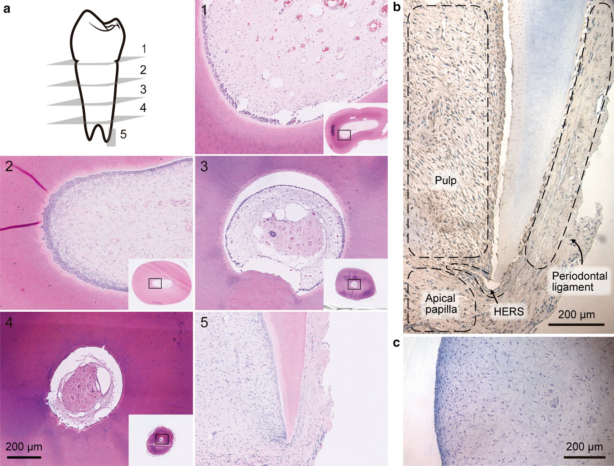
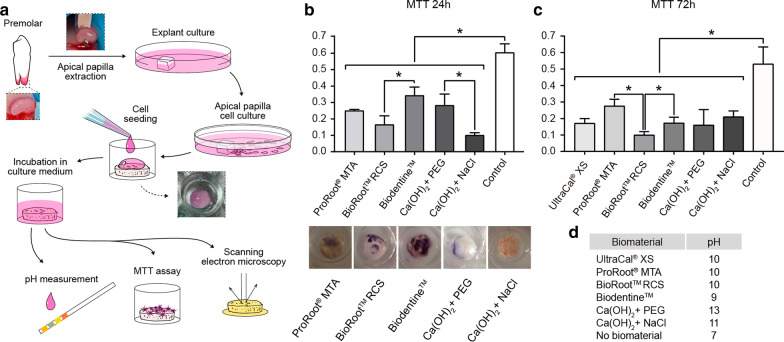
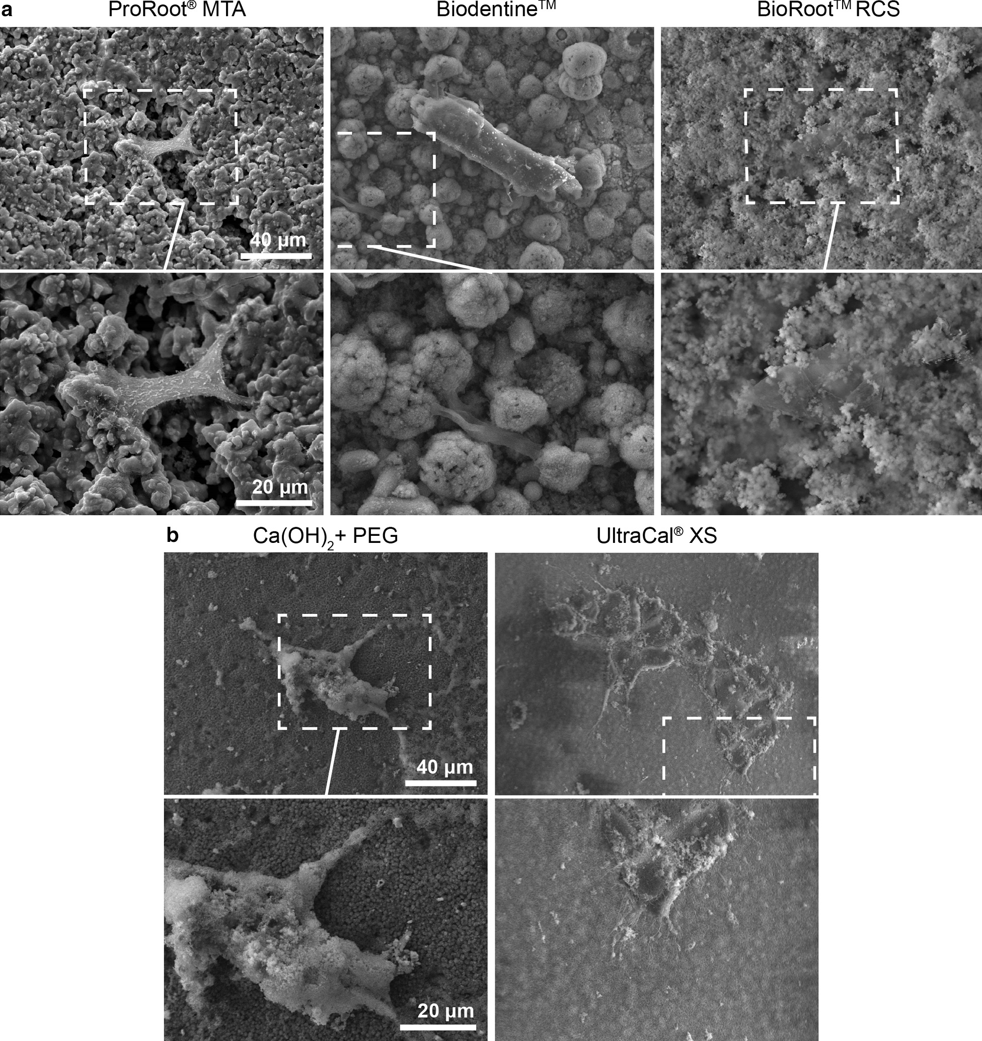
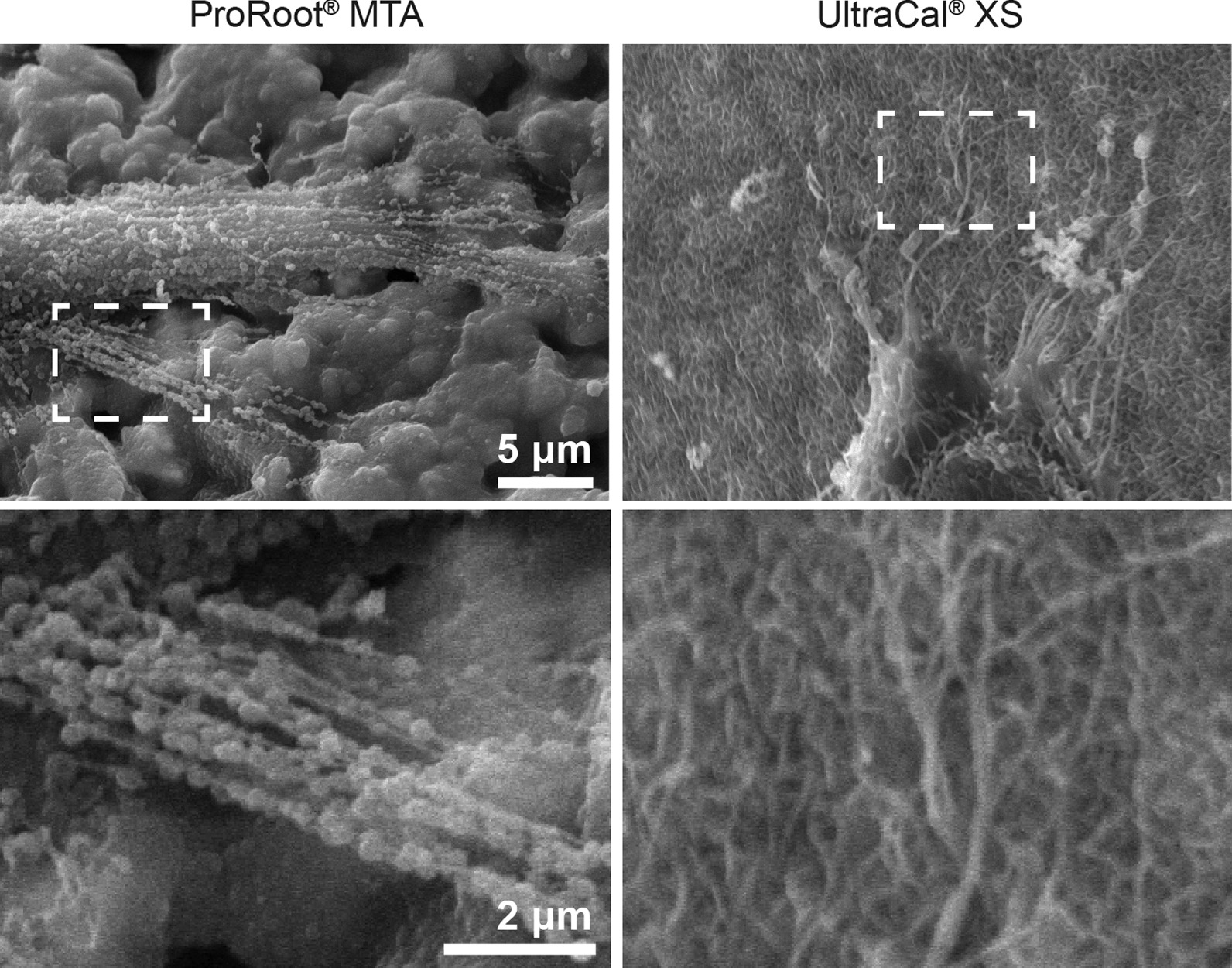
Methods: Primary cells were obtained from explants of young immature premolars. 20,000 cells adhered for 24 h over discs of Biodentine™, ProRoot®MTA, BioRoot®RCS and calcium hydroxide mixed either with sodium chloride 0.9%w/v or polyethylene glycol and UltraCal® were used to evaluate cell adhesion by scanning electron microscopy and cell viability by MTT assay.

Results: Cells adhered to ProRoot®MTA showed an increase of F-actin like protrusions, suggesting bioactivity. Cells adhered to UltraCal® show protrusion such as filopodia. On the contrary, cells adhered to BioRoot®RCS showed no signs of any cellular protrusion. Regarding viability between the materials, we found a higher percentage of viability in cells cultured over discs of Biodentine™ and ProRoot®MTA.

Conclusion: ProRoot®MTA and Biodentine™ exhibit a better cellular response of human apical papilla cells in vitro conditions compared to BioRoot® and calcium hydroxide diluted in sodium chloride.

Keywords: Apical papilla; Bioceramics; Calcium hydroxide.


Conflict of interest statement

The authors declare that they have no competing interests.

Figures

Fig. 1
Fig. 1
Histologic characteristics of immature premolar teeth. a Histological views of the longitudinal section of teeth (× 1.25 and × 7.5). b Chromogenic stain for STRO-1 in apical papilla. c Hematoxylin and eosin of extracted apical papilla (× 20) n = 5. scale bar 200um
Fig. 2
Fig. 2
Viability of apical papilla cells to calcium hydroxide and tricalcium silicate-based cements. a Cartoon showing the methodology. b MTT assay: Graph indicates average and standard error of OD at 570 nm, Photograph of formazan crystal from cells adhered to the top of ProRoot®MTA, BioRoot® RCS, Biodentine™, CaOH2 diluted in PEG and CaOH2 diluted in NaCl 0.9% w/v. Apical papilla cells adhered to a 24 well plate was used as a control. The data are shown as mean ± SD n = 6 Asterisks indicate statistically significant differences. All materials are significant differences to control cells, and between the material significant different between Biodentine™ versus BioRoot® RCS p = 0.0317 and CH diluted in NaCl is significant diffrerent to: ProRoot®MTA (p = 0.0357), Biodentine (p = 0.0357) and CaOH diluted in PEG (p = 0.0095). c Quantification of MTT assay after 72 h of adhesion to, BioRoot® RCS, Biodentine™, Ultracal, CaOH2 diluted in PEG and CaOH2 diluted in NaCl 0.9% w/v. All biomaterial were significantly different to cells adhered to plastic, ProRoot®MTA versus BioRoot® RCS p = 0.0286, Biodentine™ versus BioRoot® RCS p = 0.0286 n = 5. d Table with quantification of pH
Fig. 3
Fig. 3
Apical papilla cells adhered to calcium hydroxide and tricalcium silicate-based cement. a. Scanning electron microscopic images of apical papilla cells adhered to bioceramics: ProRoot®MTA, Biodentine™ and BioRoot® RCS, b Apical papilla cells adhered to calcium hydroxide CaOH2 dissolved in PEG and UltraCal® XS. × 700 and × 1500. n = 5
Fig. 4
Fig. 4
MTA and UltraCal® increased F-actin protrusion. A scanning electron microscopic images of apical papilla cells adhered to ProRoot®MTA, and UltraCal®. Scale bar 5 μm at × 1500. Scale bar 2 μm at × 4000. n = 5

References

    1. Nazzal H, Duggal MS. Regenerative endodontics: a true paradigm shift or a bandwagon about to be derailed? Eur Arch Paediatr Dent. 2017;18:3–15. doi: 10.1007/s40368-016-0265-5. - DOI - PMC - PubMed
    1. Brito-Júnior M, Pereira RD, Veríssimo C, Soares CJ, Faria-e-Silva AL, Camilo CC, et al. Fracture resistance and stress distribution of simulated immature teeth after apexification with mineral trioxide aggregate. Int Endod J. 2014;47:958–966. doi: 10.1111/iej.12241. - DOI - PubMed
    1. Al Ansary MAD, Day PF, Duggal MS, Brunton PA. Interventions for treating traumatized necrotic immature permanent anterior teeth: Inducing a calcific barrier & root strengthening. Dent Traumatol. 2009;25:367–379. doi: 10.1111/j.1600-9657.2009.00797.x. - DOI - PubMed
    1. Duggal M, Tong HJ, Al-Ansary M, Twati W, Day PF, Nazzal H. Interventions for the endodontic management of non-vital traumatised immature permanent anterior teeth in children and adolescents: a systematic review of the evidence and guidelines of the European Academy of Paediatric Dentistry. Eur Arch Paediatr Dent. 2017;18:139–151. doi: 10.1007/s40368-017-0289-5. - DOI - PMC - PubMed
    1. Garcia-Godoy F, Murray PE. Recommendations for using regenerative endodontic procedures in permanent immature traumatized teeth. Dent Traumatol. 2012;28:33–41. doi: 10.1111/j.1600-9657.2011.01044.x. - DOI - PubMed

Publication types

LinkOut - more resources