. 2021 Mar 9;7(1):13.
doi: 10.1038/s41421-021-00245-6.

Identification of an intraocular microbiota

Affiliations

Identification of an intraocular microbiota

Yuhua Deng et al. Cell Discov. .

Erratum in

  • Author Correction: Identification of an intraocular microbiota.
    Deng Y, Ge X, Li Y, Zou B, Wen X, Chen W, Lu L, Zhang M, Zhang X, Li C, Zhao C, Lin X, Zhang X, Huang X, Li X, Jin M, Peng GH, Wang D, Wang X, Lai W, Liang J, Li JJ, Liang Q, Yang L, Zhang Q, Li Y, Lu P, Hu X, Li X, Deng X, Liu Y, Zou Y, Guo S, Chen T, Qin Y, Yang F, Miao L, Chen W, Chan CC, Lin H, Liu Y, Lee RWJ, Wei L. Deng Y, et al. Cell Discov. 2024 May 15;10(1):51. doi: 10.1038/s41421-024-00675-y. Cell Discov. 2024. PMID: 38750045 Free PMC article. No abstract available.

Abstract

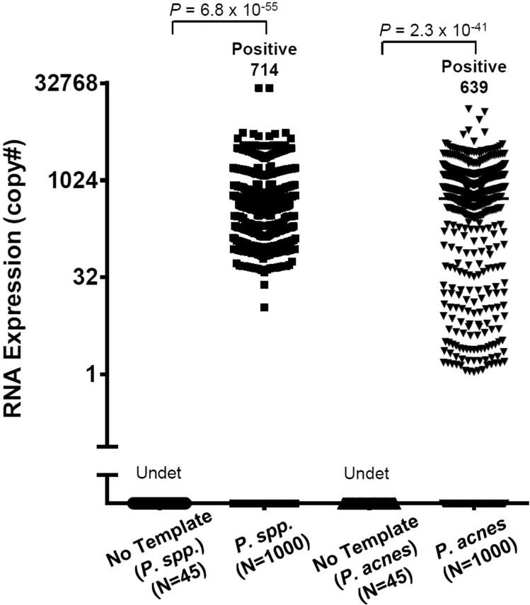
The current dogma in ophthalmology and vision research presumes the intraocular environment to be sterile. However, recent evidence of intestinal bacterial translocation into the bloodstream and many other internal organs including the eyes, found in healthy and diseased animal models, suggests that the intraocular cavity may also be inhabited by a microbial community. Here, we tested intraocular samples from over 1000 human eyes. Using quantitative PCR, negative staining transmission electron microscopy, direct culture, and high-throughput sequencing technologies, we demonstrated the presence of intraocular bacteria. The possibility that the microbiome from these low-biomass communities could be a contamination from other tissues and reagents was carefully evaluated and excluded. We also provide preliminary evidence that a disease-specific microbial signature characterized the intraocular environment of patients with age-related macular degeneration and glaucoma, suggesting that either spontaneous or pathogenic bacterial translocation may be associated with these common sight-threatening conditions. Furthermore, we revealed the presence of an intraocular microbiome in normal eyes from non-human mammals and demonstrated that this varied across species (rat, rabbit, pig, and macaque) and was established after birth. These findings represent the first-ever evidence of intraocular microbiota in humans.


Conflict of interest statement

The authors declare no competing interests.

Figures

Fig. 1
Fig. 1. Detection of P. spp. in human AH specimens.
Expression of P. spp. 16S rRNA and a P. acnes-specific gene—PPA_RS04200 (presented as the copy#) in the AH from 1000 human eyes undergoing cataract surgery was quantified using real-time PCR assays. The P value was calculated using parametric Student’s t-test. Undet, undetected.
Fig. 2
Fig. 2. Detection of intraocular bacteria in AH specimens and cultures.
a Negative staining transmission electron microscopy was used to visualize cultured P. acnes at 20,000× magnification. The negative control shows the visualization of water without AH specimen. b Negative staining transmission electron microscopy shows bacteria in minimally manipulated fresh AH specimens at 20,000× magnification. c Cultured E. coli was visualized by light microscopy. The negative control consists of sample preparation buffer without any AH inoculation. d Bacteria in cultured AH samples (examples of culture-positive and -negative samples) were visualized by light microscopy. e Representative anaerobic cultures of skin specimens and negative controls (PBS solution used to dilute specimens). f Representative anaerobic cultures of AH specimens.
Fig. 3
Fig. 3. A unique intraocular microbiota detected by metagenomic sequencing analysis.
Comparative metagenomic analysis was performed for AH, conjunctiva (CO), plasma (PL), and skin (SK) specimens from 20 patients undergoing cataract surgery. a The average percentage of human reads among total sequenced reads from AH, CO, PL, and SK samples. b Relative abundance of the major genera of bacteria in the metagenomes of AH, CO, PL, and SK samples. c The average numbers of microbial genes identified in AH, CO, PL, and SK samples. d Hierarchical clustering analysis of relative abundance of microbial metabolic pathways identified in AH, CO, PL, and SK samples. P value was calculated using ANOVA test. e Diversity of the microbial community analyzed by PCoA. The Bray Curtis distance was used and P value was calculated using PERMANOVA test.
Fig. 4
Fig. 4. The microbiota in AH from patients with cataract, AMD, and glaucoma.
a Relative abundance of the six major phyla of bacteria in the intraocular metagenomes in patients with cataract (N = 41), AMD (N = 20), or glaucoma (N = 26). b The alpha diversity (measured by Shannon index) of the intraocular metagenomes in patients with cataract, AMD, and glaucoma. The error bar represents the mean of all Shannon index within the disease group ± SEM. The statistical difference was measured between patients with cataract and each of other disease groups, respectively. Mann-Whitney U test significance levels are denoted by asterisks (*P < 0.05). c PCoA of the similarity of the intraocular metagenomes in patients with cataract (N = 30), AMD (N = 18), and glaucoma (N = 16). The Bray Curtis distance was used and P value was calculated using PERMANOVA test. d Hierarchical clustering analysis of the functional genes in the intraocular metagenomes in patients with cataract (N = 30), AMD (N = 18), and glaucoma (N = 16).
Fig. 5
Fig. 5. The intraocular metagenome in the eyes of rabbit, rat, macaque, and pig analyzed using PCoA.
a The PCoA of intraocular metagenomes in four mammals. The Bray Curtis distance was used and P value was calculated using PERMANOVA test. b The abundance of bacterial DNA in rat intraocular fluids was measured using real-time PCR assays of total 16S rDNA and represented as the relative abundance to the level of rat Actb gene. c The abundance of P. acnes DNA in rat intraocular fluids was measured using real-time PCR assays of P. acnes 16S rDNA and represented as the relative abundance to the level of rat Actb gene. The error bar represents the mean ± SEM. The statistical difference was measured using Mann-Whitney U test and the significance levels are denoted by asterisks (*P < 0.05, **P < 0.01, NS: not significant).

References

    1. Blumberg J, Kreiman G. How cortical neurons help us see: visual recognition in the human brain. J. Clin. Invest. 2010;120:3054–3063. doi: 10.1172/JCI42161. - DOI - PMC - PubMed
    1. Perez VL, Caspi RR. Immune mechanisms in inflammatory and degenerative eye disease. Trends Immunol. 2015;36:354–363. doi: 10.1016/j.it.2015.04.003. - DOI - PMC - PubMed
    1. Rooks MG, Garrett WS. Gut microbiota, metabolites and host immunity. Nat. Rev. Immunol. 2016;16:341–352. doi: 10.1038/nri.2016.42. - DOI - PMC - PubMed
    1. Manfredo Vieira S, et al. Translocation of a gut pathobiont drives autoimmunity in mice and humans. Science. 2018;359:1156–1161. doi: 10.1126/science.aar7201. - DOI - PMC - PubMed
    1. Asquith M, et al. A study of microbial translocation inan animal model of spondyloarthritis (abstract) Ann. Rheum. Dis. 2018;77:618.

LinkOut - more resources