Comparative Study
. 2021 Mar 9;11(1):5486.
doi: 10.1038/s41598-021-84762-0.

Amplification of potential thermogenetic mechanisms in cetacean brains compared to artiodactyl brains

Affiliations
Comparative Study

Amplification of potential thermogenetic mechanisms in cetacean brains compared to artiodactyl brains

Paul R Manger et al. Sci Rep. .

Abstract

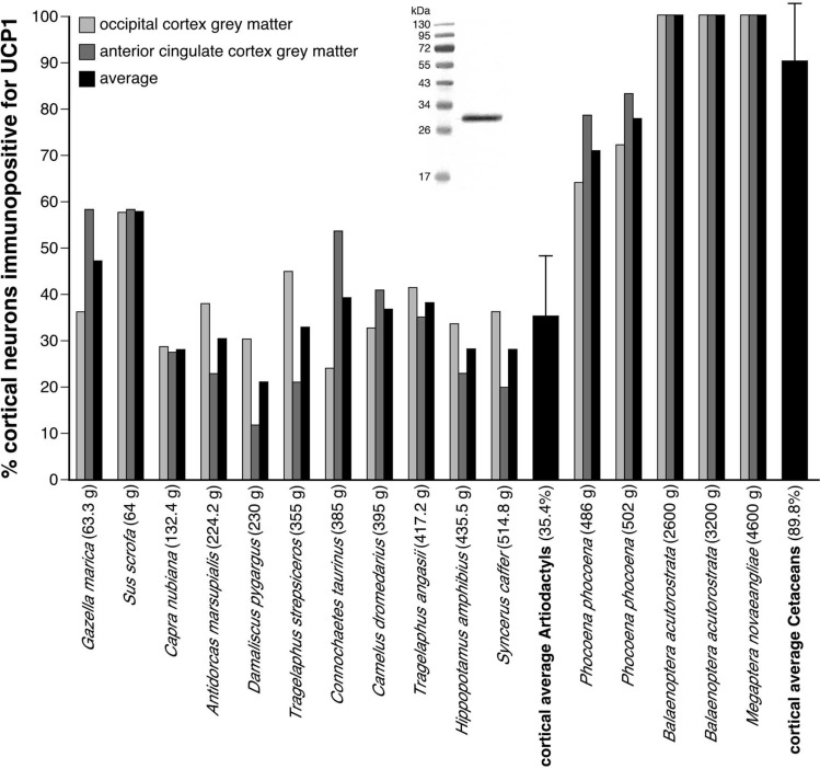
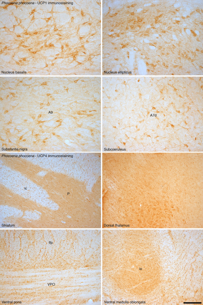
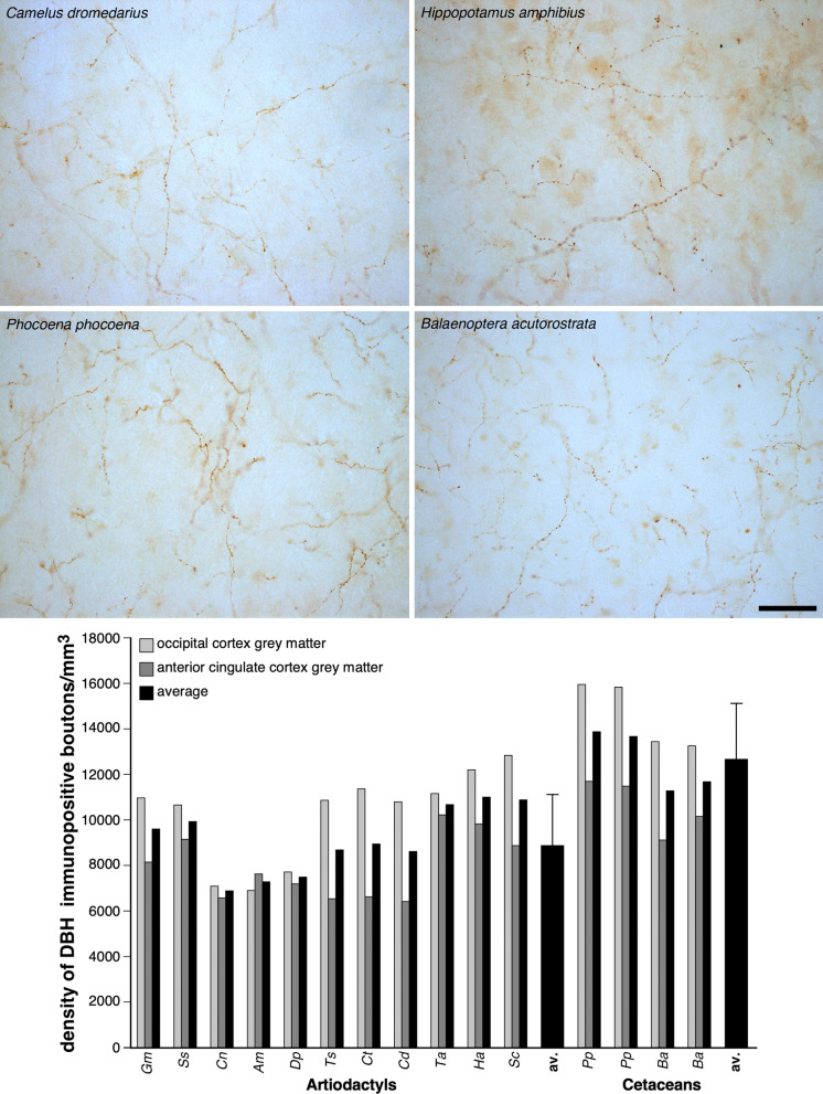
To elucidate factors underlying the evolution of large brains in cetaceans, we examined 16 brains from 14 cetartiodactyl species, with immunohistochemical techniques, for evidence of non-shivering thermogenesis. We show that, in comparison to the 11 artiodactyl brains studied (from 11 species), the 5 cetacean brains (from 3 species), exhibit an expanded expression of uncoupling protein 1 (UCP1, UCPs being mitochondrial inner membrane proteins that dissipate the proton gradient to generate heat) in cortical neurons, immunolocalization of UCP4 within a substantial proportion of glia throughout the brain, and an increased density of noradrenergic axonal boutons (noradrenaline functioning to control concentrations of and activate UCPs). Thus, cetacean brains studied possess multiple characteristics indicative of intensified thermogenetic functionality that can be related to their current and historical obligatory aquatic niche. These findings necessitate reassessment of our concepts regarding the reasons for large brain evolution and associated functional capacities in cetaceans.


Conflict of interest statement

The authors declare no competing interests.

Figures

Figure 1
Figure 1
UCP1 immunostaining in cetartiodactyl cerebral cortex. Photomicrographs of Nissl stained (purple colored images) and UCP1-immunostained (brown colored images) cortical sections in a range of artiodactyl (two left columns) and cetacean species (two right columns). Note in all cases the presence of UCP1-immunostained cortical neurons, but in the artiodactyls these are limited to the lower layers of the cortex, while almost all cortical neurons from all layers are immunopositive in the cetaceans. Scale bar in the UCP1-immunostained section of Connochaetes taurinus equals 500 µm and applies to all artiodactyl images. Scale bar in the UCP1-immunostained section of Phocoena phocoena equals 100 µm and applies to both images. Scale bar in the UCP1-immunostained section of Balaenoptera acutorostrata equals 500 µm and applies to both images. Scale bar in the UCP1-immunostained stained section of Megaptera novaeangliae equals 250 µm and applies to both images.
Figure 2
Figure 2
Quantification of UCP1 immunostaining in cetartiodactyl cerebral cortex. Graphical representation of the results of the stereological analysis of the percentage of cortical neurons immunopositive for UCP1 in the occipital and anterior cingulate cortices of the species studied. For each species the brain mass is given in grams next to the name on the x-axis. Note that the average percentage of cortical neurons immunopositive for UCP1 in the artiodactyls studied was 35.4%, while in the cetaceans studied it was 89.9% (Table 1, error bars on average bars represent one standard deviation). The Western immunoblot in the middle of the graph shows the specificity of the UCP1 antibody to brown fat taken from a laboratory rat (see Figure S6 for full-length unedited Western immunoblot).
Figure 3
Figure 3
UCP1 and UCP4 immunostaining in non-cortical regions of the harbor porpoise brain. In addition to examining the expression of UCP1 and UCP4 in the cerebral cortex of the brain of the harbor porpoise, we examined several other brain regions. In all regions we found neurons with distinct UCP1 immunoreactivity, with an intracellular staining pattern similar to that observed in the neurons of the cerebral cortex. The photomicrographs shown here depict UCP1 immunostaining in various non-cortical regions of the harbour porpoise brain, including the nucleus basalis, nucleus ellipticus, the substantia nigra (A9), and the nucleus subcoeruleus (A7d, its diffuse region). In addition, in all regions we found glial cells with distinct immunoreactivity to the UCP4 antibody. Interestingly, the density of glial cells immunopositive for UCP4 appears higher in the white matter than in the grey matter, reflecting the same proportional distribution of stained glia as when comparing the white and grey matter of the cerebral cortex. The photomicrographs shown here depict UCP4 immunostaining in various non-cortical regions of the harbour porpoise brain, including the striatum (P—putamen, ic—internal capsule), dorsal thalamus, ventral pons (VPO—ventral pontine nucleus, lfp—longitudinal fasciculus of pons) and the ventral medulla oblongata (io—portion of inferior olivary nuclear complex). Scale bar = 250 µm and applies to all.
Figure 4
Figure 4
UCP4 Western blotting and immunostaining in cetartiodactyl cerebral cortex. While UCP4 was present in the cortical grey and white matter of all species, as evidenced in the Western blot at the top of the panel from the occipital cortex of all species studied, it was only found to be immunolocalized to glial cells in the cetaceans (See Figure S6 for full-length unedited Western immunoblots). Photomicrographs of Nissl-stained (purple colored images) and UCP4-immunostained (brown colored images) from cortical and subcortical white matter sections in a range of cetacean species. Note the presence of UCP4-immunoreactivity in approximately 30% of glial cells in the cerebral cortex and approximately 60% of glial cells in the white matter in all cetacean species (Table 1). The scale bar in the UCP4-immunostained section of Megaptera novaeangliae—white matter, equals 100 µm and applies to all photomicrographs. Pp—harbor porpoise, Phocoena phocoena; Ba—minke whale, Balaenoptera acutorostrata; Mn—humpback whale, Megaptera novaeangliae; Ct—blue wildebeest, Connochaetes taurinus; Ts—greater kudu, Tragelaphus strepsiceros; Dp—blesbok, Damaliscus pygargus; Ss—domestic pig, Sus scrofa; Cd—dromedary camel, Camelus dromedarius; Gm—sand gazelle, Gazella marica; Am—springbok, Antidorcas marsupialis; Sc—African buffalo, Syncerus caffer; Ha—river hippopotamus, Hippopotamus amphibius; Cn—Nubian ibex, Capra nubiana; Ta—nyala, Tragelaphus angasii; Rn—laboratory rat, Rattus norvegicus.
Figure 5
Figure 5
Quantification of noradrenergic bouton density in cetartiodactyl cerebral cortex. Photomicrographs of dopamine-ß-hydroxylase (DBH)-immunostained axonal boutons in the occipital cortical grey matter of Camelus dromedarius, Hippopotamus amphibius, and Phocoena phocoena, and the anterior cingulate cortical grey matter of Balaenoptera acutorostrata. The scale bar = 50 µm and applies to all photomicrographs. Note the higher density of the DBH-immunoreactive boutons in the cortical grey matter of cetaceans compared to the artiodactyls as confirmed with stereological analysis (see the graph below the photomicrographs), showing that the density of DBH-immunoreactive boutons in the cortical grey matter of cetaceans is, on average, 1.4 times higher than that observed in artiodactyls (Table 1, error bars on average bars represent one standard deviation). Gm—sand gazelle, Gazella marica; Ss—domestic pig, Sus scrofa; Cn—Nubian ibex, Capra nubiana; Am—springbok, Antidorcas marsupialis; Dp—blesbok, Damaliscus pygargus; Ts—greater kudu, Tragelaphus strepsiceros; Ct—blue wildebeest, Connochaetes taurinus; Cd—dromedary camel, Camelus dromedarius; Ta—nyala, Tragelaphus angasii; Ha—river hippopotamus, Hippopotamus amphibius; Sc—African buffalo, Syncerus caffer; av. —average; Pp—harbor porpoise, Phocoena phocoena; Ba—minke whale, Balaenoptera acutorostrata.

References

    1. Marino L, et al. Cetaceans have complex brains for complex cognition. PLoS Biol. 2007;5:e139. doi: 10.1371/journal.pbio.0050139. - DOI - PMC - PubMed
    1. Marino L, et al. A claim in search of evidence: reply to Manger’s thermogenesis hypothesis of cetacean brain structure. Biol. Rev. 2008;83:417–440. - PubMed
    1. Connor RC. Dolphin social intelligence: complex alliance relationships in bottlenose dolphins and a consideration of selective environments for extreme brain size evolution in mammals. Philos. Trans. R. Soc. B. 2007;362:587–602. doi: 10.1098/rstb.2006.1997. - DOI - PMC - PubMed
    1. Kesarev VS. The inferior brain of the dolphin. Soviet Sci. Rev. 1971;1:52–58.
    1. Nikolskaya KA. Evolutionary aspects of intellect in vertebrates: Can intellect be a factor confining choice of the habitat? Invest. Russ. 2005;8:1442–1500.

Publication types

Substances