. 2021 May;28(5):265-276.
doi: 10.1038/s41434-021-00239-9. Epub 2021 Mar 10.

Functional expression of complement factor I following AAV-mediated gene delivery in the retina of mice and human cells

Affiliations

Functional expression of complement factor I following AAV-mediated gene delivery in the retina of mice and human cells

Anna K Dreismann et al. Gene Ther. 2021 May.

Abstract

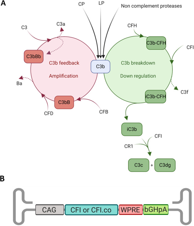
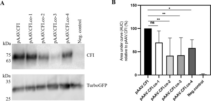
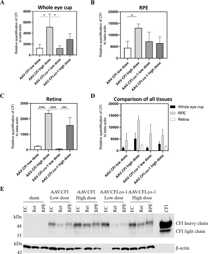
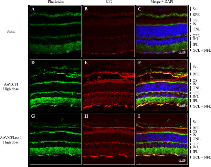
Dry age-related macular degeneration (AMD) is characterised by loss of central vision and currently has no approved medical treatment. Dysregulation of the complement system is thought to play an important role in disease pathology and supplementation of Complement Factor I (CFI), a key regulator of the complement system, has the potential to provide a treatment option for AMD. In this study, we demonstrate the generation of AAV constructs carrying the human CFI sequence and expression of CFI in cell lines and in the retina of C57BL/6 J mice. Four codon optimised constructs were compared to the most common human CFI sequence. All constructs expressed CFI protein; however, most codon optimised sequences resulted in significantly reduced CFI secretion compared to the non-optimised CFI sequence. In vivo expression analysis showed that CFI was predominantly expressed in the RPE and photoreceptors. Secreted protein in vitreous humour was demonstrated to be functionally active. The findings presented here have led to the formulation of an AAV-vectored gene therapy product currently being tested in a first-in-human clinical trial in subjects with geographic atrophy secondary to dry AMD (NCT03846193).


Conflict of interest statement

AKD and JPH are employed by Gyroscope Therapeutics Ltd, EO was employed by Gyroscope Therapeutics Ltd while engaged in the research project, PJL and REM are consultants to Gyroscope Therapeutics Ltd, MEM and ARB have no conflict of interest. AKD, JPH, PJL and REM have stock options in Gyroscope Therapeutics Limited.

Figures

Fig. 1
Fig. 1. Schematic diagram of the role of CFI in alternative pathway regulation and AAV design.
A CFI is a key regulator of the alternative pathway and degrades C3b into iC3b and C3dg in the presence of cofactor. Figure adapted with permission from [12]. B Schematic diagram of AAV2 backbone coding sequence. AAV = adeno-associated virus, bGHpA = bovine growth hormone poly adenylation sequence, bp = base pair, C3 = Complement C3, CAG = chicken b-actin promoter with a CMV enhancer and a β-globin intron, CFB = complement factor B, CFD = complement factor D, CFH = complement factor H, CFI = complement factor I, CR1 = Complement Receptor 1, WPRE = Woodchuck hepatitis virus post-transcriptional regulator element. Biorender.com was used to generate Fig. 1A, and B.
Fig. 2
Fig. 2. Immunoblot analysis of supernatants and cell lysates of pAAV.CFI, pAAV.CFI.co and pCMV.GFP co-transfected ARPE-19 cells.
A Upper immunoblot: Supernatants of transiently transfected cells. CFI and codon-optimised CFI were detected with mouse anti-human CFI. Plasmid backbone containing AAV sequences without transgene served as a negative control (representative result). Supernatant was loaded non-reduced to prevent separation of heavy and light chain. Lower immunoblot: Lysate of transiently transfected cells. GFP was detected with anti-turbo-GFP and implies that there were not large differences in transfection efficiency between the groups. B Quantification of CFI expression immunoblots by densitometry analysis using ImageJ software. Equal volumes were loaded in each experiment and summary data from 7 independent experiments is shown. Results are presented as relative density of CFI signal relative to signal intensity of AAV.CFI.wt. Data are shown as mean + standard deviation. AAV = adeno-associated virus, CFI = complement factor I, co = codon optimised. Asterisks represent significant differences between codon optimised constructs and the most common CFI sequence (reference) analysed by one-way ANOVA and Dunn’s multiple comparison test: * = P < 0.05, ** = P < 0.01, ns = not significant.
Fig. 3
Fig. 3. In vivo expression of CFI RNA and protein.
AD RT-qPCR to analyse gene expression of AAV.CFI and AAV.CFI.co-1 (n = 3 per condition) of whole eye cup (A), RPE (B), retina (C), and a comparison of all tissues analysed (D). RPE samples generated more CFI mRNA than retina (p < 0.0001) and multiple comparisons revealed significant differences between injection materials, which are described in the text. Data are shown as mean + standard deviation. E Immunoblot of whole eye cups, retina, and RPE of mice injected with AAV.CFI.wt or AAV.CFI.co-1, samples of three mice per condition were pooled for analysis. β-actin staining was performed as a loading control. AAV = adeno-associated virus, CFI = complement factor I, co = codon optimised, ec = eye cup, ret = retina, RPE = retinal pigment epithelium. Asterisks represent significant differences between constructs analysed by one-way ANOVA with Tukey’s multiple comparison: * = P < 0.05, ** = P < 0.01, *** = P < 0.001, **** = P < 0.0001.
Fig. 4
Fig. 4. Immunohistological analysis of retinal sections of AAV.CFI and AAV.CFI.co-1 injected mouse eyes.
10 µm retinal sections of sham (AC), AAV.CFI (DF) or AAV.CFI.co-1 (GI) injected mice stained with anti-phalloidin, DAPI, and anti-CFI to demonstrate that CFI protein localises to the RPE cell layer and photoreceptors. AAV = adeno-associated virus, CFI = complement factor I, co = codon optimised, GCL = ganglion cell layer, IS = inner segment of photoreceptors, NFL = nerve fibre layer; ONL = outer nuclear layer, OPL = outer plexiform layer, OS = outer segment of photoreceptors, RPE = retinal pigment epithelium, Scl = Sclera. Magnification: ×20.
Fig. 5
Fig. 5. Immunohistological analysis retinal sections of sham and AAV.CFI injected mouse eyes – higher magnification of RPE.
Immunohistological analysis retinal sections of sham (A-C) and AAV.CFI (D-F) injected mouse eyes. Retinal sections were double labelled with phalloidin to stain actin filaments (A, D) and CFI (B, E). Nuclei were stained with DAPI and are shown in merge (C, F). Vesicular staining is depicted with arrows, RPE microvilli are highlighted with asterisk. AAV = adeno-associated virus, BrM: Bruch’s membrane, CFI = complement factor I, Chr: choriocapillaris, IPM: inter photoreceptor matrix, RPE: retinal pigment epithelium, Scl: sclera. Magnification: 189x.
Fig. 6
Fig. 6. Immunohistological analysis of whole mounted RPE of AAV.CFI and AAV.CFI.co-1 injected mouse eyes.
Whole mounted RPE of sham (A-C), AAV.CFI (D-F) and AAV.CFI.co-1 (G-I) injected mice was stained with anti-phalloidin and anti-CFI. CFI is highly expressed in RPE cells of AAV.CFI.wt and AAV.CFI.gs treated mice. Abbreviations: AAV = adeno-associated virus, CFI = complement factor I, co = codon optimised. Magnification: 40x.
Fig. 7
Fig. 7. Ex vivo demonstration of CFI functional activity by a cofactor assay following subretinal administration.
C3b, the substrate, is incubated with CFH as a cofactor; in the absence of CFI no degradation occurs (lane ‘C3b + CFH’, assay negative control). As soon as C3b is incubated with CFH and CFI, degradation of C3b into iC3b occurs, visible as 68 and 43 kDa bands (lane ‘C3b + CFH + CFI, assay positive control). C3 and CFH are incubated with ‘wash’ sample derived from either sham injected or AAV.CFI injected mice as a source of CFI and degradation of the C3 alpha band into the iC3b bands is observed. Abbreviations: AAV = adeno-associated virus, C3 = complement C3, CFI = complement factor I, CFH = complement factor H.

References

    1. Heesterbeek TJ, Lorés-Motta L, Hoyng CB, Lechanteur YT, den Hollander AI. Risk factors for progression of age-related macular degeneration. Ophthal Physiol Optics. 2020;40:140–70. doi: 10.1111/opo.12675. - DOI - PMC - PubMed
    1. Lechner J, Chen M, Hogg RE, Toth L, Silvestri G, Chakravarthy U, et al. Higher plasma levels of complement C3a, C4a and C5a increase the risk of subretinal fibrosis in neovascular age-related macular degeneration. Immunity Ageing. 2016;13:1–9. doi: 10.1186/s12979-016-0060-5. - DOI - PMC - PubMed
    1. Reynolds R, Hartnett ME, Atkinson JP, Giclas PC, Rosner B, Seddon JM. Plasma complement components and activation fragments: associations with age-related macular degeneration genotypes and phenotypes. Investig Ophthalmol Vis Sci. 2009;50:5818–27. doi: 10.1167/iovs.09-3928. - DOI - PMC - PubMed
    1. Scholl HP, Issa PC, Walier M, Janzer S, Pollok-Kopp B, Börncke F, et al. Systemic complement activation in age-related macular degeneration. PLoS ONE. 2008;3:e2593. doi: 10.1371/journal.pone.0002593. - DOI - PMC - PubMed
    1. Fritsche LG, Igl W, Bailey JN, Grassmann F, Sengupta S, Bragg-Gresham JL, et al. A large genome-wide association study of age-related macular degeneration highlights contributions of rare and common variants. Nat Genet. 2016;48:134–43. doi: 10.1038/ng.3448. - DOI - PMC - PubMed

Publication types

Substances

Associated data