. 2021 Mar 22;17(3):e1009446.
doi: 10.1371/journal.pgen.1009446. eCollection 2021 Mar.

Critical role of the BAF chromatin remodeling complex during murine neural crest development

Affiliations

Critical role of the BAF chromatin remodeling complex during murine neural crest development

Kathleen Wung Bi-Lin et al. PLoS Genet. .

Abstract

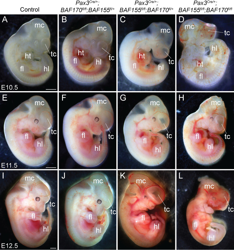
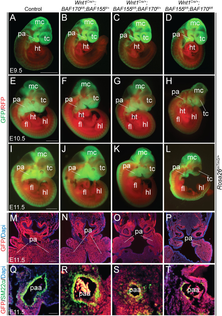
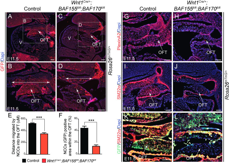
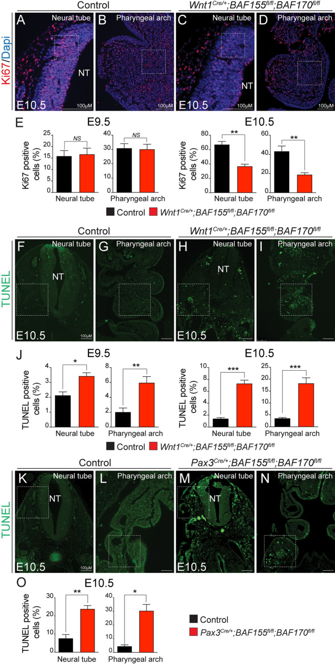
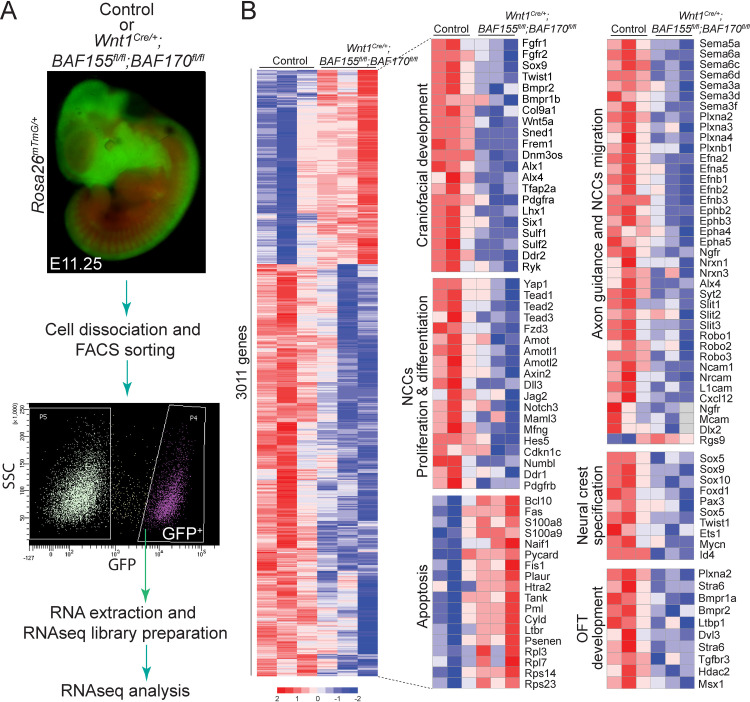
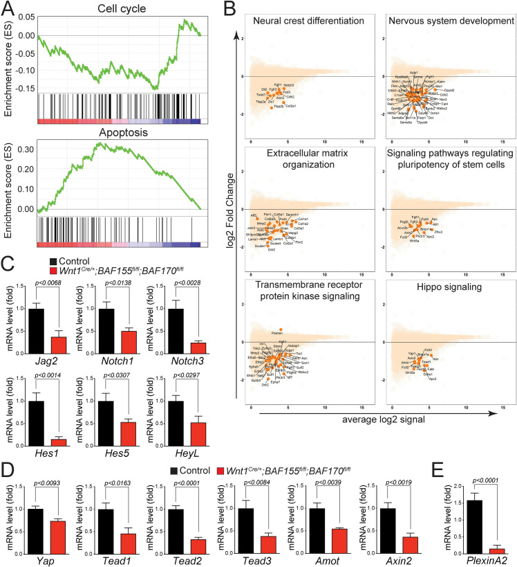
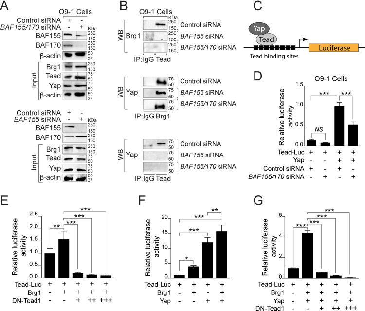
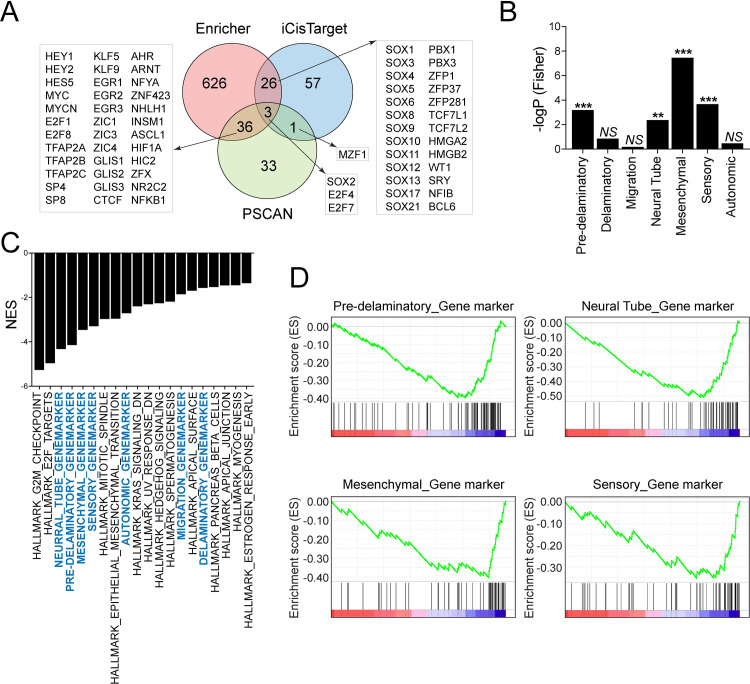
The BAF complex plays an important role in the development of a wide range of tissues by modulating gene expression programs at the chromatin level. However, its role in neural crest development has remained unclear. To determine the role of the BAF complex, we deleted BAF155/BAF170, the core subunits required for the assembly, stability, and functions of the BAF complex in neural crest cells (NCCs). Neural crest-specific deletion of BAF155/BAF170 leads to embryonic lethality due to a wide range of developmental defects including craniofacial, pharyngeal arch artery, and OFT defects. RNAseq and transcription factor enrichment analysis revealed that the BAF complex modulates the expression of multiple signaling pathway genes including Hippo and Notch, essential for the migration, proliferation, and differentiation of the NCCs. Furthermore, we demonstrated that the BAF complex is essential for the Brg1-Yap-Tead-dependent transcription of target genes in NCCs. Together, our results demonstrate an important role of the BAF complex in modulating the gene regulatory network essential for neural crest development.


Conflict of interest statement

The authors have declared that no competing interests exist.

Figures

Fig 1
Fig 1. Pax3Cre-mediated deletion of BAF155/170 leads to severe craniofacial defects and embryonic lethality.
(A-L) Phenotypic defects in control, BAF170-deficient (Pax3Cre/+;BAF170fl/fl;BAF155fl/+), BAF155-deficient (Pax3Cre/+;BAF155fl/fl;BAF170fl/+) and BAF155/170-deficient (Pax3Cre/+;BAF155fl/fl;BAF170fl/fl) embryos. Sagittal view of E10.5 (A-D), E11.5 (E-H), and E12.5 (I-L) embryos. Severe craniofacial defects in BAF155/170-deficient embryos (D, H, and L) when compared with their littermate controls (A, E, and I). Among BAF155/170-deficient embryos, 9/9 showed forebrain defects, evident by improper development of the telencephalon (D, H, and L). BAF155-deficient embryos (C, G, and K) also demonstrated abnormal development of craniofacial tissues and telencephalon when compared with their littermate controls (A, E, and I). No obvious morphological defects were observed in BAF170-deficient embryos at the embryonic stages analyzed (B, F, and J). n = 3–10 embryos were analyzed for each genotype at each given embryonic stage. Scale bars 200μM (A-D) and 500μM (E-L) fl, forelimb; hl, hindlimb; ht, heart; mc, metencephalon; tc, telencephalon.
Fig 2
Fig 2. Wnt1Cre-mediated neural crest-specific deletion of BAF155/170 leads to severe craniofacial defects and embryonic lethality.
(A-H) Phenotypic defects in control, BAF170-deficient (Wnt1Cre/+;BAF170fl/fl;BAF155fl/+), BAF155-deficient (Wnt1Cre/+;BAF155fl/fl;BAF170fl/+) and BAF155/170-deficient (Wnt1Cre/+;BAF155fl/fl;BAF170fl/fl) embryos. Sagittal view of E9.5 (A-D), E10.5 (E-H), and E11.5 (I-L) embryos. No obvious developmental defects were observed at E9.5 between different genotypes (A-D). Developmental defects in neural-crest derived tissues, including craniofacial defects in BAF155/170-deficient embryos (H and L) when compared with their littermate controls (E and I). Among BAF155/170-deficient embryos, 7/10 showed severe craniofacial defects. Hemorrhage in the forebrain (telencephalon) of E11.5 BAF155/170-deficient embryos (3/10 embryos) (L). Craniofacial defects are also present in BAF155-deficient embryos (6/10 showed craniofacial deformities, enlarged blood vessels) but less severe than the BAF155/170-deficient embryos (K). No obvious morphological defects were observed in BAF170-deficient embryos (10/10) at the embryonic stages analyzed (F and J). n = 4–10 embryos were analyzed for each genotype at each given embryonic stage. Scale bars 200μM (A-H) and 500μM (E-H). fl, forelimb; hl, hindlimb; ht, heart; mc, metencephalon; tc, telencephalon.
Fig 3
Fig 3. Defective NCCs differentiation in the pharyngeal arch arteries of neural crest-specific BAF155/170 knockout embryos.
(A-H) Lineage tracing of Wnt1Cre/+-derived NCCs using R26mTmG/+ reporter at E9.5 (A-D), E10.5 (E-H) and E11.5 (I-L). Control, BAF170-deficient (Wnt1Cre/+;BAF170fl/fl;BAF155fl/+), BAF155-deficient (Wnt1Cre/+;BAF155fl/fl;BAF170fl/+) and BAF155/170-deficient (Wnt1Cre/+;BAF155fl/fl;BAF170fl/fl) embryos were analyzed by GFP and RFP immunofluorescence (n = 4 each genotype). Merged GFP/RFP images are presented here (A-L). Scale bars 200μM (A-H) and 500μM (I-L). (M-P) Anti-GFP immunostaining on the frontal sections of E11.5 control (M), BAF170-deficient (N), BAF155-deficient (O), and BAF155/170-deficient (P) embryos showing neural crest-derived cells in the pharyngeal arches (n = 3 each genotype). Scale bar 75μM (M-P). (Q-T) Anti-SM22α immunostaining on the frontal sections of E11.5 control (Q), BAF170-deficient (R), BAF155-deficient (S), and BAF155/170-deficient (T) embryos showing differentiation of neural crest-derived cells into the smooth muscle of the 4th pharyngeal arch artery (n = 3 each genotype). Scale bar 50μM (Q-T). fl, forelimb; hl, hindlimb; ht, heart; mc, metencephalon; pa, pharyngeal arch; paa, pharyngeal arch artery; tc, telencephalon.
Fig 4
Fig 4. Impaired NCCs contribution to the developing cardiac outflow tract (OFT) of the neural crest-specific BAF155/170 mutant embryos.
(A-D) Lineage tracing of Wnt1Cre/+-derived cardiac NCCs using R26mTmG/+ reporter in E11.5 control and BAF155/170-deficient (Wnt1Cre/+;BAF155fl/fl;BAF170fl/fl) embryos. Anti-GFP immunostaining on the sagittal sections showing recruited cardiac NCCs in the OFT of control (A-B) and BAF155/170-deficient (C-D) embryos. Nuclei were visualized by Dapi staining (blue). Double head arrows show the distance of migrated cardiac NCCs in the OFT (B and D). (E) Quantification of distance migrated by NCCs into the OFT (n = 3). (F) Quantification of GFP+ neural crest cell area in the OFT (n = 3). (G-H) anti-PlexinA2 immunostaining and Dapi counterstaining on the OFT sections of control (E) and BAF155/170-deficient (F) embryos. (I-J) anti-SM22α immunostaining and Dapi counterstaining on OFT sections of control (G) and BAF155/170-deficient (H) embryos. (K-L) Double immunostaining for GFP and SM22α on OFT sections of control (K) and BAF155/170-deficient (L) embryos. (n = 3–4 each genotype). Values are reported as means ± SEM (*P < 0.05, **P < 0.01, ***P < 0.001; NS, not significant). Scale bar 75μM (A-L). OFT, outflow tract; V, ventricle.
Fig 5
Fig 5. Increased apoptosis and decreased cell proliferation in BAF155/170Wnt1-CKO embryos.
(A-D) Anti-Ki67 immunostaining and Dapi counterstaining on frontal sections with neural tube and pharyngeal arch area of E9.5 and E10.5 control and BAF155/170-deficient (Wnt1Cre/+;BAF155fl/fl;BAF170fl/fl) embryos. (E) Quantification of cell proliferation was calculated as the ratio of Ki67-positive cells to the total number of cells as determined by Dapi counterstaining in the defined area of the neural tube and pharyngeal arch (E). (n = 3–4 each genotype). (F-J) TUNEL assay and quantification was performed on E9.5 and E10.5 control and BAF155/170-deficient (Wnt1Cre/+;BAF155fl/fl;BAF170fl/fl) sections. (n = 3–4 each genotype). (K-O) TUNEL assay and quantification was performed on E10.5 control and BAF155/170-deficient (Pax3Cre/+;BAF155fl/fl;BAF170fl/fl) sections. (n = 3–4 each genotype). Values are reported as means ± SEM (*P < 0.05, **P < 0.01, ***P < 0.001; NS, not significant).
Fig 6
Fig 6. Gene expression changes in isolated NCCs due to BAF155/170 deletion.
(A) Experimental design for RNAseq analysis. Single-cell suspensions were prepared from lineage traced control (Wnt1Cre/+:BAF155flox/+:BAF170flox/+:Rosa26mTmG/+) and BAF155/170-deficient (Wnt1Cre/+:BAF155flox/flox:BAF170flox/flox:Rosa26mTmG/+) embryos for Fluorescence-activated cell sorting (FACS). NCCs that were positive for GFP were collected from control and BAF155/170-deficient embryos for RNA isolation and subsequently for library preparation and RNAseq analysis. (B) Heat map of differentially expressed transcripts (3011 genes) from RNAseq analysis of sorted control and BAF155/170-deficient NCCs. Genes regulating different aspects of neural crest biology including proliferation and differentiation were significantly down-regulated in BAF155/170-deficient NCCs compared to the controls. In contrast, genes regulating apoptosis were up-regulated in BAF155/170-deficient NCCs compared to controls. (n = 3 each genotype).
Fig 7
Fig 7. Signaling pathways regulating NCCs proliferation and differentiation are downregulated in BAF155/170 mutants.
(A) GSEA analysis showed that genes controlling cell cycle progression were negatively enriched while genes controlling apoptosis were positively enriched. (B) MA plots showing down-regulation of genes associated with critical pathways essential for NCCs proliferation, migration, and differentiation. (C-D) Relative mRNA levels of Notch (C) and Hippo (D) pathway genes in control and BAF155/170-deficient (Wnt1Cre/+;BAF155fl/fl;BAF170fl/fl) sorted NCCs. n = 3–4 each genotype. (E) Relative mRNA levels of PlexinA2 in control and BAF155/170-deficient (Wnt1Cre/+;BAF155fl/fl;BAF170fl/fl) sorted NCCs. Values are reported as means ± SEM (*P < 0.05, **P < 0.01, ***P < 0.001; NS, not significant).
Fig 8
Fig 8. BAF complex is essential for Brg1-Yap-Tead-dependent transcription of target genes in NCCs.
(A) Western blot for BAF155 and BAF170 using protein extracts from O9-1 cells transfected with control, BAF155 or BAF155/170 siRNA for 72 hours. Knockdown efficiency was evaluated by BAF155 and BAF170 western blots. Input blots for Brg1, Tead, and Yap together with β-actin controls are presented. (B) Cell extracts from control siRNA, BAF155 siRNA, and BAF155/170 siRNA transfected cells were prepared for immunoprecipitation (IP) using IgG control and anti-pan Tead antibody followed by western blotting (WB) for Brg1. IP was performed using IgG control and anti-Brg1 antibody followed by WB for Yap. Similarly, IP was performed using IgG control and anti-pan Tead antibody followed by WB for Yap. Representative blots are presented. (C) Schematic illustration of the Tead luciferase reporter construct with Tead and Yap. (D) Results of normalized luciferase reporter assays in O9-1 cells with Tead-luciferase reporter (Tead-Luc) in the presence of control siRNA or BAF155/170 siRNA. (E) Results of normalized luciferase reporter assays in HEK293T cells with Tead-Luc reporter in the presence of Brg1 alone or in combination with dominant-negative Tead1 (DN-Tead1). (F) Results of normalized luciferase reporter assays in HEK293T cells with Tead-Luc reporter in the presence of Brg1, Yap alone, or both. (G) Results of normalized luciferase reporter assays in HEK293T cells with Tead-Luc reporter in the presence of Brg1 and Yap in combination with different doses of DN-Tead1. Values are reported as means ± SD (*P < 0.05, **P < 0.01, ***P < 0.001; NS, not significant).
Fig 9
Fig 9. Altered transcriptional network due to BAF155/170 deletion in NCCs.
(A) Venn diagram on transcription factors which show significant enrichments in the BAF155/170-deficient (Wnt1Cre/+;BAF155fl/fl;BAF170fl/fl) NCCs compared to the controls. RNAseq data were analyzed via Enrichr, Pscan, and iCisTarget. (B) Fisher’s test showing enrichment of differentially expressed neural crest genes in scRNAseq clusters identified by Soldato et al., 2019. (C) GSEA analysis showed that custom pathways (with cluster genes identified by Soldato et al., 2019, highlighted with blue color) are retrieved as top GSEA hits (top 20 pathways are presented). Pathways that are significant in the Fisher test occupy a higher rank in the GSEA output. (D) GSEA was performed to analyze the custom pathways affected by BAF155/170 deletion in NCCs.

References

    1. Feiner L, Webber AL, Brown CB, Lu MM, Jia L, Feinstein P, et al.. Targeted disruption of semaphorin 3C leads to persistent truncus arteriosus and aortic arch interruption. Development. 2001;128(16):3061–70. . - PubMed
    1. Gao Z, Kim GH, Mackinnon AC, Flagg AE, Bassett B, Earley JU, et al.. Ets1 is required for proper migration and differentiation of the cardiac neural crest. Development. 2010;137(9):1543–51. Epub 2010/03/31. 10.1242/dev.047696 - DOI - PMC - PubMed
    1. Tang L, Nogales E, Ciferri C. Structure and function of SWI/SNF chromatin remodeling complexes and mechanistic implications for transcription. Prog Biophys Mol Biol. 2010;102(2–3):122–8. Epub 2010/05/25. 10.1016/j.pbiomolbio.2010.05.001 - DOI - PMC - PubMed
    1. Mashtalir N, D’Avino AR, Michel BC, Luo J, Pan J, Otto JE, et al.. Modular Organization and Assembly of SWI/SNF Family Chromatin Remodeling Complexes. Cell. 2018;175(5):1272–88 e20. Epub 2018/10/23. 10.1016/j.cell.2018.09.032 - DOI - PMC - PubMed
    1. Han D, Jeon S, Sohn DH, Lee C, Ahn S, Kim WK, et al.. SRG3, a core component of mouse SWI/SNF complex, is essential for extra-embryonic vascular development. Dev Biol. 2008;315(1):136–46. Epub 2008/01/22. 10.1016/j.ydbio.2007.12.024 . - DOI - PubMed

Publication types

MeSH terms