. 2021 Nov 22;117(13):2652-2663.
doi: 10.1093/cvr/cvab074.

Targeted delivery of protein arginine deiminase-4 inhibitors to limit arterial intimal NETosis and preserve endothelial integrity

Affiliations

Targeted delivery of protein arginine deiminase-4 inhibitors to limit arterial intimal NETosis and preserve endothelial integrity

Roberto Molinaro et al. Cardiovasc Res. .

Abstract

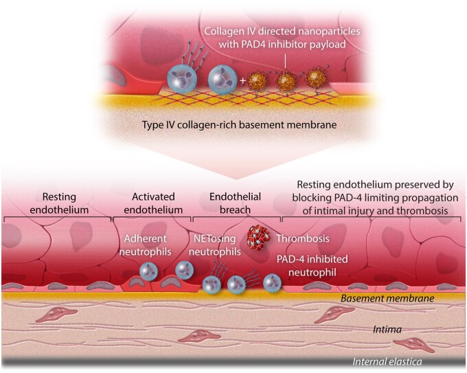
Aims: Recent evidence suggests that 'vulnerable plaques', which have received intense attention as underlying mechanism of acute coronary syndromes over the decades, actually rarely rupture and cause clinical events. Superficial plaque erosion has emerged as a growing cause of residual thrombotic complications of atherosclerosis in an era of increased preventive measures including lipid lowering, antihypertensive therapy, and smoking cessation. The mechanisms of plaque erosion remain poorly understood, and we currently lack validated effective diagnostics or therapeutics for superficial erosion. Eroded plaques have a rich extracellular matrix, an intact fibrous cap, sparse lipid, and few mononuclear cells, but do harbour neutrophil extracellular traps (NETs). We recently reported that NETs amplify and propagate the endothelial damage at the site of arterial lesions that recapitulate superficial erosion in mice. We showed that genetic loss of protein arginine deiminase (PAD)-4 function inhibited NETosis and preserved endothelial integrity. The current study used systemic administration of targeted nanoparticles to deliver an agent that limits NETs formation to probe mechanisms of and demonstrate a novel therapeutic approach to plaque erosion that limits endothelial damage.

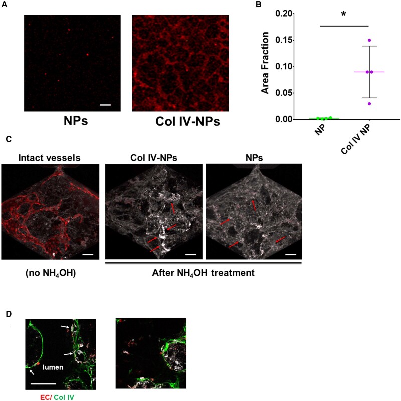
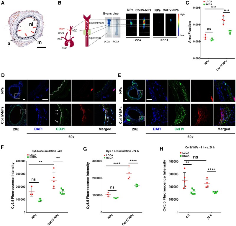
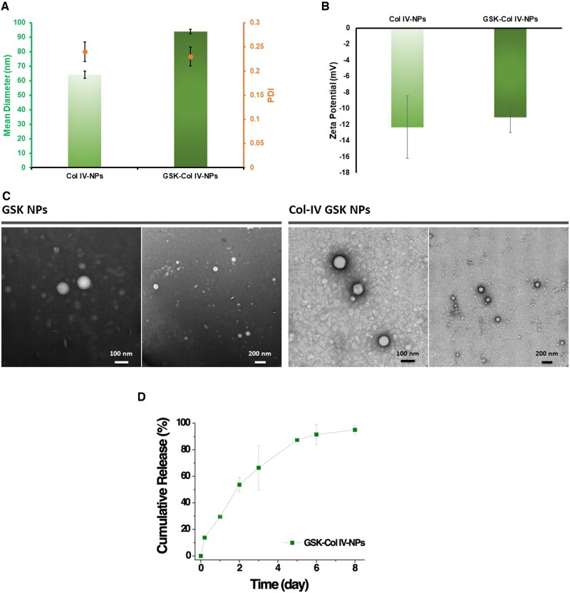
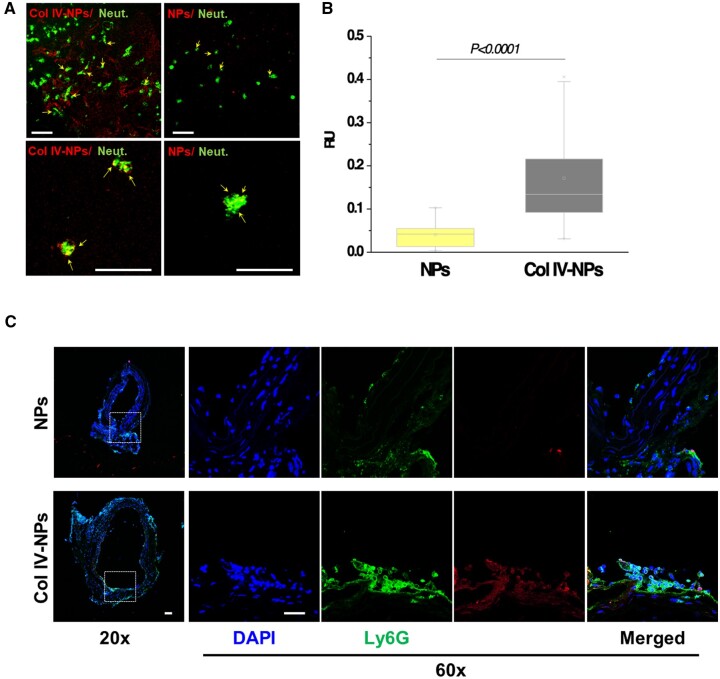
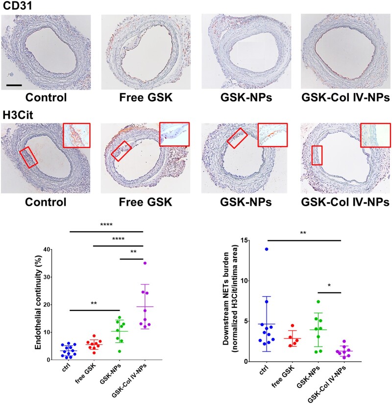
Methods and results: We developed Collagen IV-targeted nanoparticles (Col IV NP) to deliver PAD4 inhibitors selectively to regions of endothelial cell sloughing and collagen IV-rich basement membrane exposure. We assessed the binding capability of the targeting ligand in vitro and evaluated Col IV NP targeting to areas of denuded endothelium in vivo in a mouse preparation that recapitulates features of superficial erosion. Delivery of the PAD4 inhibitor GSK484 reduced NET accumulation at sites of intimal injury and preserved endothelial continuity.

Conclusions: NPs directed to Col IV show selective uptake and delivery of their payload to experimentally eroded regions, illustrating their translational potential. Our results further support the role of PAD4 and NETs in superficial erosion.

Keywords: Neutrophil extracellular traps; Atherosclerosis; Cardiovascular nanomedicine; Experimental superficial erosion; Targeted nanoparticles.


Figures

None
Graphical abstract
Figure 1
Figure 1
Collagen IV-targeting nanoparticles adhere in vitro to basement membrane produced by endothelial cells in both 2D and 3D settings. (A and B) Compared to non-targeting NPs, Col IV-NPs showed 40-fold increased adhesion to HSVEC-produced basement membrane on a glass slide (n = 4). (scale bar = 100 µm) (C) When tested in a 3D setting, we observed a tubule-shaped binding for Col IV-NPs and a more diffuse deposition for the non-targeting NPs (red arrows). (D) DsRed-labelled ECs (in red), Col IV (in green), and Col IV-NPs (in white) reveals co-localization of NPs at sites of exposed Col IV after EC removal. Mann–Whitney test. *P < 0.05.
Figure 2
Figure 2
In vivo targeting of Col IV to exposed basement membrane after induction of flow disturbance to recapitulate aspects of superficial erosion. (A) Immunohistochemical analysis of in vivo preparation of superficial erosion in mice. Microscopic observation reveals neo-intima formation (ni) and areas of denuded endothelium (red arrows) and exposed basement membrane (black dashed line). (Scale bar 100 µm). (B) A constrictive cuff was placed on previously injured and healed LCCA to perturb flow. Representative Evans Blue staining reveals increased endothelial permeability in areas of disturbed flow. Fluorescence reflectance images of RCCA (healthy carotid) and LCCA (‘eroded’ plaque) in mice injected with bare NPs and Col IV-NPs at 24h post-injection. (C) Quantitative assessment of fluorescence intensities of bare and Col IV-NPs reveals a 1.8-fold higher accumulation for Col IV-NPs to denuded endothelium compared to bare NPs (n = 3–4). (D and E) Immunofluorescent staining of CD31 (D) and Col IV (E) shows NP localization downstream of the flow perturbation. Col IV-NPs co-localize with areas of desquamation (white arrows in D) and rich in Col IV (E). (Scale bar = 25 µm). (FH) Quantitative assessment of fluorescence intensities using fluorescence reflectance imaging analysis of bare or Col IV-NPs loaded with cy5.5 shows higher payload accumulation when delivered by Col IV-NPs at both 4 and 24 h after injection (n = 3–5): Tukey’s multiple comparisons test: **P < 0.01; ****P < 0.0001.
Figure 3
Figure 3
Physicochemical characterization of Col IV-targeting NPs. (A) Dynamic light scattering (DLS) analysis of empty and GSK484 (GSK)-loaded Col IV-NPs reveals a slight increase of the hydrodynamic size after GSK encapsulation (64 and 94 nm for empty and GSK Col IV-NPs, respectively). GSK encapsulation did not significantly affect the homogeneity of the formulations, as indicated by the polydispersity index values before and after GSK encapsulation (PDI = 0.24 and 0.23, respectively). (B) Zeta potential analysis reveals a negative surface charge for both empty and GSK-loaded Col IV-NPs, without significant changes after GSK encapsulation. (C) Representative TEM images of GSK NPs and Col IV GSK NPs validates DLS analysis. (D) Cumulative release of GSK from Col IV NPs at 37°C.
Figure 4
Figure 4
In vitro evaluation of Col IV-NPs targeting to basement membrane. (A) Confocal images of bare and Col IV-NPs (in red) incubated with primary mouse neutrophils (in green) in conditions that mimic superficial erosion. Arrows show NPs internalized in neutrophils (scale bar = 100 µm). (B) Quantitative assessment of NP/neutrophil fluorescent co-localization (n = 24 per group). (C) Immunofluorescent staining of Ly6G (neutrophil marker—in green) shows NPs localization downstream of the flow perturbation. Col IV-NPs co-localize with neutrophils recruited at areas of exposed basement membrane recapitulating eroded plaques. (Scale bar = 25 µm). Mann–Whitney test. P < 0.0001.
Figure 5
Figure 5
Therapeutic efficacy of GSK484-loaded Col IV-targeted NPs in a mouse preparation of superficial erosion. Immunohistochemical analysis of previously injured LCCA subjected to flow perturbation from mice (n = 8–12 per group, representative image shown) treated with free GSK484 (GSK) and GSK-loaded into bare NPs (GSK-NPs) and Col IV-NPs (GSK-Col IV-NPs). NPs were injected in PBS as vehicle. CD31 (endothelial cell marker) and H3Cit (citrullinated histone, neutrophil extracellular traps marker) staining revealed a significantly improved therapeutic efficacy for the group treated with GSK-Col IV-NPs as gauged by preservation of endothelial continuity and reduction of NET accumulation downstream of the flow perturbation, as shown by the representative data in the graph. Inserts show higher magnification of H3cit staining. (Scale bar 100 µm). One-Way ANOVA. *P < 0.05; **P < 0.01; ****P < 0.0001.

References

    1. Stone GW, Maehara A, Lansky AJ, de Bruyne B, Cristea E, Mintz GS, Mehran R, McPherson J, Farhat N, Marso SP, Parise H, Templin B, White R, Zhang Z, Serruys PW; PROSPECT Investigators. A prospective natural-history study of coronary atherosclerosis. N Engl J Med 2011;364:226–235. - PubMed
    1. Pasterkamp G, den Ruijter HM, Libby P. Temporal shifts in clinical presentation and underlying mechanisms of atherosclerotic disease. Nat Rev Cardiol 2017;14:21–29. - PubMed
    1. Libby P, Pasterkamp G. Requiem for the ‘vulnerable plaque’. Eur Heart J 2015;36:2984–2987. - PubMed
    1. Tricot O, Mallat Z, Heymes C, Belmin JL, Lesèche G, Tedgui A. Relation between endothelial cell apoptosis and blood flow direction in human atherosclerotic plaques. Circulation 2000;101:2450–2453. - PubMed
    1. de Bont CM, Boelens WC, Pruijn GJM. NETosis, complement, and coagulation: a triangular relationship. Cell Mol Immunol 2019;16:19–27. - PMC - PubMed

Publication types

MeSH terms

LinkOut - more resources