. 2021 Apr 15;11(4):jkab067.
doi: 10.1093/g3journal/jkab067.

Transcriptomic analysis of feminizing somatic stem cells in the Drosophila testis reveals putative downstream effectors of the transcription factor Chinmo

Affiliations

Transcriptomic analysis of feminizing somatic stem cells in the Drosophila testis reveals putative downstream effectors of the transcription factor Chinmo

Lydia Grmai et al. G3 (Bethesda). .

Abstract

One of the best examples of sexual dimorphism is the development and function of the gonads, ovaries and testes, which produce sex-specific gametes, oocytes, and spermatids, respectively. The development of these specialized germ cells requires sex-matched somatic support cells. The sexual identity of somatic gonadal cells is specified during development and must be actively maintained during adulthood. We previously showed that the transcription factor Chinmo is required to ensure the male sexual identity of somatic support cells in the Drosophila melanogaster testis. Loss of chinmo from male somatic gonadal cells results in feminization: they transform from squamous to epithelial-like cells that resemble somatic cells in the female gonad but fail to properly ensheath the male germline, causing infertility. To identify potential target genes of Chinmo, we purified somatic cells deficient for chinmo from the adult Drosophila testis and performed next-generation sequencing to compare their transcriptome to that of control somatic cells. Bioinformatics revealed 304 and 1549 differentially upregulated and downregulated genes, respectively, upon loss of chinmo in early somatic cells. Using a combination of methods, we validated several differentially expressed genes. These data sets will be useful resources to the community.

Keywords: Chinmo; ovary; sex transformation; sexual identity; testis.


Figures

Figure 1
Figure 1
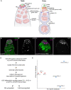
Transcriptomic profiling of chinmo-deficient CySCs. (A) Diagram of the adult Drosophila testis (left) and ovary (right). (Left) The testis niche is composed of quiescent somatic cells termed “hub” cells (green). The niche supports ∼8–12 GSCs (dark pink) and ∼13 somatic CySCs (dark blue). The GSC divides to produce a Gb (light pink) that differentiates. The Gb is ensheathed by two quiescent cyst cells (light blue), daughter cells of the CySCs. These same two cyst cells continue to ensheath the Gb as it divides four more times with incomplete cytokinesis, giving rise to spermatogonia (light pink and as “differentiating germ cells”) that enter meiosis. (Right) The ovarian niche (green) supports approximately two to three GSCs (dark pink). The GSC divides to produce a cystoblast that begins differentiation. IGS cells (light orange, also called escort cells) wrap around the cystoblast as it differentiates. Approximately one to four FSCs (dark blue) reside in the middle of the germarium and produce mitotic follicle cells (FCs) which function as epithelium that envelops the 16-cell germ cyst. (B, C) Representative confocal image of a control tj>GFP testis (B) and a tj>GFP+chinmo-RNAi testis (C) at 2 days of adulthood. In the control testis, GFP (magenta) was expressed in the nuclei of tj-expressing somatic support cells (i.e., CySCs and cyst cells) that ensheathed early germ cells (B). In the tj>GFP +chinmo-RNAi testis, GFP (magenta) was still expressed in the nuclei of tj-expressing somatic support cells (C). However, these chinmo-deficient somatic support cells were in close contact with each other and no longer properly enveloping germ cells, demonstrating that they were becoming sex-transformed. Vasa (green) marks germ cells. An asterisk marks the niche. Scale bar = 20 μm (D) Work-flow of the RNA-seq. Briefly, we purified GFP-positive cells from tj>GFP or tj>GFP+chinmo-RNAi adult testes at 2–3 days after eclosion. We isolated total RNA from these cells and generated cDNA libraries, which were used for the RNA-seq. The reads were mapped to the Drosophila genome (dm6). 1853 genes were differentially expressed (i.e., FC ≥ 2 and Padj ≤ 0.05) in tj>GFP+chinmo-RNAi cells compared with tj>GFP cells, with 304 upregulated and 1549 downregulated. (E) Principal component analysis of tj>GFP (red circles labeled “WT”) and tj>GFP+chinmo-RNAi (blue circles labeled “chinmo-RNAi”). The WT samples clustered together and were distinct from the chinmo-RNAi samples. Genotypes (B) w/Y; tj-Gal4, UAS-GFPnls/UAS-GFPnls; +/+, (C) w/Y; tj-Gal4, UAS-GFPnls/+; UAS-chinmo-RNAi/UAS-Dcr-2, (E) w/Y; tj-Gal4, UAS-GFPnls/UAS-GFPnls; +/+ (labeled “WT”) and w/Y; tj-Gal4, UAS-GFPnls/+; UAS-chinmo-RNAi/UAS-Dcr-2 (labeled “chinmo-RNAi”)
Figure 2
Figure 2
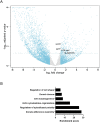
Genotype ontology and gene expression in chinmo-depleted somatic cells. (A) Scatter (Volcano) plot for genes in chinmo-depleted somatic cells (tj>GFP+chinmo-RNAi) compared with controls (tj>GFP). The x-axis is the log2 of the fold change and the y-axis is the negative log10 of the adjusted P-value (Padj). Gray line indicates Padj = 0.05. Gray circles indicate genes with log2 (FC) between −1 and 1 (corresponding to FC between 0.5 and 2). Blue circles indicate genes with log2 (FC) greater than 1 (corresponding to FC >2). The larger black circles indicate the differentially upregulated genes βH-spectrin, pyd, scrib, and DE-cad. (B) Gene Ontology. DAVID Functional Annotation Clustering analysis depicting enriched biological processes for the 100 upregulated genes with putative Chinmo binding sites. The enrichment score plotted on the y-axis is based on the Fisher’s exact P-values of each gene in the group. The higher the score, the more enriched is the group. Genotype: (A) w/Y; tj-Gal4, UAS-GFPnls/+; UAS-chinmo-RNAi/UAS-Dcr-2.
Figure 3
Figure 3
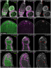
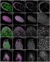
chinmo-deficient somatic cells upregulate βH-Spectrin. (A–A”) xy-section of a WT ovary shows that βH-spectrin (white) is present in somatic cells. Bracket in A” indicates the niche cells. Arrows in A” indicate apical enrichment of βH-spectrin in follicle cells. (B–B”) In a WT testis, βH-spectrin (white) is expressed strongly in niche cells (outlined by a dashed line in B”) and at very low levels in other somatic cells. (C–D”) In a chinmoST mutant testis (C–C”) or a testis somatically depleted for chinmo (D–D”), βH-spectrin (white) is ectopically expressed in feminized somatic cells. Arrows in C” and D” indicate apical enrichment of βH-spectrin in chinmo-depleted somatic cells. In (A–D), Vasa (green) marks the germline and Tj (magenta) marks cyst cells. Scale bar = 10 μm. Time point in (A–D) is 7–8 days post-eclosion. Genotypes: (A) +/+; +/+; +/+ (OregonR), (B) +/Y; +/+; +/+ (OregonR), (C) w/Y; chinmoST/chinmoST; +/+, and (D) w/Y; tj-Gal4/+; UAS-chinmo-RNAi/UAS-Dcr-2 (labeled “tj>chinmo-RNAi”)
Figure 4
Figure 4
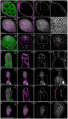
chinmo-deficient somatic cells upregulate DE-cad. (A–A’”) Middle xy-section of a WT ovary shows that DE-cad (white) is expressed at AJs (arrows in A’”) in somatic follicle cells. (B–B’”) An apical xy-section of a WT ovary shows that DE-cad (white) is apically enriched in follicle cells. (C–C”’) In a WT testis, DE-cad (white) is expressed strongly in the niche (outlined by a dashed line in C’”) and at a modest level in the somatic cells (arrow, C’”). (D–D”’) Middle xy-section of a chinmoST mutant testis shows that DE-cad (white) is ectopically expressed at AJs (arrows, D’”) in feminized somatic cyst cells. (E–E’”) An apical xy-section of a chinmoST mutant testis reveals apical enrichment of DE-cad (white) in the feminized somatic cells. (F–F’”) Middle xy-section of a tj>chinmo-RNAi testis shows that DE-cad (white) is ectopically expressed at AJs (arrows, F’”) in feminized somatic cyst cells. (G–G’”) An apical xy-section of a tj>chinmo-RNAi testis shows apical enrichment of DE-cad (white) in the feminized follicle-like cells. In (A–G), Vasa (green) marks the germline and Tj (magenta) marks cyst cells. Scale bar = 10 μm. (A’”–G”’) is a magnification of the boxed regions in (A”–G”), respectively. Time point in (A–G) is 7–8 days post-eclosion. Genotypes: (A, B) +/+; +/+; +/+ (OregonR), (C) +/Y; +/+; +/+ (OregonR), (D, E) w/Y; chinmoST/chinmoST; +/+ (F, G) w/Y; tj-Gal4/+; UAS-chinmo-RNAi/UAS-Dcr-2 (labeled “tj>chinmo-RNAi”).
Figure 5
Figure 5
chinmo-deficient somatic cells upregulate Pyd. (A–A’”) Middle xy-section of a WT ovary shows that Pyd (white) is expressed strongly in somatic follicle cells. Arrows (A’”) indicate Pyd expression in AJs. (B–B’”) An apical xy-section of a WT ovary shows the apical enrichment of Pyd (white) in the follicle cells. (C–C’”) In a WT testis, Pyd (white) is expressed at moderate levels in the niche (outlined by a dashed line in C’”) and at low levels in other somatic cells (C”’, arrows). (D–D’”) Middle xy-section of a chinmoST mutant testis shows that Pyd (white) is ectopically expressed in AJs (arrows in D’”) in feminized somatic cyst cells. (E–E’”) An apical xy-section of a chinmoST mutant testis shows apical enrichment in the feminized cells. (F–F’”) Middle xy-section of a tj>chinmo-RNAi testis shows that Pyd (white) is ectopically expressed in AJs (arrows in F’”) in feminized somatic cyst cells. (G–G’”) An apical xy-section of a tj>chinmo-RNAi testis shows apical enrichment of Pyd (white) in the feminized follicle-like cells. In (A–G), Vasa (green) marks the germline and Tj (magenta) marks cyst cells. Scale bar = 10 μm. (A’”–G”’) is a magnification of the boxed regions in (A”–G”), respectively. Time point in (A–G) is 7–8 days post-eclosion. Genotypes: (A, B) +/+; +/+; +/+ (OregonR), (C) +/Y; +/+; +/+ (OregonR), (D, E) w/Y; chinmoST/chinmoST; +/+, (F, G) w/Y; tj-Gal4/+; UAS-chinmo-RNAi/UAS-Dcr-2 (labeled “tj>chinmo-RNAi”).
Figure 6
Figure 6
chinmo-deficient somatic cells upregulate Scrib. (A–A’’’) Middle xy-section of a WT ovary shows that Scrib (white) is expressed strongly in the lateral domains (arrows in A’”) of follicle cells and at lower levels in the germline. (B–B’’’) An apical xy-section of a WT ovary shows the apical enrichment of Scrib (white) in the follicle cells. (C–C’’’) In a WT testis, Scrib (white) is expressed in the niche (outlined in C’”), at moderate levels in germ cells, and at low levels in somatic cyst cells (arrows in C’”). (D–D’’’) Middle xy-section of a tj>chinmo-RNAi testis shows that Scrib (white) is ectopically expressed in the lateral domain (arrows in D’”) of epithelial feminized somatic cells. (E–E’’’) An apical xy-section of a tj>chinmo-RNAi testis shows apical enrichment of Scrib (white) in the feminized follicle-like cells. In (A–E), Vasa (green) marks the germline and Tj (magenta) marks cyst cells. Scale bar = 10 μm. (A’”–E”’) is a magnification of the boxed regions in (A”–E”), respectively. Time point in (A–E) is 7–8 days post-eclosion. Genotypes: (A, B) w/w; +/+; scrib-GFP/scrib-GFP (C) w/Y; +/+; scrib-GFP/scrib-GFP, and (D, E) w/Y; tj-Gal4/UAS-Dcr-2; UAS-chinmo-RNAi/scrib-GFP (labeled “tj>chinmo-RNAi”).
Figure 7
Figure 7
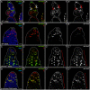
chinmo-deficient somatic cells upregulate mirr. (A–A’”) Middle xy-section of a WT germarium shows that mirr (green) is present in early FSCs (arrows in A’–A’”). (B–B’”) In a WT testis, mirr (green) is expressed strongly in niche cells (outlined in B””) and at a modest level in other somatic cells. (C–C’”) In a chinmoST mutant testis, mirr (green) is ectopically expressed in feminized somatic cells (bracket in C” and C’”). (D–D’”) mirr (green) is ectopically expressed in the feminized somatic cells (brackets in D” and D’”) in a tj>chinmo-RNAi testis. In (A–D), Vasa (blue) marks the germline and Tj (red) marks cyst cells. Scale bar = 10 μm. Time point in (A–D) is 7–8 days post-eclosion. Genotypes: (A) w/w; +/+; mirr-lacZ/TM6B, Tb, (B) w/Y; +/+; mirr-lacZ/TM6B, Tb, (C) w/Y; chinmoST/chinmoST; mirr-lacZ/TM6B, Tb, (D) w/Y; tj-Gal4/UAS-Dcr-2; UAS-chinmo-RNAi/mirr-lacZ (labeled “tj>chinmo-RNAi”).

Similar articles

Cited by

References

    1. Albertson R, Chabu C, Sheehan A, Doe CQ.. 2004. Scribble protein domain mapping reveals a multistep localization mechanism and domains necessary for establishing cortical polarity. J Cell Sci. 117:6061–6070. - PubMed
    1. Ambrosini G, Groux R, Bucher P.. 2018. Pwmscan: A fast tool for scanning entire genomes with a position-specific weight matrix. Bioinformatics. 34:2483–2484. - PMC - PubMed
    1. Amoyel M, Sanny J, Burel M, Bach EA.. 2013. Hedgehog is required for CySC self-renewal but does not contribute to the GSC niche in the Drosophila testis. Development. 140:56–65. - PMC - PubMed
    1. Amoyel M, Simons BD, Bach EA.. 2014. Neutral competition of stem cells is skewed by proliferative changes downstream of Hh and Hpo. EMBO J. 33:2295–2313. - PMC - PubMed
    1. Anders S, Pyl PT, Huber W.. 2015. Htseq–a python framework to work with high-throughput sequencing data. Bioinformatics. 31:166–169. - PMC - PubMed

Publication types