Review
. 2021 May;78(10):4487-4505.
doi: 10.1007/s00018-021-03796-9. Epub 2021 Mar 9.

The complement system in age-related macular degeneration

Affiliations
Review

The complement system in age-related macular degeneration

Angela Armento et al. Cell Mol Life Sci. 2021 May.

Abstract

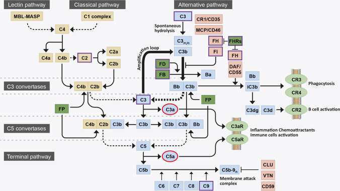
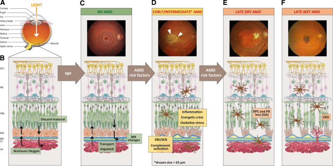
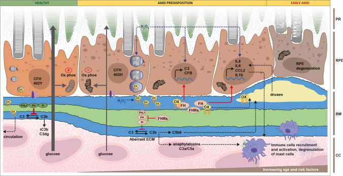
Age-related macular degeneration (AMD) is a chronic and progressive degenerative disease of the retina, which culminates in blindness and affects mainly the elderly population. AMD pathogenesis and pathophysiology are incredibly complex due to the structural and cellular complexity of the retina, and the variety of risk factors and molecular mechanisms that contribute to disease onset and progression. AMD is driven by a combination of genetic predisposition, natural ageing changes and lifestyle factors, such as smoking or nutritional intake. The mechanism by which these risk factors interact and converge towards AMD are not fully understood and therefore drug discovery is challenging, where no therapeutic attempt has been fully effective thus far. Genetic and molecular studies have identified the complement system as an important player in AMD. Indeed, many of the genetic risk variants cluster in genes of the alternative pathway of the complement system and complement activation products are elevated in AMD patients. Nevertheless, attempts in treating AMD via complement regulators have not yet been successful, suggesting a level of complexity that could not be predicted only from a genetic point of view. In this review, we will explore the role of complement system in AMD development and in the main molecular and cellular features of AMD, including complement activation itself, inflammation, ECM stability, energy metabolism and oxidative stress.

Keywords: Age-related macular degeneration; Ageing; Complement system; Genetics; Ophthalmology; Retinal biology.


Conflict of interest statement

The authors declare that they have no conflict of interest.

Figures

Fig. 1
Fig. 1
Flow diagram of the complement system activation. The complement system is initiated by three activation pathways: the lectin pathway, the classical pathway (both in yellow) and the alternative pathway (indicated in blue). They all converge in the formation of a C3 convertase, responsible for the breakdown (dotted line) of circulating C3 into C3b. During activation via the alternative pathway, FB binds a spontaneously hydrolysed form of C3 [termed C3(H2O)] and is cleaved by FD to form a distinct C3 convertase, C3bBb. This C3 convertase continuously cleaves C3 into C3b, exposing an internal thioester bond within the protein that allows C3b to become covalently attached to local surfaces. Deposited C3b, a potent opsonin, forms the starting block of a surface bound C3 convertase, that cleaves yet more C3 into C3b and contributes to the amplification loop of complement (thick arrows). C3b is also necessary for the formation of a downstream C5 convertase, responsible for the breakdown (dotted line) of C5 into C5b and the subsequent recruitment of C6, C7, C8 and polyC9 into the C5b-9n complex, also known as the membrane attack complement (MAC). The MAC forms a pore on pathogen/cell surfaces and leads to lysis. Complement system activation is tightly controlled by negative (light red) and positive (dark green) regulators: FI and its cofactors FH, CR1 and MCP promote proteolytic cleavage and inactivation of C3b (iC3b) and FH and DAF promote the disassembly of the C3bBb C3 convertase; CLU, VTN and CD59 inhibit the formation of C5b-9n; FP stabilises C3 and C5 convertase; and the FHR proteins compete with FH for C3b binding and inhibit FI-mediated C3b cleavage. C3b breakdown fragments bind membrane-bound receptors (light green): iC3b binds CR3 and CR4 and mediates phagocytosis; C3dg and C3d bind CR2 and mediate B cell activation. Anaphylatoxins C3a and C5a (highlighted in red), generated with the breakdown of C3 and C5, bind membrane-bound receptors (light green) C3aR and C5aR to promote inflammation and immune cells activation. AMD-associated genetic risk variants mostly occur in the genes of the proteins involved in the alternative pathway of complement (highlighted in violet)
Fig. 2
Fig. 2
Schematic of the human eye in health, age and AMD. a Diagram highlighting the anatomical features of the human eye. b Schematic of a healthy human retina with its cell layers and transport of nutrients across Bruch’s membrane, where; GCL ganglion cell layer, INL inner nuclear layer, ONL outer nuclear layer, RPE retinal pigmented epithelium, BM Bruch’s membrane, CC choriocapillaris, and CH choroid. c–f Progression of AMD shown by fundus images and schematic changes within the retinal cell layers. In older patients without AMD c, fundus imaging is normal and the macula (black circle), parafovea (black dotted circle) and fovea (white dotted circle) appear intact. On a structural level, BM is altered and transport properties are becoming impaired. Additional AMD risk factors d promote inflammation, oxidative stress, energetic crisis, complement activation and drusen formation. Drusen can be visualised in fundus images as yellow spots (white arrows). Progression to later stages of AMD can lead to e geographic atrophy (GA) characterised by defined areas of RPE cell death (termed dry AMD) and f choroidal neovascularisation (CNV) into the retina (termed wet AMD), ultimately leading to photoreceptors (PR) cell death. Fundus images were obtained from the Macula Reading Centre, University of Tübingen (UKT)
Fig. 3
Fig. 3
Involvement of complement system in AMD predisposition. In healthy conditions, RPE cells, exposed to healthy Bruch’s membrane (BM) and carrying the FH 402Y polymorphism, transport efficiently glucose from the Choroid/choriocapillaris (CC) through BM to the photoreceptors (PR), phagocyte POS and eliminate oxidised lipids (ox) into the circulation. Integrins (violet) anchor the RPE cells to BM, FI and cofactors FH and FHL1 (green) inhibit complement activation and mitochondria respiration (ox phos) is intact. AMD predisposition is provided by the combination of genetic risk, increasing age and external risk factors. RPE cells carrying the AMD-associated FH 402H polymorphism show reduced mitochondrial function, activity of phagolysosomes (L) and integrin interactions with BM. With increasing age and external risk factors, the BM ECM becomes even more altered and glucose transport is impaired. At this stage complement turnover is stimulated by aberrant ECM and a lack of effective inhibition due to the presence of high-risk variants in FI, FH, FHL1 (red) and the accumulation of FH antagonists, such as the FHR proteins (red). Products of complement turnover accumulate in the intercapillary septa and within BM itself. Additionally, oxidative stress is increased, which provides metabolic stress to the RPE cells and can stimulate inflammation. Hydrogen peroxide (H2O2) and products of lipid peroxidation (ox, yellow) accumulate in the altered BM, promote the upregulation of inflammatory cytokines and the high-risk FH 402H variant and FHR accumulation amplify this effect. In early AMD, continuous accumulation of oxidised lipids, complement activation products and MAC accumulation in drusen further promotes inflammation. Local inflammation, together with the release of anaphylatoxins C3a and C5a, causes the recruitment and activation of immune cells and degranulation of mast cells into the BM/RPE/retina interface, aggravating retina homeostasis: RPE cells degenerate and are unable to support rod photoreceptor cells, which start to show signs of damage

References

    1. van Lookeren Campagne M, et al. Mechanisms of age-related macular degeneration and therapeutic opportunities. J Pathol. 2014;232(2):151–164. doi: 10.1002/path.4266. - DOI - PubMed
    1. Schramm EC, et al. Genetic variants in the complement system predisposing to age-related macular degeneration: a review. Mol Immunol. 2014;61(2):118–125. doi: 10.1016/j.molimm.2014.06.032. - DOI - PMC - PubMed
    1. Priya RR, Chew EY, Swaroop A. Genetic studies of age-related macular degeneration: lessons, challenges, and opportunities for disease management. Ophthalmology. 2012;119(12):2526–2536. doi: 10.1016/j.ophtha.2012.06.042. - DOI - PMC - PubMed
    1. Ajana S et al. (2020) Predicting progression to advanced age-related macular degeneration from clinical, genetic and lifestyle factors using machine learning. Ophthalmology. - PubMed
    1. Bhutto I, Lutty G. Understanding age-related macular degeneration (AMD): relationships between the photoreceptor/retinal pigment epithelium/Bruch’s membrane/choriocapillaris complex. Mol Aspects Med. 2012;33(4):295–317. doi: 10.1016/j.mam.2012.04.005. - DOI - PMC - PubMed

Substances