. 2021 Jun;178(12):2533-2546.
doi: 10.1111/bph.15438. Epub 2021 May 5.

KY19382, a novel activator of Wnt/β-catenin signalling, promotes hair regrowth and hair follicle neogenesis

Affiliations

KY19382, a novel activator of Wnt/β-catenin signalling, promotes hair regrowth and hair follicle neogenesis

Yeong Chan Ryu et al. Br J Pharmacol. 2021 Jun.

Abstract

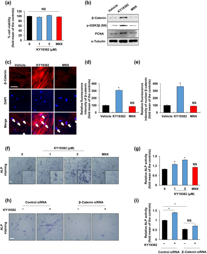
Background and purpose: The promotion of hair regeneration and growth heavily depends on the activation of Wnt/β-catenin signalling in the hair follicle, including dermal papilla (DP). KY19382, one of the newly synthesized analogues of indirubin-3'-monoxime (I3O), was identified as a Wnt/β-catenin signalling activator via inhibition of the interaction between CXXC-type zinc finger protein 5 (CXXC5) and dishevelled (Dvl). Given the close relationship between the Wnt/β-catenin signalling and hair regeneration, we investigated the effect of KY19382 on hair regrowth and hair follicle neogenesis.

Experimental approach: In vitro hair induction effects of KY19382 were performed in human DP cells. The hair elongation effects of KY19382 were confirmed through the human hair follicle and vibrissa culture system. In vivo hair regeneration abilities of KY19382 were identified in three models: hair regrowth, wound-induced hair follicle neogenesis (WIHN) and hair patch assays using C57BL/6 mice. The hair regeneration abilities were analysed by immunoblotting, alkaline phosphatase (ALP) and immunohistochemical staining.

Key results: KY19382 activated Wnt/β-catenin signalling and elevated expression of ALP and the proliferation marker PCNA in DP cells. KY19382 also increased hair length in ex vivo-cultured mouse vibrissa and human hair follicles and induced hair regrowth in mice. Moreover, KY19382 significantly promoted the generation of de novo hair follicles as shown by WIHN and hair patch assays.

Conclusion and implications: These results indicate that KY19382 is a potential therapeutic drug that exhibits effective hair regeneration ability via activation of the Wnt/β-catenin signalling for alopecia treatments.

Keywords: CXXC5; GSK-3β; Wnt/β-catenin signalling; dermal papilla cells; neogenesis.


Conflict of interest statement

K‐Y.C. is the CEO of CK Biotech. Inc. (Seoul, Korea), which has a licence to develop and use the compounds disclosed in the publication. The authors have no further conflicts of interest to declare.

Figures

FIGURE 1
FIGURE 1
KY19382 increases hair induction activity via activation of the Wnt/β‐catenin pathway. (a) Cell viability of human dermal papilla (DP) cells treated with vehicle (0.1% [v/v] DMSO), 100‐μM minoxidil (MNX) or the shown concentrations of KY19382 for 48 h (n = 6). (b, c) Human DP cells were treated with vehicle (0.1% DMSO), 5‐μM KY19382 or 100‐μM minoxidil for 48 h. (b) Immunoblotting analyses for β‐catenin, α‐tubulin, p‐GSK3β and PCNA were analysed using human DP cells. (c) Immunocytochemical staining for β‐catenin. Nuclei were counterstained with DAPI. Arrows indicate nuclei. (d, e) Quantitative measurements of the total and nuclear β‐catenin intensities of human DP cells (n = 12 taken in three different images). (f, g) Human DP cells were treated with vehicle (0.1% DMSO), 100‐μM minoxidil or the marked concentrations of KY19382 for 48 h. (f) Alkaline phosphatase (ALP) staining and (g) ALP activity test (n = 5). (h, i) Human DP cells were transfected with β‐catenin siRNA or negative control for 12 h. After transfection, human DP cells were incubated with vehicle (0.1% DMSO) or 5‐μM KY19382 for 48 h. (h) Transfected cells were subjected to ALP staining. (i) ALP activity was quantified (n = 6). Scale bars = 100 μm. Values are expressed as means ± SEM. * P < .05, significantly different from vehicle, control or as indicated; NS, not significant
FIGURE 2
FIGURE 2
KY19382 accelerates mouse vibrissa and human hair follicle elongation. (a–d) Mouse vibrissa follicles were cultured with vehicle (0.1% DMSO), 5‐μM KY19382 or 100‐μM minoxidil (MNX) for 6 days (n = 5). (a) The length of the vibrissa follicles was measured at 6 days after treatment. The elongation rate of mouse vibrissa follicles was evaluated as the difference in the length of vibrissa follicles, wherein the vibrissa follicle length in the control group at Day 6 was considered 100%. Scale bars = 200 μm. (b) Immunohistochemistry (IHC) analyses of hair bulb of mouse vibrissa follicle for Ki67 and β‐catenin. Dashed lines mean mouse vibrissa follicles. (c) Mouse vibrissa follicles were subjected to alkaline phosphatase (ALP) staining. Arrow indicates ALP‐positive region. (d) X‐gal staining of vibrissa follicles from Axin2 lacZ/+ mice. Arrow indicates X‐gal‐positive region. (b–d) Scale bars = 100 μm. (e) Human hair follicles were incubated with vehicle (0.1% DMSO), 5‐μM KY19382 or 100‐μM minoxidil for 3 days. The length of human hair follicles was evaluated at 3 days after treatment with 5‐μM KY19382 or 100‐μM minoxidil. The elongation rate of human follicles was calculated as the difference in the length of human follicles wherein the human follicle length in the control group at Day 3 was considered 100% (n = 15). Scale bars = 200 μm. Values are expressed as means ± SEM. * P < .05, significantly different from vehicle
FIGURE 3
FIGURE 3
KY19382 stimulates hair regrowth in vivo. C57BL/6N mice were treated with Vehicle 1 (PEG400), Vehicle 2 (50% [v/v] ethanol, 30% water and 20% propylene glycol), 0.5‐mM KY19382 or 100‐mM minoxidil (MNX) for 14 or 28 days (n = 5). (a) The gross image showed hair regrowth in mice treated with each drug for 28 days. (b) Quantitative measurements of the weight of regrown hairs. (c) Haematoxylin and eosin (H&E) staining to evaluate the hair follicles of skins with different treatments. (d, e) Quantitative analyses of the relative number of hair follicles and dermal thickness of H&E staining images (n = 5). (f) Immunohistochemistry (IHC) staining for keratin 15, β‐catenin and Ki67 using the dorsal skin of mice treated with each drug for 14 days. Lines show keratin 15‐positive bulge stem cells region. (g, h) Quantitative estimations of the total and nuclear β‐catenin intensities in bulge stem cells (n = 15 taken in five different images). (i) Alkaline phosphatase (ALP) staining was performed to evaluate the ALP activity of hair follicles with different treatments. Arrow points to the ALP‐positive region. (j) Immunoblotting analyses for β‐catenin, PCNA and ERK. Scale bars = 100 μm. Values are expressed as means ± SEM. * P < .05, significantly different as indicated ; NS, not significant
FIGURE 4
FIGURE 4
KY19382 stimulates WIHN in vivo. C57BL/6N mouse wounds were treated with 0.5‐mM KY19382, 100‐mM minoxidil (MNX), Vehicle 1 (PEG400) or Vehicle 2 (50% [v/v] ethanol, 30% water and 20% propylene glycol) for 14, 25 or 40 days (n = 6). (a) Alkaline phosphatase (ALP) staining to evaluate the neogenic follicles of mice treated with each drug for 25 days. (b) Quantitative measurements of ALP‐positive neogenic follicles. Dashed lines show ALP‐positive neogenic hair follicles. (c) H&E staining to evaluate newly formed hair follicles in mice treated with each drug for 14 days. Dashed lines mean the boundary between the epidermis and dermis. Arrows show newly formed follicles. (d) Immunohistochemistry (IHC) staining for Fgf9, keratin 17, β‐catenin and Ki67. (e, f) Quantitative measurement of the nuclear and total β‐catenin intensities of neogenic hair follicle cells (n = 15 taken in five different images). (g) Quantitative analyses of Fgf9‐positive dermal cells and (h) Ki67‐positive newly formed hair follicle cells (each n = 5). (i) Gross images showed newly formed hair in mice after treatment for 40 days. (j) Immunoblotting analyses for Fgf9, keratin 17, β‐catenin, PCNA and ERK for 14 days. Scale bars = 100 μm. Values are expressed as means ± SEM. * P < .05, significantly different as indicated; NS, not significant)
FIGURE 5
FIGURE 5
KY19382 induces hair follicle neogenesis in hairless mice. Mouse dermal cells were cultured with vehicle (0.1% DMSO) or 5‐μM KY19382 for 72 h and then subcutaneously injected with epidermal cells into hairless mice. The hair follicle neogenesis was analysed at 14 days after transplantation (n = 5). (a) Hair follicle neogenesis at the injected area of hairless mice. Arrow indicates the hair regeneration area on the skin. (b) Magnified image of area exhibiting hair follicle neogenesis. (c) Quantitative analyses of regenerated hair follicles. (d) Immunohistochemistry (IHC) analyses for β‐catenin and Ki67. (e, f) Quantitative analyses of the nuclear β‐catenin intensity and Ki67‐positive cells of regenerated hair follicles. Dashed lines mean hair follicles. Scale bars = 50 μm. Values are expressed as means ± SEM. * P < .05, significantly different from control (0 μM KY19382)

References

    1. Alexander, S. P. H. , Fabbro, D. , Kelly, E. , Mathie, A. , Peters, J. A. , Veale, E. L. , Armstrong, J. F. , Faccenda, E. , Harding, S. D. , Pawson, A. J. , Sharman, J. L. , Southan, C. , Davies, J. A. , & CGTP Collaborators . (2019). The concise guide to PHARMACOLOGY 2019/20: Enzymes. British Journal of Pharmacology, 176, S297–S396. 10.1111/bph.14752 - DOI - PMC - PubMed
    1. Alexander, S. P. H. , Roberts, R. E. , Broughton, B. R. S. , Sobey, C. G. , George, C. H. , Stanford, S. C. , Cirino, G. , Docherty, J. R. , Giembycz, M. A. , Hoyer, D. , Insel, P. A. , Izzo, A. A. , Ji, Y. , MacEwan, D. J. , Mangum, J. , Wonnacott, S. , & Ahluwalia, A. (2018). Goals and practicalities of immunoblotting and immunohistochemistry: A guide for submission to the British Journal of Pharmacology. British Journal of Pharmacology, 175(3), 407–411. 10.1111/bph.14112 - DOI - PMC - PubMed
    1. Alonso, L. , & Fuchs, E. (2006). The hair cycle. Journal of Cell Science, 119(Pt 3), 391–393. 10.1242/jcs02793 - DOI - PubMed
    1. Andl, T. , Reddy, S. T. , Gaddapara, T. , & Millar, S. E. (2002). WNT signals are required for the initiation of hair follicle development. Developmental Cell, 2(5), 643–653. 10.1016/s1534-5807(02)00167-3 - DOI - PubMed
    1. Botchkarev, V. A. , & Kishimoto, J. (2003). Molecular control of epithelial–mesenchymal interactions during hair follicle cycling. The Journal of Investigative Dermatology. Symposium Proceedings, 8(1), 46–55. 10.1046/j.1523-1747.2003.12171.x - DOI - PubMed

Publication types