. 2022 Feb;16(3):813-829.
doi: 10.1002/1878-0261.12940. Epub 2021 Mar 22.

LncRNA-ZXF1 stabilizes P21 expression in endometrioid endometrial carcinoma by inhibiting ubiquitination-mediated degradation and regulating the miR-378a-3p/PCDHA3 axis

Affiliations

LncRNA-ZXF1 stabilizes P21 expression in endometrioid endometrial carcinoma by inhibiting ubiquitination-mediated degradation and regulating the miR-378a-3p/PCDHA3 axis

Deshui Kong et al. Mol Oncol. 2022 Feb.

Abstract

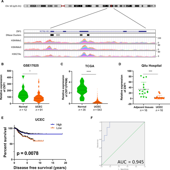
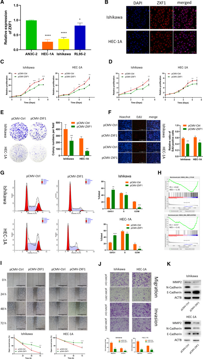
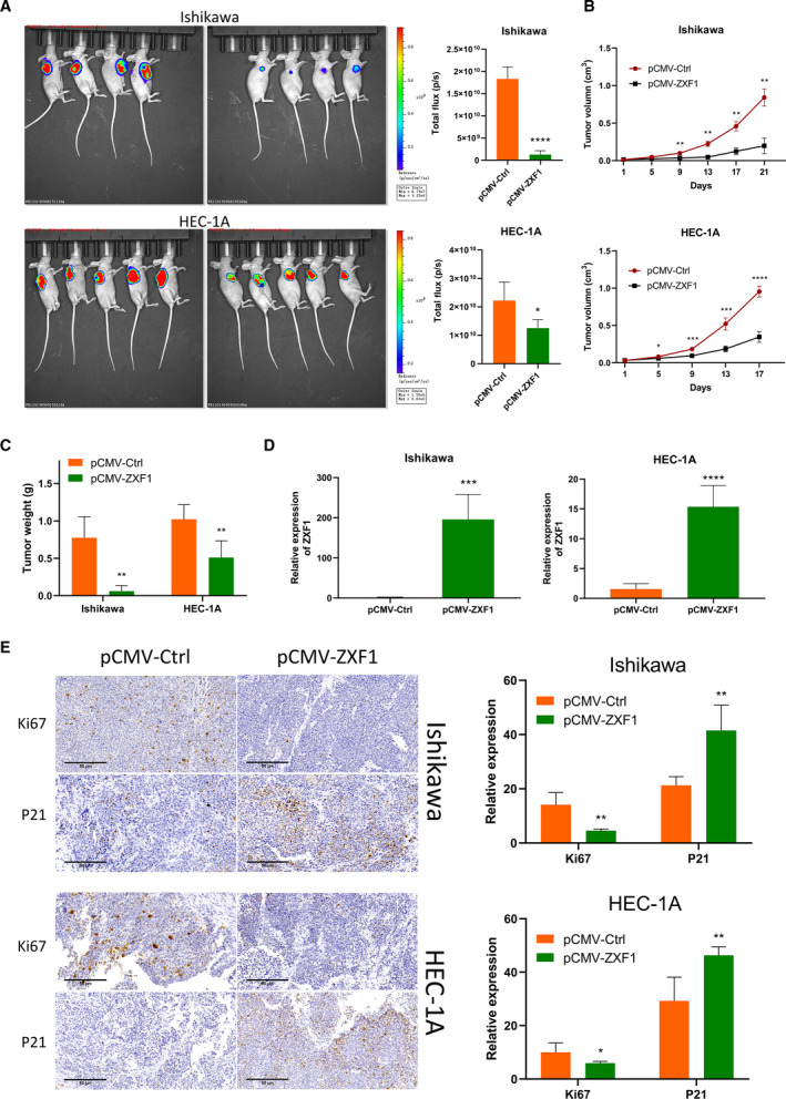
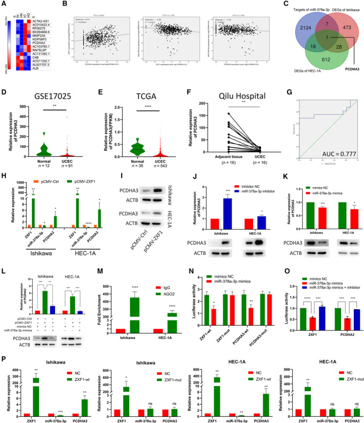
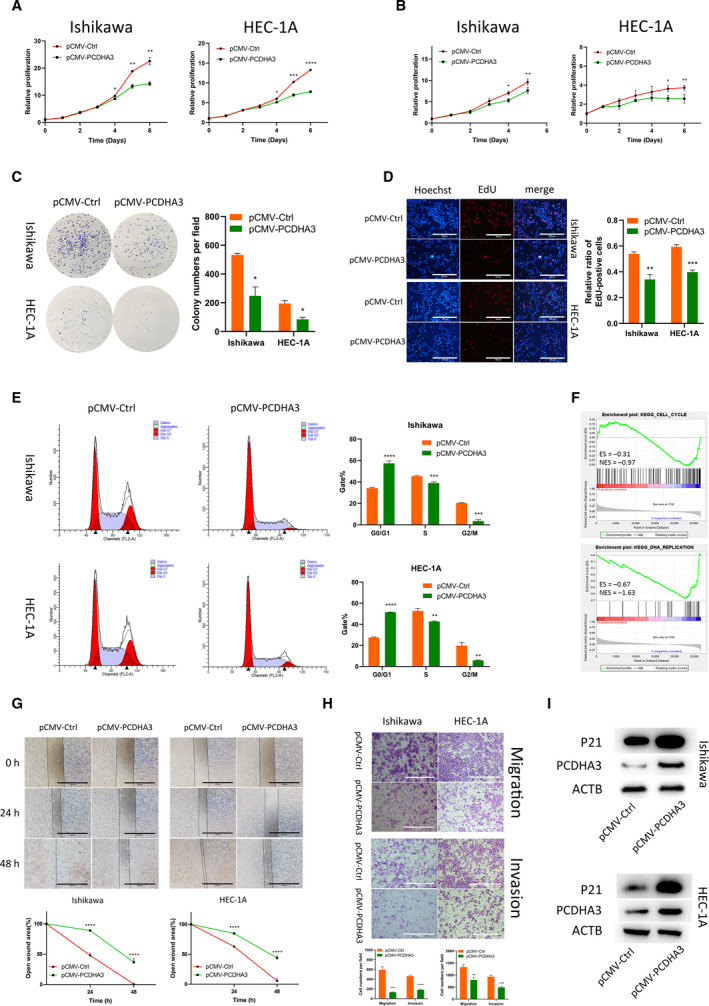
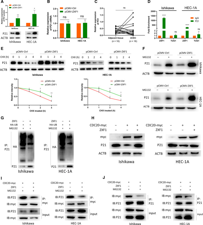
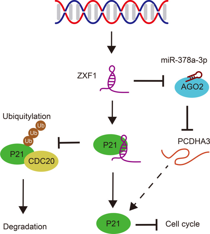
Long noncoding RNAs (lncRNAs) have a profound effect on biological processes in various malignancies. However, few studies have investigated their functions and specific mechanisms in endometrial cancer. In this study, we focused on the role and mechanism of lncRNA-ZXF1 in endometrial cancer. Bioinformatics and in vitro and in vivo experiments were used to explore the expression and function of lncRNA-ZXF1. We found that lncRNA-ZXF1 altered the migration and invasion of endometrioid endometrial cancer (EEC) cells. Furthermore, our results suggest that lncRNA-ZXF1 regulates EEC cell proliferation. This regulation may be achieved by the lncRNA-ZXF1-mediated alteration in the expression of P21 through two mechanisms. One is that lncRNA-ZXF1 functions as a molecular sponge of miR-378a-3p to regulate PCDHA3 expression and then modulate the expression of P21. The other is that lncRNA-ZXF1 inhibits CDC20-mediated degradation of ubiquitination by directly binding to P21. To the best of our knowledge, this study is the first to explore lncRNA-ZXF1 functioning as a tumor-suppressing lncRNA in EEC. LncRNA-ZXF1 may become therapeutic, diagnostic, and prognostic indicator in the future.

Keywords: P21; cell cycle; endometrioid endometrial cancer; lncRNA-ZXF1; ubiquitination.


Conflict of interest statement

The authors declare no conflict of interest.

Figures

Fig. 1
Fig. 1
LncRNA‐ZXF1 has diagnostic and prognostic potential in UCEC. (A) ZXF1 is located on chromosome 10 (q23.31). ENCODE and UCEC data predict the transcriptional regulation of ZXF1 and indicate the ZXF1 promoter region. H3K27Ac mark (often found near regulatory elements) in 7 cell lines and DNase I hypersensitivity peak clusters from ENCODE (95 cell lines). (B and C). In the normalized GSE17025 (B) and TCGA (C) data, ZXF1 expression was reduced in tumor tissues compared with control samples. The P‐value was obtained from the Mann–Whitney U‐test. The violin plot displays the overall distribution of the data, including the medians and quartile numbers. (D) Sixteen pairs of endometrial cancer and adjacent tissues were collected. The qRT‐PCR results showed that ZXF1 expression was also lower in tumor samples. The P‐value was obtained from the Mann–Whitney U‐test. (E) A Kaplan–Meier analysis was performed to determine prognostic value of ZXF1. The median ZXF1 expression in TCGA data was used to divide the samples into two groups. Half of the patients with high ZXF1 expression had a longer disease‐free survival time. The P‐value was calculated using the log‐rank test. (F) The ROC curve was used to show the diagnostic value of ZXF1 in samples from Qilu Hospital.
Fig. 2
Fig. 2
LncRNA‐ZXF1 overexpression reduces the viability and invasion of EEC cells. (A) Comparison of ZXF1 expression in the EEC cell lines using qRT‐PCR. (B) Fluorescence in situ hybridization of ZXF1 was performed in EEC cell lines. ZXF1 was detected in both the cytoplasm and nucleus. Scale bars, 50 μm. (C) The Celltiter‐Glo curves showed that ZXF1 reduced the viability of EECs. Comparisons among multiple groups are analyzed using two‐way ANOVA. (D) The MTT assay showed that ZXF1 inhibited the proliferation of endometrial tumor cells. Comparisons among multiple groups are analyzed using two‐way ANOVA. (E) Colony formation experiments proved that ZXF1 reduces the number of colonies formed from single EEC cells. (F) DNA replication in the nucleus was detected using the EdU incorporation assay. After ZXF1 overexpression, DNA replication was inhibited. Scale bars, 100 μm. (G) Flow cytometry showed that ZXF1 arrested the cell cycle in Ishikawa and HEC‐1A cells. (H) All samples were divided into two groups according to the expression of ZXF1 in TCGA, and then, a GSEA was performed to enrich the cell cycle and DNA replication results. (I) Overexpression of ZXF1 in tumor cells used in the wound healing experiment inhibited migration. Scale bars, 100 μm. (J) Transwell migration and invasion assays were performed using Ishikawa and HEC‐1A cells transfected with pCMV‐ZXF1. The presence of ZXF1 weakened the migration and invasion capabilities of tumor cells. Scale bars, 100 μm. (K) Protein expression levels of invasion‐related markers in Ishikawa and HEC‐1A cells transfected with ZXF1 and control vectors. All data are mean ± SD. Significance calculated using the unpaired t‐test. *P < 0.05, **P < 0.01, ***P < 0.001,****P < 0.0001. Representative data are from three independent experiments.
Fig. 3
Fig. 3
ZXF1 inhibits the growth of subcutaneous EEC tumors in vivo. (A) Subcutaneous inoculation of EEC cells expressing luciferase was performed in the armpits of mice to induce tumorigenesis. Total flux calculations showed the tumor suppressor function of ZXF1 in vivo. (B) Growth of subcutaneous tumors over time. Comparisons among multiple groups are analyzed using two‐way ANOVA. (C) After the mice were sacrificed, the comparison of tumor weights suggested that ZXF1 expression was not conducive to the growth of subcutaneous tumors. (D) Expression of ZXF1 in mouse tumor samples. e. Relative expression of the Ki67 and P21 proteins in mouse tumor samples. Scale bars, 50 μm. All data are mean ± SD. Significance calculated using the unpaired t‐test. *P < 0.05, **P < 0.01, ***P < 0.001,****P < 0.0001. Representative data are from three independent experiments.
Fig. 4
Fig. 4
LncRNA‐ZXF1 modulates the miR‐378a‐3p/PCDHA3 axis as a ceRNA. (A) Heatmap of DEGs sequenced after ZXF1 overexpression in endometrial cancer cells. (B) The correlations among ZXF1, miR‐378a‐3p, and PCDHA3 were analyzed using TCGA data. (C) Venn diagram of miR‐378a‐3p target molecules and sequenced DEGs in two cell lines. (D) and (E) PCDHA3 expression in GSE17025 and TCGA data. The GSE17025 (D) and TCGA (E) data revealed lower PCDHA3 expression in cancer tissues than in the control group. The P‐value was obtained from the Mann–Whitney U‐test. (F) Among the 16 pairs of clinical samples collected from Qilu Hospital, PCDHA3 expression was higher in tumor adjacent tissues than in tumor tissues. The P‐value was obtained from the Mann–Whitney U‐test. (G) The ROC curve of the PCDHA3 mRNA expression level in Qilu Hospital samples indicated that this molecule has potential as a prognostic indicator. (H) After ZXF1 was overexpressed in Ishikawa and HEC‐1A cells, it was detected that miR‐378a‐3p decreased and the PCDHA3 mRNA increased. (I) The PCDHA3 protein raised in EEC cells after ZXF1 overexpression. (J) and (K). The miR‐378a‐3p inhibitor and mimics were transfected into EEC cell lines. The miR‐378a‐3p inhibitor increased the expression of the PCDHA3 mRNA and protein (J), while the mimics produced the opposite result (K). (L) The expression of the PCDHA3 mRNA was restored by miR‐378a‐3p mimics in ZXF1‐overexpressing cells, and the same trend was observed at the protein level. (M) RIP experiments for miR‐378a‐3p were performed in Ishikawa and HEC‐1A cells. MiR‐378a‐3p was more enriched by AGO2 than IgG. (N). Dual‐luciferase assays were used to verify the direct binding of ZXF1 and PCDHA3 to miR‐378a‐3p. Compared with the ZXF1 mutant, miR‐378a‐3p mimics reduced the luciferase activity of wild‐type ZXF1. PCDHA3 produced a similar effect. (O) The luciferase activity of wild‐type ZXF1 was attenuated by miR‐378a‐3p mimics, and this decrease was reversed by the miR‐378a‐3p inhibitor. PCDHA3 produced a similar effect. (P) In the two EEC cells, wild‐type ZXF1 reduced the expression of miR‐378a‐3p and increased PCDHA3, while mutant‐type ZXF1 cannot. All data are mean ± SD. Significance calculated using the unpaired t‐test. *P < 0.05, **P < 0.01, ***P < 0.001,****P < 0.0001. Representative data are from three independent experiments.
Fig. 5
Fig. 5
PCDHA3 inhibits the malignant phenotypes of endometrial cancer cells. (A) CellTiter activity was used to detect the proliferation of Ishikawa and HEC‐1A cells. The proliferation of EEC cell lines was inhibited after PCDHA3 overexpression. Comparisons among multiple groups are analyzed using two‐way ANOVA. (B) After pCMV‐PCDHA3 was transfected into endometrial cancer cells, MTT assays proved that PCDHA3 inhibited the proliferation of EEC cells. Comparisons among multiple groups are analyzed using two‐way ANOVA. (C). The amplification of single EEC cells transfected with pCMV‐PCDHA3 in colony formation experiments was suppressed. (D) PCDHA3 overexpression suppressed nuclear DNA replication in Ishikawa and HEC‐1A cells. Scale bars, 100 μm. (E) The cell cycle was detected using flow cytometry. PCDHA3 increased the proportion of EEC cells in G0/G1 phase and decreased the proportion of EEC cells in S and G2/M phase. (F) TCGA data were divided into two groups according to the median expression of PCDHA3. The GSEA using these two datasets showed that cell cycle and DNA replication were enriched. (G) Wound healing experiments were used to explore cell migration capabilities. Detection of the healing rate of the scratches at different time points showed that PCDHA3 prevented the healing of the scratch wounds. Scale bars, 100 μm. (H) The Transwell system was used to detect the migration and invasion capabilities of EEC cells. Compared with the control group, fewer cells transfected with pCMV‐PCDHA3 migrated and invaded. Scale bars, 100 μm. (I) PCDHA3 overexpression increased P21 protein expression in Ishikawa and HEC‐1A cells. All data are mean ± SD. Significance calculated using the unpaired t‐test. *P < 0.05, **P < 0.01, ***P < 0.001, ****P < 0.0001. Representative data are from three independent experiments.
Fig. 6
Fig. 6
LncRNA‐ZXF1 stabilizes P21 by preventing P21 ubiquitination. (A) ZXF1 overexpression in Ishikawa and HEC‐1A cells increased P21 protein expression. (B) In Ishikawa and HEC‐1A cells overexpressing ZXF1, P21 mRNA expression did not change significantly. (C) No significant difference in the expression of the P21 mRNA was observed between 16 UCEC tissues and adjacent tissues. (D) RIP experiments verified the direct binding of P21 and ZXF1 in Ishikawa and HEC‐1A cells. Compared with IgG, the specific antibody against P21 resulted in a greater amount of ZXF1 bound. LncRNA‐Tug1 served as a negative control. (E) EEC cells were treated with CHX to examine protein degradation. P21 protein degradation was slower in EEC cells transfected with pCMV‐ZXF1 than in the control group. Comparisons among multiple groups are analyzed using two‐way ANOVA. (F) After blocking the function of the proteasome, ZXF1 increased P21 accumulation. (G) The presence of ZXF1 obstructed the ubiquitination of P21. (H) ZXF1 restored the degradation of P21 by CDC20. (I) and (J) ZXF1 prevented the binding of P21 and CDC20 in Ishikawa and HEC‐1A cells. All data are mean ± SD. Significance calculated using the unpaired t‐test. *P < 0.05, **P < 0.01, ****P < 0.0001. Representative data are from three independent experiments.
Fig. 7
Fig. 7
LncRNA‐ZXF1 regulated P21 protein expression and endometrial cancer progression through two mechanisms: inhibiting CDC20‐mediated ubiquitination of P21 by directly binding to P21 and engaging the ZXF1/miR‐378a‐3p/PCDHA3 axis to regulate P21 expression.

Similar articles

Cited by

References

    1. Rebecca LS, Kimberly DM & Ahmedin J (2020) Cancer statistics, 2020. CA Cancer J Clin 70, 7–30. - PubMed
    1. Rebecca LS, Kimberly DM & Ahmedin J (2019) Cancer statistics, 2019. CA Cancer J Clin 69, 7–34. - PubMed
    1. Kimberly DM, Leticia N, Angela BM, Julia HRK, Robin Y, Catherine MA, Ahmedin J, Joan LK & Rebecca LS (2019) Cancer treatment and survivorship statistics, 2019. CA Cancer J Clin 65, 363–385.
    1. Lheureux S & Oza AM (2016) Endometrial cancer‐targeted therapies myth or reality Review of current targeted treatments. Eur J Cancer 59, 99–108. - PubMed
    1. Brooks RA, Fleming GF, Lastra RR, Lee NK, Moroney JW, Son CH, Tatebe K & Veneris JL (2019) Current recommendations and recent progress in endometrial cancer. CA Cancer J Clin 69, 258–279. - PubMed

Publication types

LinkOut - more resources