. 2021 Mar 22;18(1):77.
doi: 10.1186/s12974-021-02126-x.

Inhibition of TLR4 signaling protects mice from sensory and motor dysfunction in an animal model of autoimmune peripheral neuropathy

Affiliations

Inhibition of TLR4 signaling protects mice from sensory and motor dysfunction in an animal model of autoimmune peripheral neuropathy

Oladayo Oladiran et al. J Neuroinflammation. .

Abstract

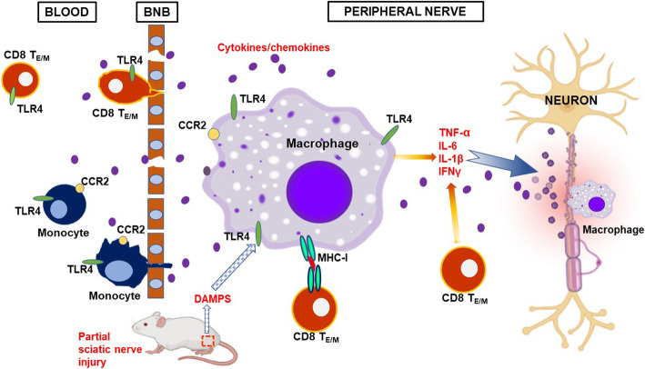
Background: While the etiology remains elusive, macrophages and T cells in peripheral nerves are considered as effector cells mediating autoimmune peripheral neuropathy (APN), such as Guillain-Barre syndrome. By recognizing both pathogen-associated molecular patterns (PAMPs) and damage-associated molecular patterns (DAMPs) signals, TLRs play a central role in the initiation of both innate and adaptive immune responses. In this study, we aimed to understand the involvement of TLR4 in the pathogenesis of APN and explore the potential of TLR4 as a drug target for therapeutic use.

Methods: APN was induced by a partial ligation on one of the sciatic nerves in B7.2 (L31) transgenic mice which possess a predisposed inflammatory background. APN pathology and neurological function were evaluated on the other non-injured sciatic nerve.

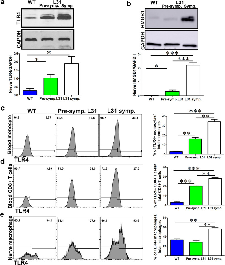
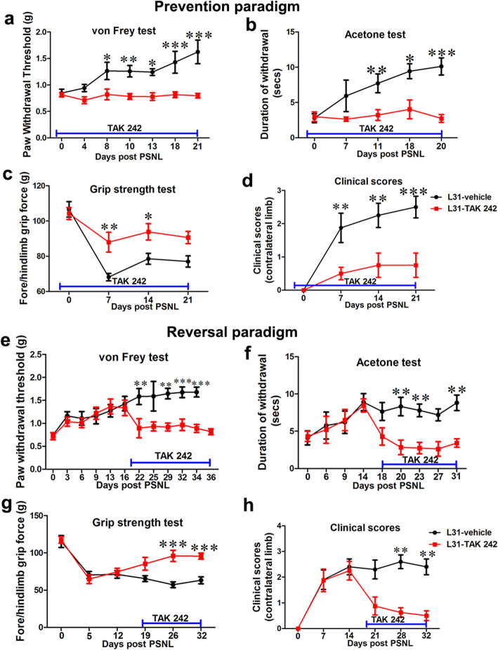
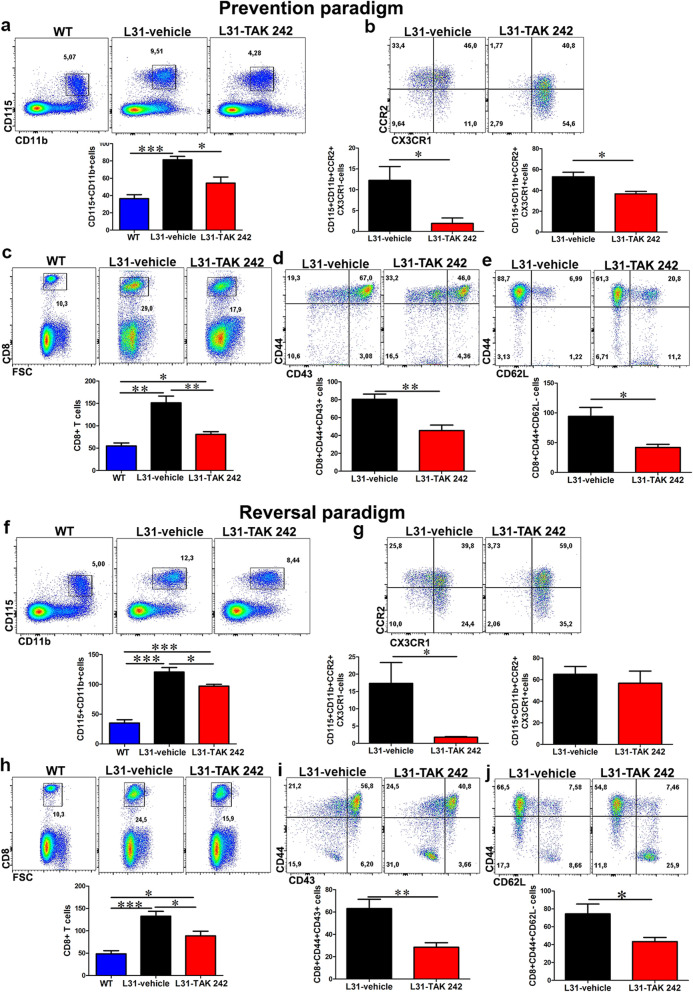
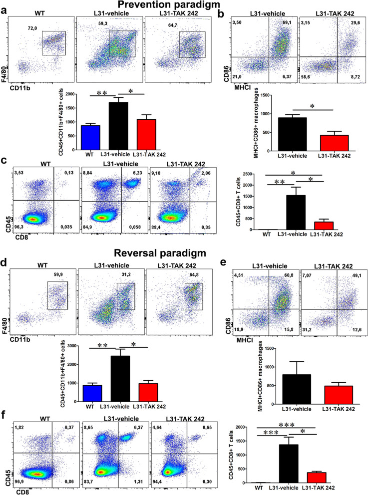
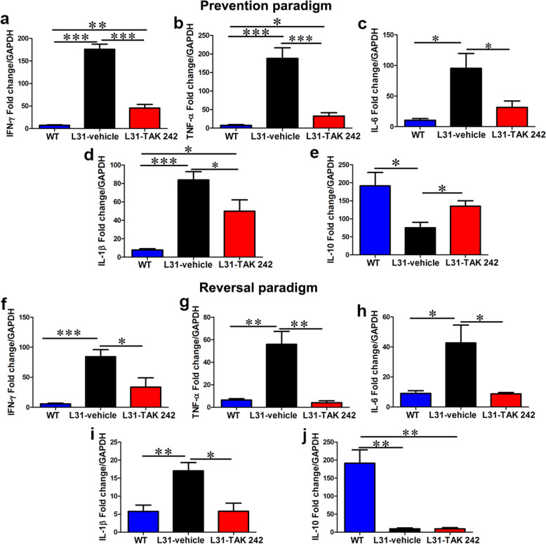
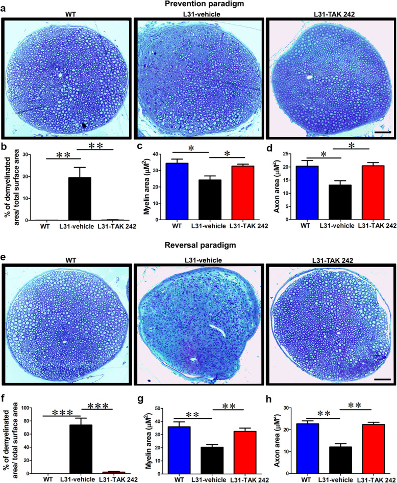
Results: TLR4 and its endogenous ligand HMGB1 were highly expressed in L31 mice, in circulating immune cells and in peripheral nerves. Enhanced TLR4 signaling was blocked with TAK 242, a selective TLR4 inhibitor, before and after disease onset. Intraperitoneal administration of TAK 242 not only inhibited monocyte, macrophage and CD8+ T cell activation, but also reduced the release of pro-inflammatory cytokines. TAK 242 protected mice from severe myelin and axonal loss, resulting in a remarkable improvement in mouse motor and sensory functions. TAK 242 was effective in alleviating the disease in both preventive and reversal paradigms.

Conclusion: The study identified the critical contribution of TLR4-mediated macrophage activation in disease course and provided strong evidence to support TLR4 as a useful drug target for treating inflammatory autoimmune neuropathy.

Keywords: Autoimmunity; CD8+ T cells; DAMPs; Demyelination; Inflammation; Macrophages; TLR4.


Conflict of interest statement

The authors declare no competing financial interests.

Figures

Fig. 1
Fig. 1
The expression of TLR4 and HMGB1 in the blood and nerve of L31 mice. a A representative western blot image showed the expression of TLR4 in the nerve of WT and L31 mice. TLR4 is significantly increased in L31 mice, with the highest increase found in L31 symptomatic mice. b A representative western blot image depicted the expression of HMGB1 in L31 mice. HMGB1 expression was increased mainly in the nerve of L31-symptomartic mice. c A representative flow cytometry histogram showed monocytes TLR4 expression in the blood. A quantitative analysis showed that TLR4 is robustly expressed in L31 mice with the highest increase found in L31-symptomatic mice. d TLR4 is expressed on blood CD8+ T cells in L31 mice with a significant increase in symptomatic mice. Quantification (c, d) depicted the number of cells per μl blood. e TLR4 is expressed on sciatic nerve macrophages in WT and L31 mice. Quantitative analysis showed a significant increase in expression in L31 symptomatic mice. Quantification (e) depicted the number of cells per a segment of 2-cm-long sciatic nerve. n = 4–5/group, *p < 0.05; **p < 0.01; ***p < 0.001
Fig. 2
Fig. 2
The effect of TLR4 signaling deficiency on sensory and motor deficits/ recovery in L31 mice. In the prevention paradigm (ad), a compared to mice in the vehicle group, paw withdrawal thresholds assessed using von Frey test maintained to similar level of baseline for mice in treated group, indicating that TAK 242 prevented disease associated numbness/ mechanical hyposensitivity. b Thermal sensitivity examined by acetone test was also normal in treated mice. c Quantitative data from grip strength showed that muscle strength was greater in treated group compared to vehicle group. d Clinical scores showed no appearance of neurological symptoms such as tail weakness and weakness of hind limb in treated group. In the reversal paradigm, treatment started between D17 post-PSNL when sensory and motor deficits were established (eh). e TAK 242 reversed mechanical hyposensitivity in treated group to pre-PSNL level. f Increased paw withdrawal duration in acetone test was equally reversed in treated mice. g Quantitative data from grip strength showed that the decreased muscle strength was reversed in treatment group, although not to baseline level. h Clinical score showed that the incidence of motor deficits was reduced in treated group. n = 10–12/group; *p < 0.05; **p < 0.01; ***p < 0.001
Fig. 3
Fig. 3
The effect of TLR4 signaling inhibition on monocyte and CD8+ T cells activation status in the blood of L31 mice. In the prevention paradigm (ae), a representative flow cytometry dot plot and quantification analysis showed a decrease in the number of CD115+CD11b+ monocytes in the blood of treated mice following TLR4 inhibition. b CCR2+ monocytes (pro-inflammatory) subset which includes CCR2+/CX3CR1 and CCR2+/CX3CR1+ were significantly reduced in treated group. c Similarly, the frequency and number of CD8+ T cells were reduced in the blood of treated mice. d The majority of the reduced CD8+ T cells in the treated group were of the activated subset (CD44+CD43+). e The frequency and number of CD8+ T cells with effector memory phenotype (CD44+CD62L) were significantly reduced in the blood of treated mice. In the reversal paradigm (fj), f frequency and total number of CD115+CD11b+ monocytes was significantly reduced in treated group. g The inflammatory subset (CCR2+CX3CR1) group was significantly reduced in treated mice as shown by representative flow and quantitative data. CCR2+/CX3CR1+ number appeared similar in both groups. h CD8+ T cells frequency and absolute number reduced in the blood following TLR4 signaling inhibition. i Majority of the reduced CD8+ T cells were of the activated (CD44+CD43+) and j effector memory phenotype (CD44+CD62L-). All quantitative analyses are shown as number of cells per μl blood. n = 6/group; *p < 0.05; **p < 0.01; ***p < 0.001
Fig. 4
Fig. 4
The effect of TLR4 signaling inhibition on macrophage and CD8+ T cells number and activation status in the nerve of L31 mice. In the prevention paradigm (ad), a quantitative analysis from flow cytometry showed a reduced number of macrophages (CD45+CD11b+F4/80+) in treated group at the end of the experiment. (b) The number of activated macrophages (MHCI+CD86+) were significantly reduced in treated group. c Frequency and absolute number of infiltrating CD8+ T cells were significantly diminished in treated group. In the reversal paradigm (df), d quantitative analysis from flow cytometry showed that TAK 242 favored the reduction of nerve macrophages (CD45+CD11b+F4/80+). e A quantitative analysis indicated a reduced number of activated macrophages (MHCI+CD86+). Majority of the reduced macrophages in treated group were those committed to antigen-presenting function (MHCI+CD86+). f A representative flow cytometry dot plot showed a reduced frequency and number of infiltrating CD8+ T cells in the sciatic nerve of treated mice. All quantitative analyses are shown as number of cells per a segment of 2-cm-long sciatic nerve. n = 6/group; *p < 0.05; **p < 0.01; ***p < 0.001
Fig. 5
Fig. 5
The effect of TLR4 signaling inhibition on cytokine expression in L31 mice. In the prevention paradigm (ae), real-time quantitative PCR showed a significantly reduced expression of pro-inflammatory molecules a IFN-γ, b TNF-α, c IL-6, and d IL-1β, in the TAk 242-treated group. e The mRNA expression of the anti-inflammatory molecule, IL-10 was significantly enhanced in the treated group. In the reversal paradigm (fj), the mRNA expression levels of f IFN-γ, g TNF-α, h IL-6, and i IL-1β, in the peripheral nerve were significantly reduced by TAK 242 treatment. j The mRNA level of IL-10 was unchanged across both groups in the reversal paradigm. n = 6/group; *p < 0.05; **p < 0.01; ***p < 0.001
Fig. 6
Fig. 6
The effect of TLR4 signaling inhibition on axon and myelin integrity in L31-symp mice. In the prevention paradigm (ad), a representative micrographs of sciatic nerve cross-sections showed partial axonal damage and myelin loss in vehicle group, which was not obvious in TAK 242-treated mice. Nerve structure in TAK 242 remain comparable to WT mice. b Semi quantitative analysis revealed a partial axonal and myelin loss (20%) in vehicle-treated mice, which was prevented in TAK 242 mice. c, d Assessment of myelin and axon area on randomly selected 100 myelinated axons revealed that in the vehicle group, even in the area where nerve structure seems preserved, myelin and axon area was reduced indicating that myelin could become thinner and axons could be atrophic, which were prevented by TAK 242. In the reversal paradigm (eh), e representative micrographs of sciatic nerve cross sections showed massive demyelination and axonal loss in the vehicle group, TAK 242-treated mice showed an almost intact nerve structure which was comparable to control mice. f More than 70% of nerve structure was destroyed in vehicle-treated mice, which was completely rescued in TAK 242-treated mice. g, h Assessment of myelin and axon area on randomly selected 100 myelinated axons revealed that in the vehicle group, even in the remaining area where nerve structure seems preserved, myelin and axon area was significantly reduced indicating that myelin might become thinner and axons could be atrophic, which were rescued by TAK 242. n = 4–6/group; *p < 0.05; **p < 0.01; ***p < 0.001
Fig. 7
Fig. 7
Proposed cascade of TLR4-mediated mechanisms leading to inflammatory peripheral neuropathy in L31 mice. See text for detailed explanation

Similar articles

Cited by

References

    1. van den Berg B, Walgaard C, Drenthen J, Fokke C, Jacobs BC, van Doorn PA. Guillain-Barre syndrome: pathogenesis, diagnosis, treatment and prognosis. Nat Rev Neurol. 2014;10(8):469–482. doi: 10.1038/nrneurol.2014.121. - DOI - PubMed
    1. van den Berg B, Bunschoten C, van Doorn PA, Jacobs BC. Mortality in Guillain-Barre syndrome. Neurology. 2013;80(18):1650–1654. doi: 10.1212/WNL.0b013e3182904fcc. - DOI - PubMed
    1. Ubogu EE. Chemokine receptors as specific anti-inflammatory targets in peripheral nerves. Endocr Metab Immune Disord Drug Targets. 2011;11(2):141–153. doi: 10.2174/187153011795564124. - DOI - PubMed
    1. Bourque PR, Chardon JW, Massie R. Autoimmune peripheral neuropathies. Clin Chim Acta. 2015;449:37–42. doi: 10.1016/j.cca.2015.02.039. - DOI - PubMed
    1. Dalakas MC. Pathogenesis of immune-mediated neuropathies. Biochim Biophys Acta. 2015;1852(4):658–666. doi: 10.1016/j.bbadis.2014.06.013. - DOI - PubMed

MeSH terms

Substances