. 2021 Mar 22;12(1):201.
doi: 10.1186/s13287-021-02271-3.

Human placenta mesenchymal stem cell-derived exosomes delay H2O2-induced aging in mouse cholangioids

Affiliations

Human placenta mesenchymal stem cell-derived exosomes delay H2O2-induced aging in mouse cholangioids

Wenyi Chen et al. Stem Cell Res Ther. .

Abstract

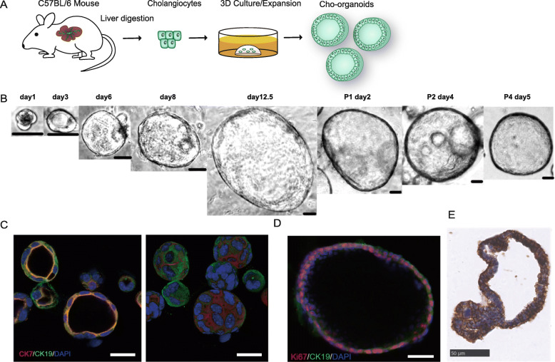
Background: Cholangiocyte senescence is an important pathological process in diseases such as primary sclerosing cholangitis (PSC) and primary biliary cirrhosis (PBC). Stem cell/induced pluripotent stem cell-derived exosomes have shown anti-senescence effects in various diseases. We applied novel organoid culture technology to establish and characterize cholangiocyte organoids (cholangioids) with oxidative stress-induced senescence and then investigated whether human placenta mesenchymal stem cell (hPMSC)-derived exosomes exerted a protective effect in senescent cholangioids.

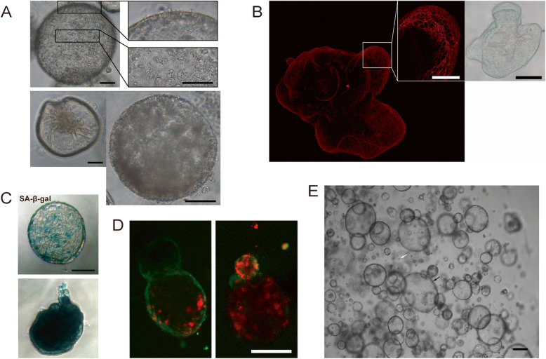
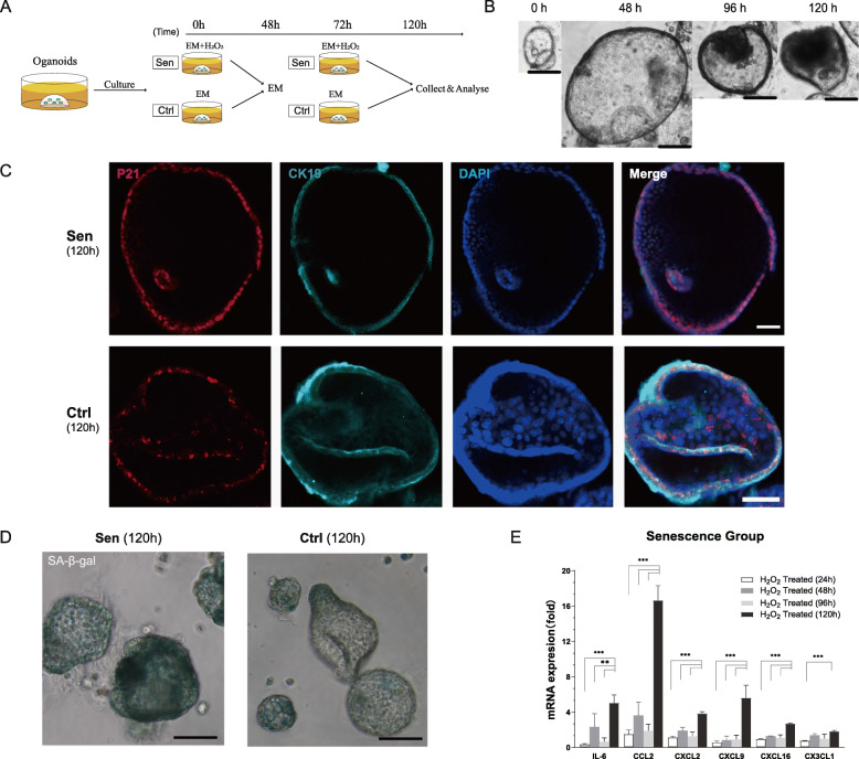
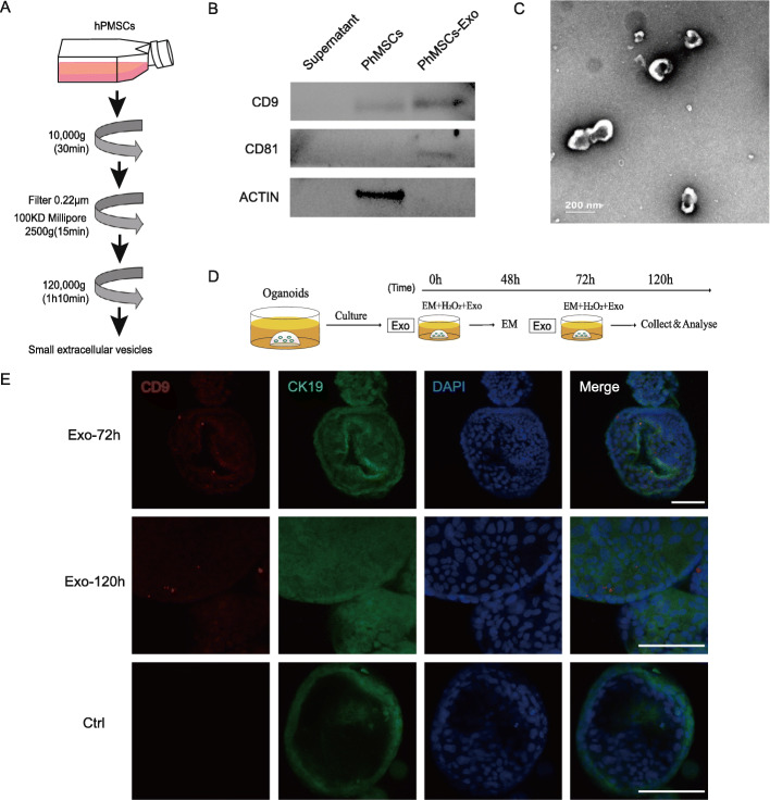
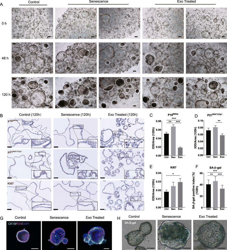
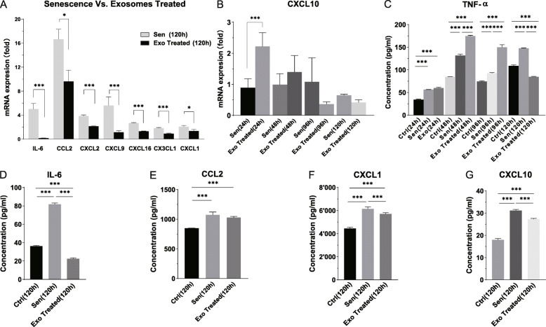
Methods: We identified the growth characteristics of cholangioids by light microscopy and confocal microscopy. Exosomes were introduced concurrently with H2O2 into the cholangioids. Using immunohistochemistry and immunofluorescence staining analyses, we assessed the expression patterns of the senescence markers p16INK4a, p21WAF1/Cip1, and senescence-associated β-galactosidase (SA-β-gal) and then characterized the mRNA and protein expression levels of chemokines and senescence-associated secretory phenotype (SASP) components.

Results: Well-established cholangioids expressed cholangiocyte-specific markers. Oxidative stress-induced senescence enhanced the expression of the senescence-associated proteins p16INK4a, p21WAF1/Cip1, and SA-β-gal in senescent cholangioids compared with the control group. Treatment with hPMSC-derived exosomes delayed the cholangioid aging progress and reduced the levels of SASP components (i.e., interleukin-6 and chemokine CC ligand 2).

Conclusions: Senescent organoids are a potential novel model for better understanding senescence progression in cholangiocytes. hPMSC-derived exosomes exert protective effects against senescent cholangioids under oxidative stress-induced injury by delaying aging and reducing SASP components, which might have therapeutic potential for PSC or PBC.

Keywords: Cholangiocytes; Exosomes; Organoids; Primary sclerosing cholangitis; Senescence.


Conflict of interest statement

The authors declare no competing interests.

Figures

Fig. 1
Fig. 1
Murine cholangiocyte organoids originating from mature cholangiocytes. a Schematic of cholangioid development. Primary cholangiocytes collected from wild-type C57BL/6 mice by liver duct cell digestion were embedded in Matrigel and covered with expansion medium, allowing them to grow into organoids. b Representative image of long-term cultured cholangioids (P1, passage 1; P2, passage 2; P4, passage 4) (scale bar, 50 μm). c Confocal images of cholangioids expressing specific cholangiocyte markers CK19 (green) and CK7 (red), with blue DAPI staining (scale bar, 20 μm). Left: organoids in two-dimensional form; right: organoids in 3D form. d CK19-positive (green) cholangioids expressing proliferating protein Ki67 (red) (scale bar, 50 μm). e Representative immunohistochemistry images of epithelial cell adhesion molecule (EpCAM) in cholangioids (scale bar, 50 μm)
Fig. 2
Fig. 2
Features of long-term cultured cholangioids. a Organoids (P1 day 12) observed by light microscopy. Cells scattered inside and outside the organoid lumen (top, right bottom); shrunken organoids (left bottom) (scale bar, 100 μm). b CK7-positive cholangiocytes within organoids (P0 day 11). Cells remained close together and developed into more than one layer, as observed by confocal laser scanning microscopy (scale bar: left, 50 μm; right, 100 μm). c SA-β-gal staining of cholangioids (P1 day 12), some senescent cholangiocytes (green, top) and completely senescent cholangioids (bottom) (scale bar, 100 μm). d Viability/cytotoxicity assay of organoids (P1 day 4). Dead cells (red) aggregated inside the organoid lumen (green) (scale bar, 50 μm). e Organoids (P1 day 2) observed by light microscopy. White arrow indicates single cholangiocyte, and black arrow indicates organoids grown from single cholangiocytes (scale bar, 200 μm)
Fig. 3
Fig. 3
Establishment of senescent cholangioids by H2O2 treatment. a Schematic of the treatment of expansion medium (EM) containing 50 nM H2O2 was classified into the Sen group. The Ctrl group was treated with EM only. Medium was changed at intervals of 24 or 48 h. b Representative light microscopy image of H2O2-stimulated organoids, showing cytological features characteristic of senescence (reduced size, darkened spheroids) (scale bar, 200 μm). c Typical organoids in the Sen and Ctrl groups expressing cell cycle arrest protein p21WAF1/Cip1 (red) after exposure to oxidative stress for 120 h; CK19 protein was stained cyan and cell nuclei were stained blue (scale bar, 50 μm). d SA-β-gal-positive organoids (green) in the Sen and Ctrl groups following oxidative stress induction for 120 h, observed by light microscopy (scale bar, 100 μm). e mRNA expression levels of SASP components and chemokines (IL-6, CCL2, CXCL2, CXCL16, and CX3CL1) increased significantly at 120 h, compared with 24, 48, and 96 h. Data are presented as mean ± standard deviation (SD) (***P < 0.001, **P < 0.01, n = 4; ordinary one-way ANOVA)
Fig. 4
Fig. 4
Exosomes were derived from hPMSCs and co-cultured with organoids. a Schematic representation of exosome collection. b Western blotting: hPMSC-derived exosomes expressing exosome-specific markers CD9 and CD81. c hPMSC-derived exosomes observed by transmission electron microscopy (scale bar, 200 nm). d Schematic of exosome treatment. Organoids treated with expansion medium (EM) containing 50 nM H2O2 and 0.1 μg/ml exosomes were classified into the Exo group. Cholangioids were collected and analyzed at 48, 72, and 120 h. e Presence of the exosome-specific marker CD9 (red) in cholangioids (CK19, green) of the Exo group after culture for 72 and 120 h, demonstrated by immunofluorescence analysis (scale bar, 100 μm)
Fig. 5
Fig. 5
hPMSC-derived exosomes have a protective effect in senescent cholangioids. a Comparison of organoids among the Ctrl, Sen, and Exo groups at 0 h (top), 48 h (middle), and 120 h (bottom). Typical senescent organoids at 120 h are indicated by white circles (scale bar, 200 μm). b Comparison of the immunohistochemical staining results for proliferation marker Ki67, as well as the senescence-associated markers p16INK4a and p21WAF1/Cip1 among the Ctrl, Sen, and Exo groups at 120 h (× 40 objective, scale bar, 200 μm; rectangle indicates × 80 objective). ce Semi-quantitative analysis of p16INK4a, p21WAF1/Cip1, and Ki67 immunohistochemical staining results at 120 h. These data were acquired with a × 20 objective, and Image-Pro Plus software was used to analyze 10 randomly selected areas (two-tailed t test, mean ± standard error of the mean, n = 10, ***P < 0.001, **P < 0.01, *P < 0.05). f Percentages of SA-β-gal-positive organoids in 30 randomly selected areas, counted under a × 20 objective with bright field illumination (two-tailed t test, mean ± SD, n = 30, ***P < 0.001, **P < 0.01). g Representative CK19-positive cholangioids (cyan) in 3D form. The area of p21WAF1/Cip1-positive staining was greater in the Sen group than in the other two groups, while the Exo group showed the smallest area (scale bar, 100 μm). h SA-β-gal-positive cholangioids. Organoids were collected and analyzed at 120 h (green, scale bar, 50 μm)
Fig. 6
Fig. 6
Exosomes reduced the expression of SASP components and chemokines in senescent cholangioids. a mRNA expression levels of SASP components (IL-6, CCL2, CXCL2, CXCL9, CXCL16, CX3CL1, and CXCL1), which decreased significantly at 120 h in the Exo group compared with the Sen group. Data are presented as mean ± SD (n = 4). b Fold changes of CXCL10 mRNA expression in organoids at different time points. Data are presented as mean ± SD (n = 4). c Tumor necrosis factor-α protein concentrations in culture supernatant at 24, 48, 96, and 120 h. Data are presented as mean ± SD (n = 3). dg IL-6, CCL2, CXCL1, and CXCL10 protein concentrations in culture supernatant at 120 h, which were significantly decreased in the Exo group compared with the Ctrl and Sen groups. Data are presented as mean ± SD (n = 3). One-way ANOVA, ***P < 0.001, **P < 0.01, *P < 0.05

Similar articles

Cited by

References

    1. Campisi J, d’Adda di Fagagna F. Cellular senescence: when bad things happen to good cells. Nat Rev Mol Cell Biol 2007;8(9):729–740, DOI: 10.1038/nrm2233. - PubMed
    1. Meng L, Quezada M, Levine P, Han Y, McDaniel K, Zhou T, Lin E, Glaser S, Meng F, Francis H, Alpini G. Functional role of cellular senescence in biliary injury. Am J Pathol. 2015;185(3):602–609. doi: 10.1016/j.ajpath.2014.10.027. - DOI - PMC - PubMed
    1. Pinto C, Giordano DM, Maroni L, Marzioni M. Role of inflammation and proinflammatory cytokines in cholangiocyte pathophysiology. Biochimica et biophysica acta Molecular basis of disease. 2018;1864(4 Pt B):1270–1278. - PubMed
    1. Ferreira-Gonzalez S, Lu WY, Raven A, Dwyer B, Man TY, O’Duibhir E, et al. Paracrine cellular senescence exacerbates biliary injury and impairs regeneration. Nat Commun. 2018;9(1):1020. doi: 10.1038/s41467-018-03299-5. - DOI - PMC - PubMed
    1. Chung BK, Karlsen TH, Folseraas T. Cholangiocytes in the pathogenesis of primary sclerosing cholangitis and development of cholangiocarcinoma. Biochimica et biophysica acta Molecular basis of disease. 2018;1864(4 Pt B):1390–1400. - PubMed

Publication types

Substances

LinkOut - more resources