. 2021 Mar 22;23(1):93.
doi: 10.1186/s13075-021-02463-5.

Diet-induced obesity leads to behavioral indicators of pain preceding structural joint damage in wild-type mice

Affiliations

Diet-induced obesity leads to behavioral indicators of pain preceding structural joint damage in wild-type mice

Geoffrey J Kerr et al. Arthritis Res Ther. .

Abstract

Introduction: Obesity is one of the largest modifiable risk factors for the development of musculoskeletal diseases, including intervertebral disc (IVD) degeneration and back pain. Despite the clinical association, no studies have directly assessed whether diet-induced obesity accelerates IVD degeneration, back pain, or investigated the biological mediators underlying this association. In this study, we examine the effects of chronic consumption of a high-fat or high-fat/high-sugar (western) diet on the IVD, knee joint, and pain-associated outcomes.

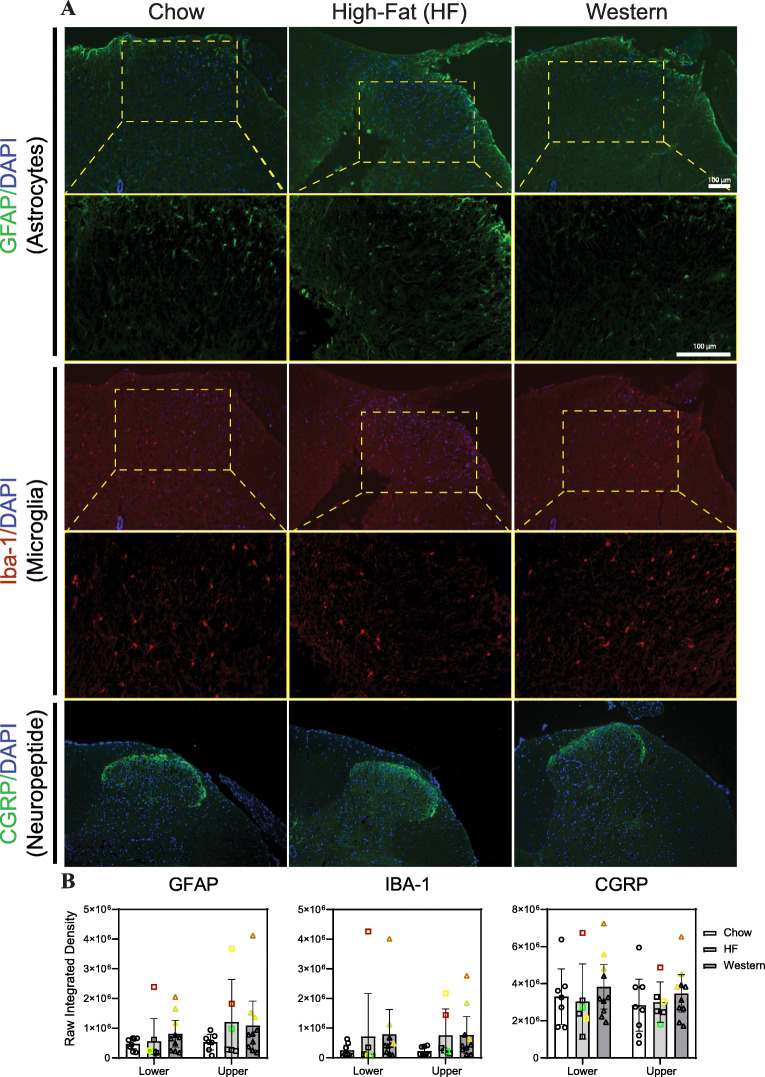
Methods: Male C57BL/6N mice were randomized into one of three diet groups (chow control; high-fat; high-fat, high-sugar western diet) at 10 weeks of age and remained on the diet for 12, 24, or 40 weeks. At endpoint, animals were assessed for behavioral indicators of pain, joint tissues were collected for histological and molecular analysis, serum was collected to assess for markers of systemic inflammation, and IBA-1, GFAP, and CGRP were measured in spinal cords by immunohistochemistry.

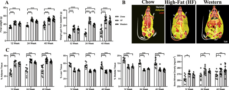
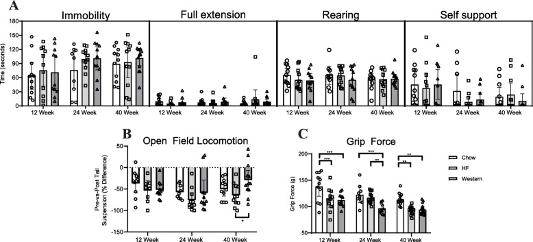
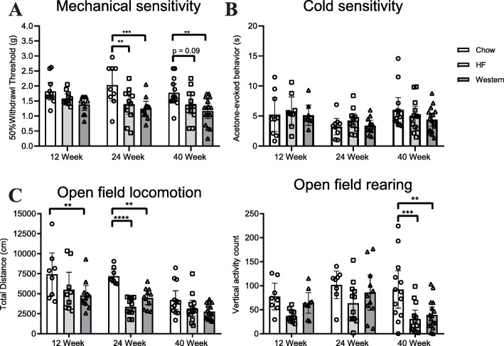
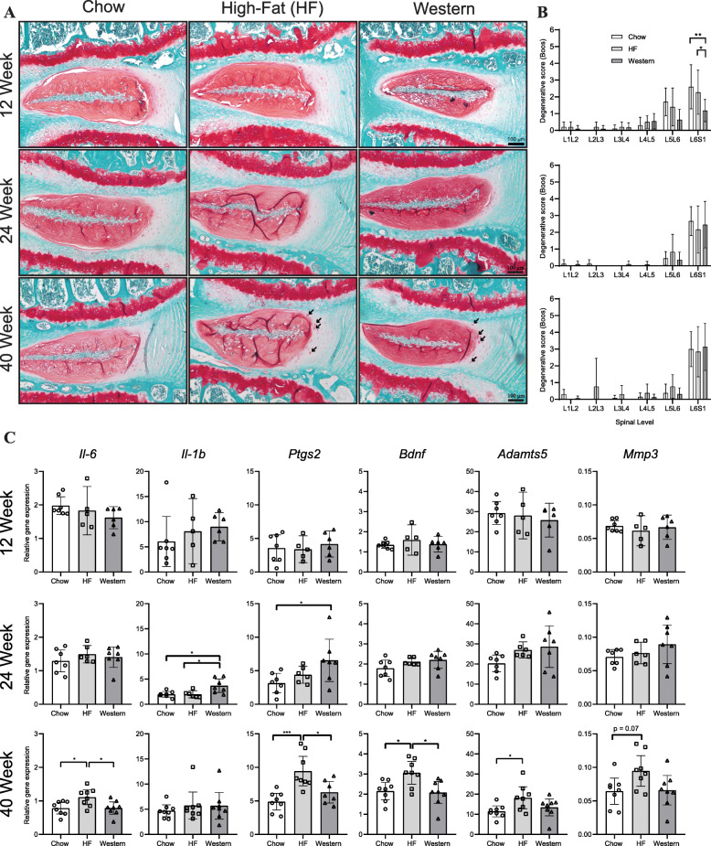
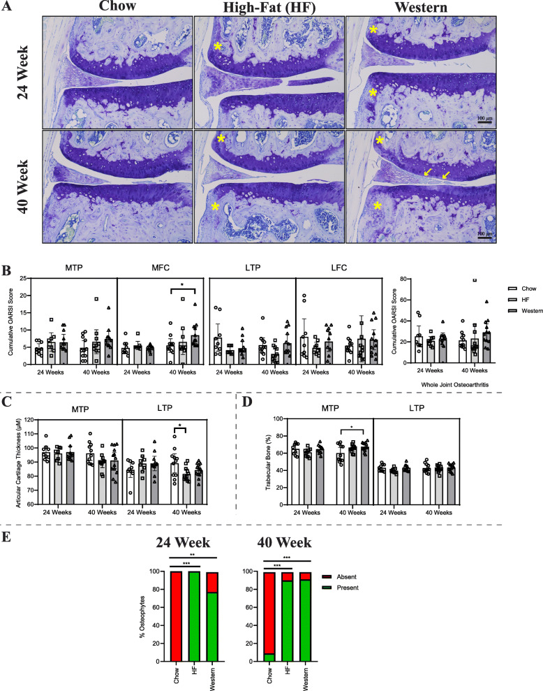
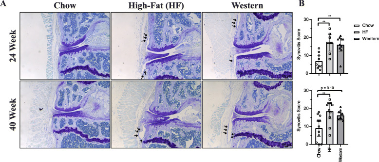
Results: Animals fed obesogenic (high-fat or western) diets showed behavioral indicators of pain beginning at 12 weeks and persisting up to 40 weeks of diet consumption. Histological indicators of moderate joint degeneration were detected in the IVD and knee following 40 weeks on the experimental diets. Mice fed the obesogenic diets showed synovitis, increased intradiscal expression of inflammatory cytokines and circulating levels of MCP-1 compared to control. Linear regression modeling demonstrated that age and diet were both significant predictors of most pain-related behavioral outcomes, but not histopathological joint degeneration. Synovitis was associated with alterations in spontaneous activity.

Conclusion: Diet-induced obesity accelerates IVD degeneration and knee OA in mice; however, pain-related behaviors precede and are independent of histopathological structural damage. These findings contribute to understanding the source of obesity-related back pain and the contribution of structural IVD degeneration.

Keywords: Behavioral measures of pain; High-fat diet; Intervertebral disc degeneration; Obesity; Osteoarthritis; Western diet.


Conflict of interest statement

The authors declare no competing interests.

Figures

Fig. 1
Fig. 1
Chronic consumption of the high-fat and western diets increases adiposity in C57BL/6N mice. a At all timepoints, mice fed the high-fat and western diets showed significant increases in overall weight and in weight-gain from baseline compared to age-matched chow-fed controls. b Representative reconstructed micro-CT images of mice following 40 weeks of experimental diets. Isotropic surface-rendering of skeletal tissue (indicated in white) is overlaid with a mid-coronal slice where lean tissue is indicated in red and adipose tissue is indicated in yellow. c Quantitative micro-CT analysis of whole-body composition showed a significant increase in adiposity and significant decreases in both percentage of lean and skeletal and in mice fed the high-fat and western diet mice compared to age-matched chow-fed controls at all time points. A significant increase was also seen in bone mineral density in mice following consumption of the high-fat and western diets at the 24- and 40-week timepoints. n = 9–16 mice per timepoint, per diet. Data are displayed as mean ± 95% CI; data points for each mouse are graphed within each group. *P < 0.05, **P < 0.01, ***P < 0.001, ****P < 0.0001 by 2-way ANOVA
Fig. 2
Fig. 2
Diet-induced obesity reduces grip strength but does not alter behavior in tail suspension. a During tail suspension, the duration of time spent by mice immobile, in full extension, rearing or self-supported was quantified. No significant differences were detected between diet groups at any of the time points assessed. b Stretch-evoked discomfort was assessed using the open field assay, in which the total distance covered in 5 min immediately before (pre) and after (post) the 3 min tail suspension assay was quantified. Obesity induced by the high-fat and western diets did not alter behavior of mice in open field compared to age-matched chow-fed controls at any of the time points assessed. However, a significant difference was seen between mice fed a high-fat and western diet at the 40-week timepoint. c Grip force during axial stretch was reduced in obese mice. Mice fed the high-fat diet showed a significant decrease in grip force at the 12- and 40-week time points compared to age-matched chow-fed controls. Mice fed the western diet showed a significant decrease in grip force compared to age-matched chow-fed controls at all time points. n = 9–16 animals per timepoint, per diet. Data are plotted mean ± 95% CI; data points for each mouse are graphed within each group. *P < 0.05, **P < 0.01, ***P < 0.001 by 2-way ANOVA
Fig. 3
Fig. 3
Diet-induced obesity increases sensitivity to mechanical stimulation and alters spontaneous locomotion. a Mechanical sensitivity of the hind paw was assessed by manual application of Von Frey filaments using the Chaplin up-down method. Mice fed the western diet showed a significant decrease in withdrawal threshold at the 24- and 40-week time points compared to age-matched chow-fed controls, indicative of increased mechanical sensitivity. Mice fed a high-fat diet showed a significant decrease in withdrawal threshold at the 24-week timepoint compared to control. b Sensitivity to cold was assessed by measuring the time spent in behavior evoked by evaporative cooling of acetone (flicking, stamping, or licking of ventral surface of the paw) during the first 40 s following application of acetone to the ventral surface of the hind paw. No significant differences were seen between the diet groups at any timepoint. c Spontaneous locomotor activity was recorded over three 2-h sessions and averaged. Mice fed the western diet showed a significant decrease in the total distance traveled at the 12- and 24-week timepoint compared to age-matched chow-fed controls, while mice fed the high-fat diet showed a decrease at the 24-week timepoint. The number of rearing events was significantly decreased in mice fed the high-fat and western diets compared to controls at the 40-week timepoint. n = 9–16 animals per timepoint, per diet. Data are plotted mean ± 95% CI; data points for each mouse are graphed within each group. *P < 0.05, **P < 0.01, ***P < 0.001, ****P < 0.0001 by 2-way ANOVA
Fig. 4
Fig. 4
Effect of diet-induced obesity on the intervertebral disc. a Representative histological sections of the L6/S1 spinal level of the lumbar spine stained with safranin-o/fast green from mice fed control chow, high-fat, or western diet for 12, 24, or 40 weeks. The accumulation of hypertrophic cells was detected within the inner annulus fibrosus of mice fed the high-fat and western diets for 40 weeks (highlighted by black arrows). b Evaluation of the grade of histopathological IVD degeneration using the modified Boos scoring system showed no significant differences between mice fed the control chow, high-fat, or western diets at the 24- and 40-week timepoints. At the 12-week timepoint, mice fed the western diet showed a significant decrease in the degenerative score compared to mice fed chow and high-fat diets. n = 9–16 animals per timepoint, per diet. Data are analyzed by Kruskal-Wallis test. c SYBR-based qPCR of intact thoracic intervertebral discs showed no significant difference between mice fed a chow, high-fat, or western diet at the 12-week timepoint for any genes investigated. At the 24-week timepoint, significant increases in Il-1b and Ptgs2 expression were seen in mice fed the western diet compared to control. By 40 weeks, significant increases in Il-6, Ptgs2, Bdnf, and Adamts5 expression were seen in mice fed the high-fat diet compared to control. n = 5–8 animals per diet/per timepoint. Analyzed by one-way ANOVA. All data are plotted mean ± 95% CI; data points for each mouse are graphed within each group. *P < 0.05, ***P < 0.001
Fig. 5
Fig. 5
Effect of diet-induced obesity on the knee joint. a Representative histological coronal sections of the medial knee compartment stained with toluidine blue from mice fed either control chow, high-fat, or western diet for 24 or 40 weeks. Images are oriented with the medial femoral condyle (MFC) located superiorly, and the medial tibial plateau (MTP) inferiorly. White arrows indicate a loss of proteoglycan staining and focal fibrillation of the cartilage, and yellow asterisks indicate osteophyte formation. b Histopathological grading of the knee joints using the murine Osteoarthritis Research Society International (OARSI) scale corresponding to MTP, MFC, lateral tibial plateau (LTP), and lateral femoral condyle (LFC), combined to generate the summed score for the whole joint. Mice fed the western diet for 40 weeks showed a significant increase in the degenerative score in the MFC compared to those fed the control chow. However, no difference was seen in the whole joint score between any of the groups at either timepoint. c Average articular cartilage thickness. After 40 weeks on the high-fat diet, mice presented with decreased articular cartilage thickness on the LTP. No other differences were seen in any other joint compartment. Data analyzed by Kruskal-Wallis test. d Percent trabecular bone in the medial and lateral subchondral compartments of the tibia. Mice fed  the western diet for 40 weeks exhibited significantly more trabecular bone in the medial compartment for the tibia. Analyzed by one-way ANOVA. e Presence and absence of osteophytes was assessed in all mice. Mice fed the high-fat and western diets showed increased osteophyte formation compared to chow-fed controls at both timepoints. Analyzed by Kruskal-Wallis test. n = 9–16 animals per diet/per timepoint. All data are plotted mean ± 95% CI, *P < 0.05, **P < 0.01, ***P < 0.001
Fig. 6
Fig. 6
Effect of diet-induced obesity on synovial inflammation and hyperplasia. a Representative coronal sections of the medial compartment of the knee stained with toluidine blue from mice fed either control chow, high-fat, or western diets for 24 or 40 weeks. Images are oriented with medial femoral condyle (MFC) located superiorly, and the medial tibial plateau (MTP) inferiorly. Black arrows indicate synovial hyperplasia. b Histopathological scoring assessed indicators of synovial hyperplasia and inflammatory infiltration. Summed scores represent the sum of the medial and lateral compartments scores across 7 serial sections. Mice fed a high-fat diet showed a significant increase in synovial inflammation and hyperplasia compared to chow-fed control at both the 24 and 40 week timepoint, while mice fed a western diet showed a significant increase in synovitis at only the 24-week timepoint. n = 8–12 animals per diet/per timepoint. Analyzed by Kruskal-Wallis test. All data are plotted mean ± 95% CI, **P < 0.01
Fig. 7
Fig. 7
Effect of diet-induced obesity on neuroplastic changes within the lumbar spinal cord. a Representative images showing transverse sections of the dorsal horn of the lumbar spinal cord used for immunohistochemical analysis of neuroplastic changes associated with chronic pain. Tissues were isolated from mice following 40 weeks of experimental diet. Slides were stained for glial fibrillary acidic protein (GFAP), ionized calcium-binding adapter molecule 1 (IBA1), and calcitonin gene-related peptide (CGRP). Yellow boxes indicate the region of interest for high magnification images (second row) for GFAP and IBA-1. (b) The fluorescence intensity was averaged across the region of interest (lamellae 1-4 of the dorsal horn) of the upper (L1-L2) and lower (L3-L6) lumbar spinal cords. Mice fed a high-fat or western diet for 40 weeks showed no significant differences in immunoreactivity for any of the proteins investigated. n = 6–8 animals/group. Individual data points of the same color indicate the same animal. Data are plotted mean ± 95% CI

Similar articles

Cited by

References

    1. Blüher M. Obesity: global epidemiology and pathogenesis. Nat Rev Endocrinol. 2019;15(5):288-98. - PubMed
    1. De Gonzalez AB, Hartge P, Cerhan JR, Flint AJ, Hannan L, MacInnis RJ, et al. Body-mass index and mortality among 1.46 million white adults. N Engl J Med. 2010;363(23):2211-9. - PMC - PubMed
    1. Taylor VH, Forhan M, Vigod SN, McIntyre RS, Morrison KM. The impact of obesity on quality of life. Best Pract Res. 2013;27(2):139-46. - PubMed
    1. Samartzis D, Karppinen J, Cheung JP, Lotz J. Disk degeneration and low back pain: are they fat-related conditions? Glob Spine J. 2013;3(3):133–44. - PMC - PubMed
    1. Shiri R, Karppinen J, Leino-Arjas P, Solovieva S, Viikari-Juntura E. The association between obesity and low back pain: a meta-analysis. Am J Epidemiol. 2010;171(2):135–54. - PubMed

Publication types