. 2021 Mar 22;12(1):202.
doi: 10.1186/s13287-021-02237-5.

Improvement of ECM-based bioroot regeneration via N-acetylcysteine-induced antioxidative effects

Affiliations

Improvement of ECM-based bioroot regeneration via N-acetylcysteine-induced antioxidative effects

Jiayu Zhang et al. Stem Cell Res Ther. .

Abstract

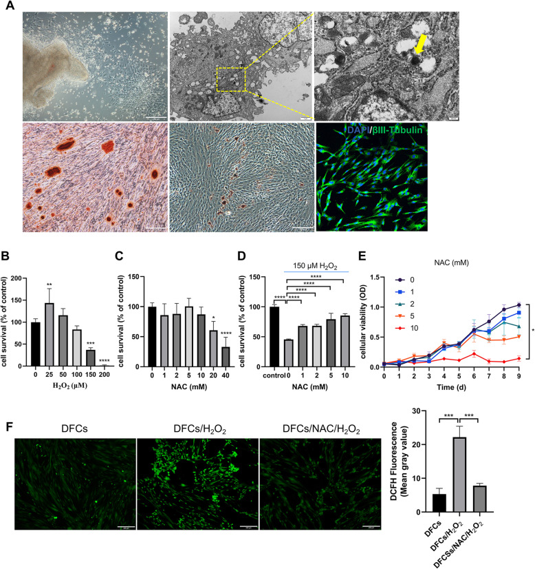
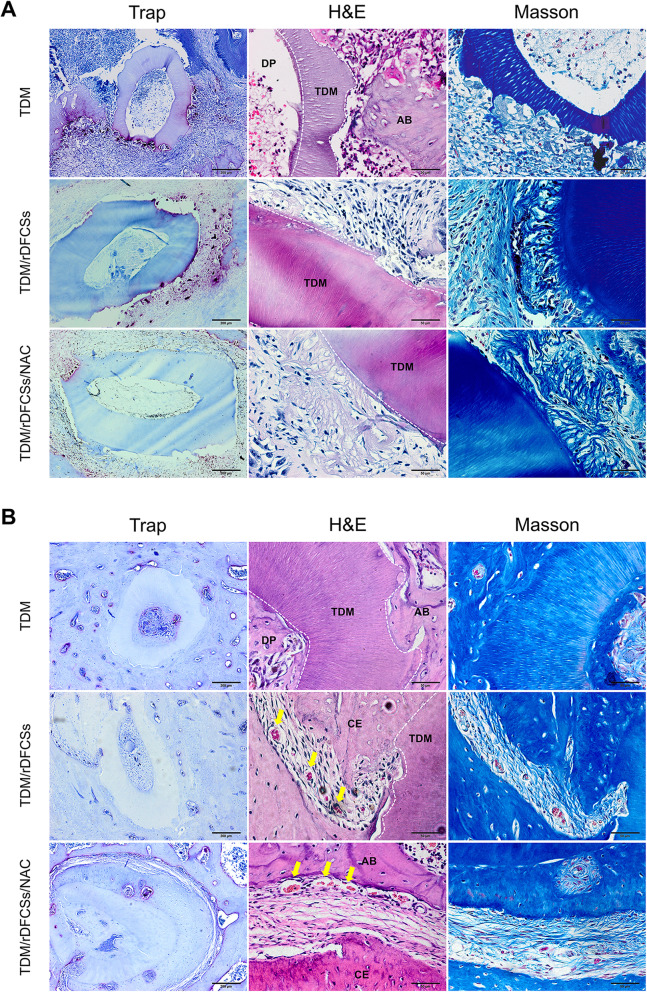
Background: The low survival rate or dysfunction of extracellular matrix (ECM)-based engineered organs caused by the adverse effects of unfavourable local microenvironments on seed cell viability and stemness, especially the effects of excessive reactive oxygen species (ROS), prompted us to examine the importance of controlling oxidative damage for tissue transplantation and regeneration. We sought to improve the tolerance of seed cells to the transplant microenvironment via antioxidant pathways, thus promoting transplant efficiency and achieving better tissue regeneration.

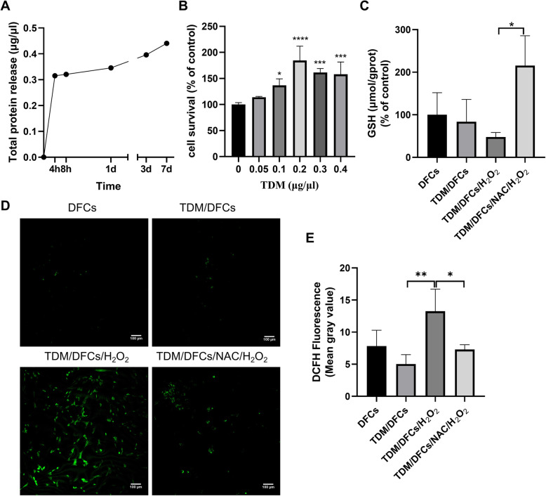
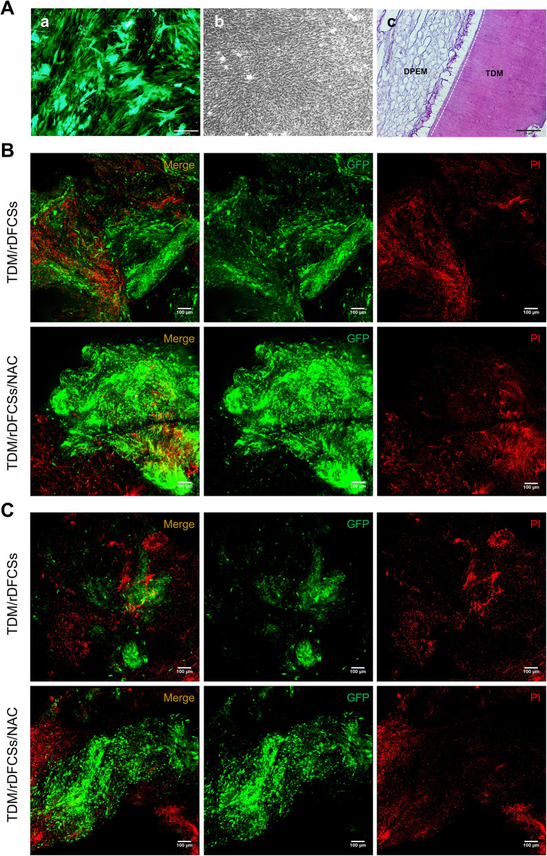
Methods: We improved the antioxidative properties of ECM-based bioroots with higher glutathione contents in dental follicle stem cells (DFCs) by pretreating cells or loading scaffolds with the antioxidant NAC. Additionally, we developed an in situ rat alveolar fossa implantation model to evaluate the long-term therapeutic effects of NAC in bioroot transplantation.

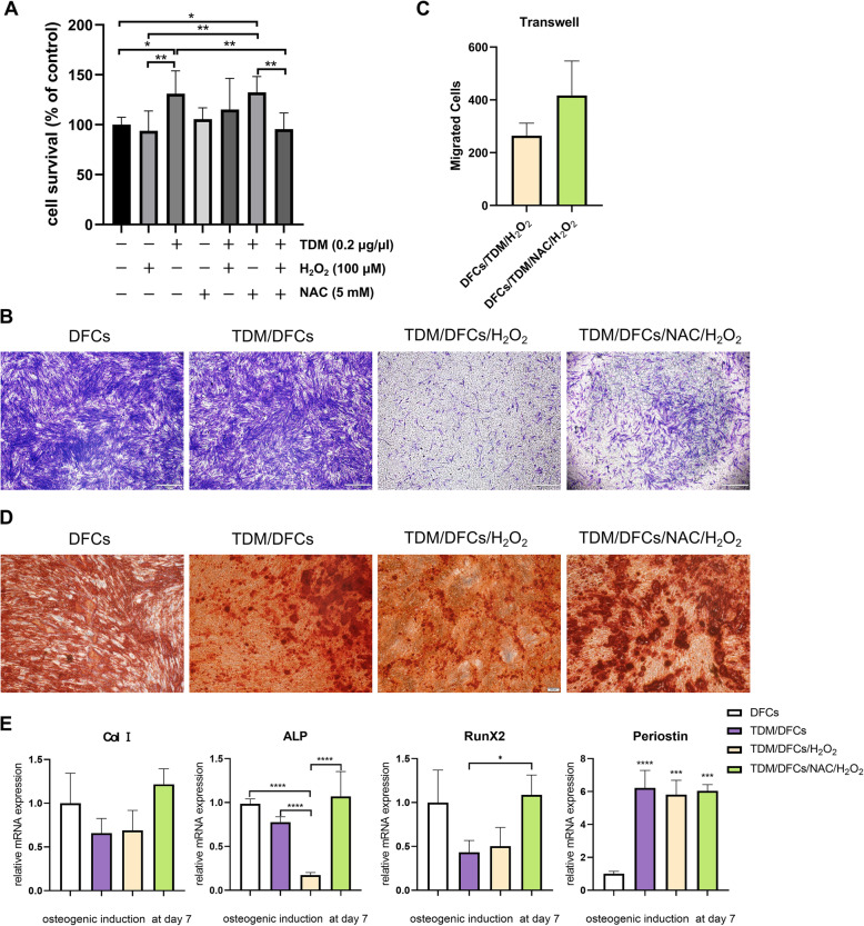
Results: The results showed that NAC decreased H2O2-induced cellular damage and maintained the differentiation potential of DFCs. The transplantation experiments further verified that NAC protected the biological properties of DFCs by repressing replacement resorption or ankylosis, thus facilitating bioroot regeneration.

Conclusions: The following findings suggest that NAC could significantly protect stem cell viability and stemness during oxidative stress and exert better and prolonged effects in bioroot intragrafts.

Keywords: Bioroot regeneration; Dental follicle stem cell; N-Acetylcysteine; Oxidative stress; Treated dentin matrix.


Conflict of interest statement

The authors declare that they have no competing interests.

Figures

Fig. 1
Fig. 1
Characterization of DFCs, establishment of H2O2-induced oxidative stress model, and determination of particular NAC treatment dose. a Characterization of DFCs. Primary DFCs were adherent to plastic and showed colony-forming abilities. TEM evaluations showed homogeneous electron-dense granules without membranous structures (indicated by yellow arrow). After being cultured in osteogenesis and adipogenesis-inducing media for 14 days and in neuronal-inducing medium for 2 h, the cultured DFCs showed mineralized nodules, lipid clusters, and positive βIII-tubulin expression, respectively. b Effect of different concentrations of H2O2 on the viability of hDFCs. c Effect of different concentrations of NAC on the viability of hDFCs. d Effect of NAC on the viability of hDFCs under oxidative stress conditions induced by 150 μm H2O2. e Effect of NAC on the proliferation of hDFCs for 1 week. f Assessment of ROS levels using fluorescence microscopy and statistical analysis of DCFH fluorescence from three experiments. Asterisks indicate statistically significant differences compared with the control group. ∗P < 0.05, ∗∗P < 0.01, and ∗∗∗P < 0.001
Fig. 2
Fig. 2
The effect of NAC and the H2O2-induced intracellular oxidative stress status on TDM/DFCs system. a Protein release of the TDM scaffold. b Effect of different concentrations of hTDM protein on the viability of hDFCs. c GSH level. d The level of ROS in hDFCs was measured by DCFH-DA with a fluorescence microscope. e statistical analysis of (d)
Fig. 3
Fig. 3
The effect of NAC on the proliferation, migration and osteogenic differentiation of hDFCs under H2O2-induced oxidative stress. a Effect of combination drug treatment on the viability of hDFCs. b After 24 h of culture in Transwell plates, crystal violet staining was performed. c Pretreatment with NAC led to increased hDFC migration to the opposite side of the membrane compared with that observed in the H2O2-treated groups. d The effect of NAC on the osteogenic differentiation of hDFCs stimulated with H2O2, as determined by Alizarin Red staining. e RT-PCR analysis of Collagen I, ALP, RunX2, and Periostin mRNA expression. ∗P < 0.05, ∗∗P < 0.01, and ∗∗∗P < 0.001
Fig. 4
Fig. 4
Allogeneic transplantation of bioroot composites in a Sprague-Dawley rat bone defect model for 8 weeks. A (a) GFP-labelled rDFCs. A (b) Construction of cell sheets. A (c) Histological section of the TDM scaffold combined with intrinsic fibre three-dimensional dental pulp extracellular matrix, as determined by haematoxylin and eosin (HE) staining. Images of PI staining to detect the survival of grafted cells on B day 1 postimplantation and C day 3 postimplantation. TDM, treated dentin matrix; DPEM, dental pulp extracellular matrix
Fig. 5
Fig. 5
Allogeneic transplantation of bioroot composites in the alveolar fossa of Sprague-Dawley rats for 8 weeks. Images of TRAP, HE, and Masson staining to detect the efficiency of transplantation at a 1 week postimplantation and b 2 months postimplantation. TDM, treated dentin matrix; AB, alveolar bone; CE, cementum; DP, dental pulp; margin of the TDM scaffold (white dashed line); neovascularization (yellow arrows)

Similar articles

Cited by

References

    1. Gentili C, Cancedda R. Cartilage and bone extracellular matrix. Curr Pharm Des. 2009;15(12):1334–1348. doi: 10.2174/138161209787846739. - DOI - PubMed
    1. Farag A, Hashimi SM, Vaquette C, Volpato FZ, Hutmacher DW, Ivanovski S. Assessment of static and perfusion methods for decellularization of PCL membrane-supported periodontal ligament cell sheet constructs. Arch Oral Biol. 2018;88:67–76. doi: 10.1016/j.archoralbio.2018.01.014. - DOI - PubMed
    1. Farag A, Hashimi SM, Vaquette C, Bartold PM, Hutmacher DW, Ivanovski S. The effect of decellularized tissue engineered constructs on periodontal regeneration. J Clin Periodontol. 2018;45:586–596. doi: 10.1111/jcpe.12886. - DOI - PubMed
    1. Tao C, Nie X, Zhu W, Iqbal J, Xu C, Wang DA. Autologous cell membrane coatings on tissue engineering xenografts for suppression and alleviation of acute host immune responses. Biomaterials. 2020;258:120310. doi: 10.1016/j.biomaterials.2020.120310. - DOI - PubMed
    1. Yamada M, Ishihara K, Ogawa T, Sakurai K. The inhibition of infection by wound pathogens on scaffold in tissue-forming process using N-acetyl cysteine. Biomaterials. 2011;32:8474–8485. doi: 10.1016/j.biomaterials.2011.07.074. - DOI - PubMed

Publication types