. 2021 May;133(1):83-93.
doi: 10.1016/j.ymgme.2021.03.005. Epub 2021 Mar 11.

Hypoxia ameliorates brain hyperoxia and NAD+ deficiency in a murine model of Leigh syndrome

Affiliations

Hypoxia ameliorates brain hyperoxia and NAD+ deficiency in a murine model of Leigh syndrome

Robert M H Grange et al. Mol Genet Metab. 2021 May.

Abstract

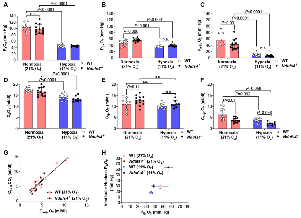
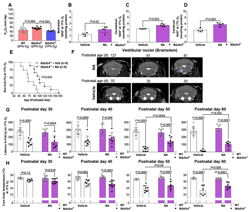
Leigh syndrome is a severe mitochondrial neurodegenerative disease with no effective treatment. In the Ndufs4-/- mouse model of Leigh syndrome, continuously breathing 11% O2 (hypoxia) prevents neurodegeneration and leads to a dramatic extension (~5-fold) in lifespan. We investigated the effect of hypoxia on the brain metabolism of Ndufs4-/- mice by studying blood gas tensions and metabolite levels in simultaneously sampled arterial and cerebral internal jugular venous (IJV) blood. Relatively healthy Ndufs4-/- and wildtype (WT) mice breathing air until postnatal age ~38 d were compared to Ndufs4-/- and WT mice breathing air until ~38 days old followed by 4-weeks of breathing 11% O2. Compared to WT control mice, Ndufs4-/- mice breathing air have reduced brain O2 consumption as evidenced by an elevated partial pressure of O2 in IJV blood (PijvO2) despite a normal PO2 in arterial blood, and higher lactate/pyruvate (L/P) ratios in IJV plasma revealed by metabolic profiling. In Ndufs4-/- mice, hypoxia treatment normalized the cerebral venous PijvO2 and L/P ratios, and decreased levels of nicotinate in IJV plasma. Brain concentrations of nicotinamide adenine dinucleotide (NAD+) were lower in Ndufs4-/- mice breathing air than in WT mice, but preserved at WT levels with hypoxia treatment. Although mild hypoxia (17% O2) has been shown to be an ineffective therapy for Ndufs4-/- mice, we find that when combined with nicotinic acid supplementation it provides a modest improvement in neurodegeneration and lifespan. Therapies targeting both brain hyperoxia and NAD+ deficiency may hold promise for treating Leigh syndrome.

Keywords: A-V difference; Arterial-venous difference; Arteriovenous difference; Brain; Hypoxia; Leigh syndrome; Metabolism; Metabolomics; NAD; Ndufs4; Niacin; Nicotinamide adenine dinucleotide; Nicotinic acid; O(2); Oxygen.


Conflict of interest statement

Declaration of Competing Interest V.K.M. and W.M.Z. are co-inventors on a patent application submitted by Massachusetts General Hospital on the use of hypoxia as a therapy. V.K.M. owns equity stake in Raze Therapeutics and is a paid advisor for Janssen Pharmaceuticals and 5 AM Ventures. R.S. holds equity in BlueBird Bio.

Figures

Figure 1.
Figure 1.. Partial pressure and blood O2 content in simultaneously sampled arterial and internal jugular venous blood collected from anesthetized and ventilated Ndufs4−/− and WT mice breathing air or after 4-weeks of 11% O2.
Partial pressure of O2 was measured in (A) arterial (PaO2) and (B) internal jugular venous (PijvO2) blood. (C) Arterial-internal jugular venous PO2 difference (Pa-ijvO2). Blood O2 content was measured in (D) arterial (CaO2) and (E) internal jugular venous (CijvO2) blood. (F) Arterial-internal jugular venous O2 content difference (Ca-ijvO2). (G) Pearson correlation between the arterial-internal jugular venous O2 content difference (Ca-ijvO2) and the internal jugular venous-arterial CO2 content difference (Cijv-aCO2) in Ndufs4−/− mice (r =0.80, P<0.002) and WT (r =0.97, P<0.0001) mice breathing air. (H) PijvO2 plotted against values previously reported in the vestibular nucleus brain tissue PO2 (PbO2) of Ndufs4−/− and WT mice breathing either air or 11% O2 [8]. Statistical significance was determined using two-way ANOVA with Sidak’s multiple comparisons test. Data are mean ± SD.
Figure 2.
Figure 2.. Lactate/pyruvate ratios and nicotinate levels measured in simultaneously sampled arterial and internal jugular venous plasma, brain tissue concentration of NAD+ and Naprt mRNA expression, collected from Ndufs4−/− and WT mice breathing air or after 4-weeks of 11% O2.
Pearson correlation between arterial and internal jugular venous plasma lactate/pyruvate ratios in (A) Ndufs4−/− (r=0.78, P=0.012) and WT (r=0.77, P=0.016) mice breathing air or in Ndufs4−/− (r=0.98, P<0.0001) and WT (r=0.97, P=0.005) mice breathing 4-weeks of 11% O2. (C) Volcano plot of the arterial-internal jugular venous (A-IJV) metabolite level difference depicting on the X-axis log2fold-changes (log2FC) and on the Y-axis statistical significance in Condition (Hypoxia vs. Normoxia) as -log10 P-values in both Ndufs4−/− and WT mice breathing 4-weeks of 11% O2 (Hypoxia) compared to both Ndufs4−/− and WT mice breathing air (Normoxia). Tissue concentration of NAD+ measured in the (D) brainstem, (E) cerebellum and (F) cerebrum. Tissue mRNA expression of Naprt measured in the (G) brainstem, (H) cerebellum and (I) cerebrum. Statistical significance was determined using two-way ANOVA with Sidak’s multiple comparisons test. All P-values generated from metabolic profiling were corrected for multiple hypothesis testing using the Benjamini-Hochberg procedure and false-discovery rate (FDR) <0.05 considered statistically significant. For Pearson correlation the ROUT method (Q = 0.5%) was used to identify and exclude 1 outlier [40]. n.s.= non-significant. Data are mean ± SD.
Figure 3.
Figure 3.. Nicotinic acid treatment provides a modest increase in lifespan and delay in the time of onset of neurological symptoms and neurodegenerative brain lesions of Ndufs4−/− mice breathing 17% O2.
(A) The partial pressure of internal jugular venous blood (PijvO2) in Ndufs4−/− mice breathing 17% O2 for 3-weeks compared to the same PijvO2 values for Ndufs4−/−and WT mice breathing air (21% O2) taken from Figure 1. Tissue concentration of NAD+ measured in the (B) brainstem, (C) cerebellum and (D) cerebrum of Ndufs4−/− mice breathing 17% O2 for 3-weeks and treated with either vehicle or nicotinic acid (NA; 240 mg/kg twice daily by intraperitoneal injection). Brain tissue was harvested 12 h after the previous dose. (E) Survival duration, (F) MRI images of the vestibular nuclei (arrow used to indicate a lesion), (G) latency to fall from an accelerating rotarod, and (H) core body temperature of Ndufs4−/− and WT mice breathing 17% O2 and treated with either vehicle or NA. For a single comparison an unpaired, two-tailed, Students T-test was used. For multiple comparisons statistical significance was determined using two-way ANOVA with Sidak’s multiple comparisons test. Log-rank (Mantel-Cox) test was used to compare Kaplan-Meier survival curves. Data are mean ± SD.

References

    1. Lake NJ, Compton AG, Rahman S, and Thorburn DR, Leigh syndrome: One disorder, more than 75 monogenic causes. Ann Neurol, 2016. 79(2): p. 190–203. - PubMed
    1. Rahman S TD Nuclear Gene-Encoded Leigh Syndrome Overview. 2015. [cited 1993–2020 Jan 27 2020]; Available from: https://www.ncbi.nlm.nih.gov/books/NBK320989/.
    1. Sofou K, De Coo IF, Isohanni P, Ostergaard E, Naess K, De Meirleir L, Tzoulis C, Uusimaa J, De Angst IB, Lonnqvist T, Pihko H, Mankinen K, Bindoff LA, Tulinius M, and Darin N, A multicenter study on Leigh syndrome: disease course and predictors of survival. Orphanet J Rare Dis, 2014. 9: p. 52. - PMC - PubMed
    1. Lake NJ, Bird MJ, Isohanni P, and Paetau A, Leigh syndrome: neuropathology and pathogenesis. J Neuropathol Exp Neurol, 2015. 74(6): p. 482–92. - PubMed
    1. Kruse SE, Watt WC, Marcinek DJ, Kapur RP, Schenkman KA, and Palmiter RD, Mice with mitochondrial complex I deficiency develop a fatal encephalomyopathy. Cell Metab, 2008. 7(4): p. 312–20. - PMC - PubMed

Publication types

LinkOut - more resources