. 2021 Sep:117:62-74.
doi: 10.1016/j.semcdb.2021.03.008. Epub 2021 Mar 19.

Kinetochore assembly throughout the cell cycle

Affiliations

Kinetochore assembly throughout the cell cycle

Alexandra P Navarro et al. Semin Cell Dev Biol. 2021 Sep.

Abstract

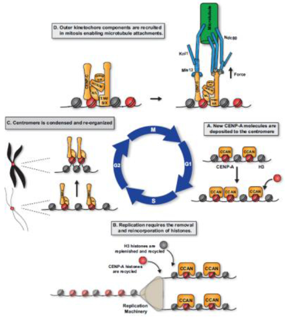
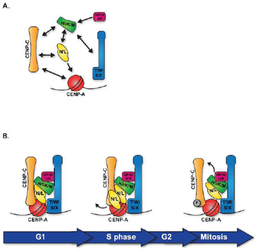
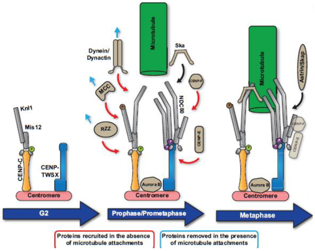
The kinetochore plays an essential role in facilitating chromosome segregation during cell division. This massive protein complex assembles onto the centromere of chromosomes and enables their attachment to spindle microtubules during mitosis. The kinetochore also functions as a signaling hub to regulate cell cycle progression, and is crucial to ensuring the fidelity of chromosome segregation. Despite the fact that kinetochores are large and robust molecular assemblies, they are also highly dynamic structures that undergo structural and organizational changes throughout the cell cycle. This review will highlight our current understanding of kinetochore structure and function, focusing on the dynamic processes that underlie kinetochore assembly.

Keywords: Centromere; Kinetochore; Mitosis.


Conflict of interest statement

Declarations of interest: none

Figures

Figure 1.
Figure 1.. Dynamic events throughout the cell cycle affect centromere structure.
A. In G1, new CENP-A molecules are deposited at centromeres. This process is tightly regulated to ensure this only happens once each cell division cycle. B. During S phase, centromere DNA must be replicated. Because no new molecules of CENP-A are deposited, this involves the distribution of previously deposited CENP-A molecules such that CENP-A molecules are diluted and H3.3 histones are incorporated as placeholders to fill in gaps. Throughout S-phase, the CCAN remains assembled upon centromeric CENP-A and helps to retain CENP-A centromere following passage of the replication fork. C. In late G2, chromatin is condensed to form the distinct chromosome structures observed in mitosis. In addition, centromere DNA must reorganize to orient the centromere proteins to recruit of outer kinetochore components and binding to spindle microtubules. D. In mitosis, following nuclear envelope breakdown and phosphorylation by CDK1, outer kinetochore proteins are recruited to the centromere completing the assembly of the kinetochore and enable kinetochores to interact directly with spindle microtubules and facilitate chromosome segregation.
Figure 2.
Figure 2.. Organization of the Inner Kinetochore.
A. The Inner kinetochore is made up of 5 distinct groups: CENP-C, CENP-N/L, CENP-H/I/K/M, CENP-T/W/S/X, and CENP-O/P/Q/U/R. These sub complexes are largely interdependent for their localization to the centromere. Direct interactions between inner kinetochore sub-complexes, as defined by biochemical reconstitutions and functional dependencies, are represented by arrows. B. Although all components of the CCAN are present at centromeres constitutively, these proteins undergo reorganization and changes throughout the cell cycle. This model represents snapshots of inner kinetochore organization at distinct cell cycle phases.
Figure 3.
Figure 3.. Organization and assembly of the outer kinetochore.
Recruitment of outer kinetochore components occurs in a step-wise manner. In late G2, Mis12/Knl1 are recruited through their interaction with CENP-C. This interaction is enabled by Aurora B phosphorylation of the Mis12 complex. Ndc80 is then recruited to the kinetochore following entry into mitosis. Once in mitosis, additional proteins are transiently recruited to the kinetochore to enable the multiple kinetochore functions in mitosis. In the absence of a microtubule interaction, proteins involved in the spindle assembly checkpoint (MCC) and the fibrous corona (RZZ, spindly, CENP-E, CENP-F, dynein/dynactin) are recruited to kinetochore’s As kinetochore-microtubule interactions are established, the Ska1 complex and Astrin/SKAP are recruited promote Ndc80 binding to microtubules. Once stable kinetochore-microtubule interactions are established and are properly bi-oriented, the spindle assembly checkpoint is satisfied and chromosome segregation occurs. CENP-E and CENP-F perform additional roles in chromosome segregation and remain at the kinetochore until anaphase.

Similar articles

Cited by

References

    1. Cheeseman IM, The kinetochore, Cold Spring Harb Perspect Biol 6(7) (2014) a015826. - PMC - PubMed
    1. Musacchio A, Desai A, A Molecular View of Kinetochore Assembly and Function, Biology (Basel) 6(1) (2017). - PMC - PubMed
    1. de la Cruz J, Karbstein K, Woolford JL Jr., Functions of ribosomal proteins in assembly of eukaryotic ribosomes in vivo, Annu Rev Biochem 84 (2015) 93–129. - PMC - PubMed
    1. McKinley KL, Cheeseman IM, The molecular basis for centromere identity and function, Nat Rev Mol Cell Biol 17(1) (2016) 16–29. - PMC - PubMed
    1. Maddox PS, Oegema K, Desai A, Cheeseman IM, “Holo”er than thou: Chromosome segregation and kinetochore function in C. elegans, Chromosome Res 12(6) (2004) 641–53. - PubMed

Publication types

MeSH terms

Substances

LinkOut - more resources