. 2021 Mar 22;10(3):bio058528.
doi: 10.1242/bio.058528.

Cex1 is a component of the COPI intracellular trafficking machinery

Affiliations

Cex1 is a component of the COPI intracellular trafficking machinery

Ludovic Enkler et al. Biol Open. .

Abstract

COPI (coatomer complex I) coated vesicles are involved in Golgi-to-ER and intra-Golgi trafficking pathways, and mediate retrieval of ER resident proteins. Functions and components of the COPI-mediated trafficking pathways, beyond the canonical set of Sec/Arf proteins, are constantly increasing in number and complexity. In mammalian cells, GORAB, SCYL1 and SCYL3 proteins regulate Golgi morphology and protein glycosylation in concert with the COPI machinery. Here, we show that Cex1, homologous to the mammalian SCYL proteins, is a component of the yeast COPI machinery, by interacting with Sec27, Sec28 and Sec33 (Ret1/Cop1) proteins of the COPI coat. Cex1 was initially reported to mediate channeling of aminoacylated tRNA outside of the nucleus. Our data show that Cex1 localizes at membrane compartments, on structures positive for the Sec33 α-COP subunit. Moreover, the Wbp1 protein required for N-glycosylation and interacting via its di-lysine motif with the Sec27 β'-COP subunit is mis-targeted in cex1Δ deletion mutant cells. Our data point to the possibility of developing Cex1 yeast-based models to study neurodegenerative disorders linked to pathogenic mutations of its human homologue SCYL1.

Keywords: Arc1; COPI coat; Cex1; SCYL1; Trafficking.


Conflict of interest statement

Competing interestsThe authors declare no competing or financial interests.

Figures

Fig. 1.
Fig. 1.
CEX1 is not genetically linked to ARC1. (A) Immunodetection of Arc1 in a WT (BY4742), cex1Δ and cex1Δ arc1Δ strains. Coomassie Blue staining was also performed to highlight protein levels in each lane. (B) Drop test of the WT, cex1Δ, cex1Δ arc1Δ and cex1Δ+pRS316-CEX1 strains on rich media in the presence of Geneticin (YPD+G418), and on glucose containing synthetic selective media (SC-Glc, SC-H, SC-U). Growth was performed at 30°C for 2 days.
Fig. 2.
Fig. 2.
Cex1 interacts with members of COPI-coat vesicles. (A) Volcano plot derived from data obtained by IP using Cex1-HA as bait. Three independent IPs were done and proteins were incubated with HA-beads or MYC-beads as control. Hits with at least two peptides, a log2fold over 1.5 and with an adjusted P-value below 1% were kept as positive. The graph shows the Cex1 bait (colored in red), together with five components of the COPI coat that co-purified with the bait (colored in blue). (B) Table summarizing Cex1 interactants involved in trafficking. The number of spectral counts detected in each of the three replicates for each protein is shown. MW, molecular weight in Dalton; DB, yeast database. (C) GO term ‘Cellular compartment’ enrichment generated form data obtained in A. Total enrichment was calculated by normalizing positive targets from the IP to the entire proteome of S. cerevisiae (obtained from the Saccharomyces Genome Database). (D) Comparison of yeast (Cex1) and mammalian (SCYL1) interactome. Proteins from COPI vesicles are shown in orange, Golgi proteins in blue, proteins from plasma membrane are in pink and proteins from the nuclear pore or from the bud neck are in grey and light grey, respectively. Genes genetically linked to CEX1 based on the work of Costanzo and colleagues (2006) are circled. (E) Interaction between Sec27-Myc and Cex1-HA. The β′-COP Sec27-Myc immunoprecipitation was performed on cex1Δ cells expressing Cex1-HA grown in fermentation conditions. Nup116 (nuclear pore complex), Sec27 and Cex1 proteins were immunodetected (indicated by an arrow), and a Ponceau staining of the blot was done as loading control. The negative control IP was done by using IgG instead of anti-Myc antibodies. The input (total protein extract, prior IP), flow-through (FT) and IP fractions were analyzed.
Fig. 3.
Fig. 3.
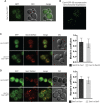
Cex1-GFP colocalizes with the α-COP Sec33-DsRED. (A) Epifluorescence microscopy of Cex1-GFP expressed in a cex1Δ strain. (B) 3D reconstitution of the Cex1-GFP construct when expressed at its genomic locus. (C) The Sec33 (Cop1/Ret1) COPI component was tagged with DsRED and its intracellular localization analyzed upon Cex1-GFP expression. cex1Δ cells expressing GFP alone were used as control. The colocalization of both markers were measured by Mander's coefficient (right panel). (D) Sec7-DsRED was used as a trans-Golgi marker together with Cex1-GFP. Their colocalization was measured by Mander's coefficient (right panel). cex1Δ cells expressing GFP alone were used as control. Scale bars: 5 µm. Representative micrographs from analyses done on different independent clones are shown.
Fig. 4.
Fig. 4.
Cex1 is associated to membrane fractions, and its deletion leads to defective trafficking of Wbp1, an ER protein having a di-lysine COPI sorting signal. (A) Subcellular fractionation of WT (SEY6210) and cex1Δ mutant strain, expressing the Cex1-HA fusion protein. Immunodetection of Cex1-HA in each fraction was done using anti-HA antibodies. Presence of cytosolic proteins in the different fraction was assessed by detecting the soluble Pgk1 protein, and ER as well as ER-Golgi vesicles resident protein by the integral membrane protein Emp47. Protein loading was controlled by TCE staining. (B) Cex1 protein levels detected in the P13, P100 and S100 fractions in A were measured using the total lysate (S5) as standard for each strain. Three independent replicates (n=3) were used. Standard deviation is shown. Statistical analyses using the t-test was done; **P<0.01; ***P<0.001. (C) Intracellular localization of the HDEL-DsRED reporter localized at the ER via COPI trafficking was assessed by fluorescence microscopy. Scale bar: 5 µm. Representative micrographs are shown from analyses done on independent clones. (D) Trafficking of Wbp1, an effector of N-glycosylation bearing a KK COPI retrieval motif, was assessed by a dot-blot assay on WT, sec27-1, cex1Δ and cex1Δ+Cex1-GFP cells grown in fermentation conditions (SC medium, at 25°C). Statistical analysis from data obtained from three independent clones was done using the t-test; **P=0.01.

References

    1. Amano, G., Matsuzaki, S., Mori, Y., Miyoshi, K., Han, S., Shikada, S., Takamura, H., Yoshimura, T. and Katayama, T. (2020). SCYL1 arginine methylation by PRMT1 is essential for neurite outgrowth via Golgi morphogenesis. Mol. Biol. Cell 31, 1963-1973. 10.1091/mbc.E20-02-0100 - DOI - PMC - PubMed
    1. Barlowe, C. and Schekman, R. (1993). SEC12 encodes a guanine-nucleotide-exchange factor essential for transport vesicle budding from the ER. Nature 365, 347-349. 10.1038/365347a0 - DOI - PubMed
    1. Blondeau, F., Ritter, B., Allaire, P. D., Wasiak, S., Girard, M., Hussain, N. K., Angers, A., Legendre-Guillemin, V., Roy, L., Boismenu, D.et al. (2004). Tandem MS analysis of brain clathrin-coated vesicles reveals their critical involvement in synaptic vesicle recycling. Proc. Natl Acad. Sci. USA 101, 3833-3838. 10.1073/pnas.0308186101 - DOI - PMC - PubMed
    1. Borner, G. H. H., Rana, A. A., Forster, R., Harbour, M., Smith, J. C. and Robinson, M. S. (2007). CVAK104 is a novel regulator of Clathrin–mediated SNARE sorting. Traffic 8, 893-903. 10.1111/j.1600-0854.2007.00576.x - DOI - PMC - PubMed
    1. Burman, J. L., Bourbonniere, L., Philie, J., Stroh, T., Dejgaard, S. Y., Presley, J. F. and McPherson, P. S. (2008). Scyl1, mutated in a recessive form of spinocerebellar neurodegeneration, regulates COPI-mediated retrograde traffic. J. Biol. Chem. 283, 22774-22786. 10.1074/jbc.M801869200 - DOI - PubMed

Publication types

MeSH terms

LinkOut - more resources