

Unusual cause of acute upper airway obstruction: spontaneous pharyngeal haematoma
- PMID: 33753397
- PMCID: PMC7986675
- DOI: 10.1136/bcr-2021-242061
Unusual cause of acute upper airway obstruction: spontaneous pharyngeal haematoma
Abstract
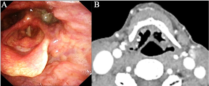
Spontaneous pharyngeal haematoma is a rare but life-threatening cause of acute upper airway obstruction, and the clinical manifestation may mimic haemoptysis. A 65-year-old man presented to our emergency department with symptoms of sore throat and haemoptysis. He had no medical history. At arrival, O2 saturation was 95% on 5 L/min of oxygen with a mask. Results of a blood examination including a coagulation test were normal. Laryngoscopy showed enlargement of the left pharynx and a narrowed airway. Contrast-enhanced CT showed extravascular leakage of contrast medium inside the left pharyngeal haematoma. Fortunately, the haematoma did not lead to airway obstruction, and it decreased spontaneously. We finally diagnosed this case as spontaneous pharyngeal haematoma. When we examine a patient with a symptom of haemoptysis accompanied by sore throat, it is necessary to consider pharyngeal haematoma and to prepare emergency airway protection for acute upper airway obstruction.
Keywords: ear; emergency medicine; nose and throat/otolaryngology.
© BMJ Publishing Group Limited 2021. No commercial re-use. See rights and permissions. Published by BMJ.
Conflict of interest statement
Competing interests: None declared.
Figures

Similar articles
-
Pharyngeal haematoma and partial airway obstruction caused by interaction between warfarin and topical miconazole gel.BMJ Case Rep. 2021 Mar 2;14(3):e239999. doi: 10.1136/bcr-2020-239999. BMJ Case Rep. 2021. PMID: 33653857 Free PMC article.
-
Laryngopyocoele: an unusual cause of a sore throat.Am J Emerg Med. 2012 Oct;30(8):1655.e1-2. doi: 10.1016/j.ajem.2011.07.018. Epub 2011 Oct 24. Am J Emerg Med. 2012. PMID: 22030175
-
Retropharyngeal Hematoma as an Unusual Presentation of Myelodysplastic Syndrome: A Case Report.Am J Case Rep. 2018 Aug 17;19:969-972. doi: 10.12659/AJCR.909502. Am J Case Rep. 2018. PMID: 30115902 Free PMC article.
-
Spontaneous retropharyngeal hemorrhage causing airway obstruction: a case report with a review of the literature.S D Med. 2006 Jul;59(7):295-7, 299. S D Med. 2006. PMID: 16895052 Review.
-
Laryngopyocele in a case of bilateral mixed laryngocele: an impending airway emergency.BMJ Case Rep. 2019 Aug 30;12(8):e229450. doi: 10.1136/bcr-2019-229450. BMJ Case Rep. 2019. PMID: 31473632 Free PMC article. Review.
References
Publication types
MeSH terms
LinkOut - more resources
Full Text Sources
Other Literature Sources
Medical