. 2021 May 17;89(6):e00026-21.
doi: 10.1128/IAI.00026-21. Print 2021 May 17.

Lymphotoxin β Receptor: a Crucial Role in Innate and Adaptive Immune Responses against Toxoplasma gondii

Affiliations

Lymphotoxin β Receptor: a Crucial Role in Innate and Adaptive Immune Responses against Toxoplasma gondii

Anne Tersteegen et al. Infect Immun. .

Abstract

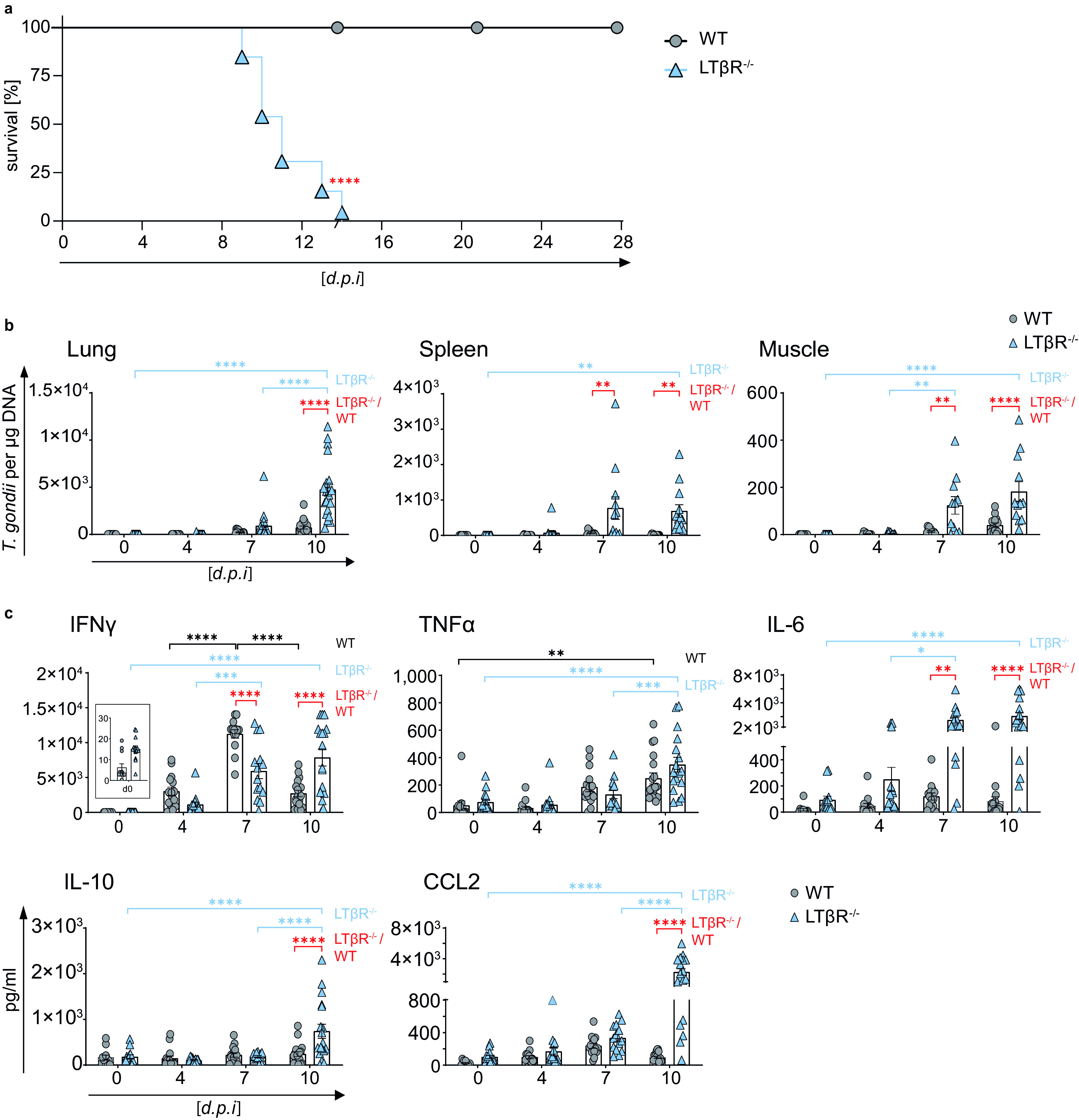
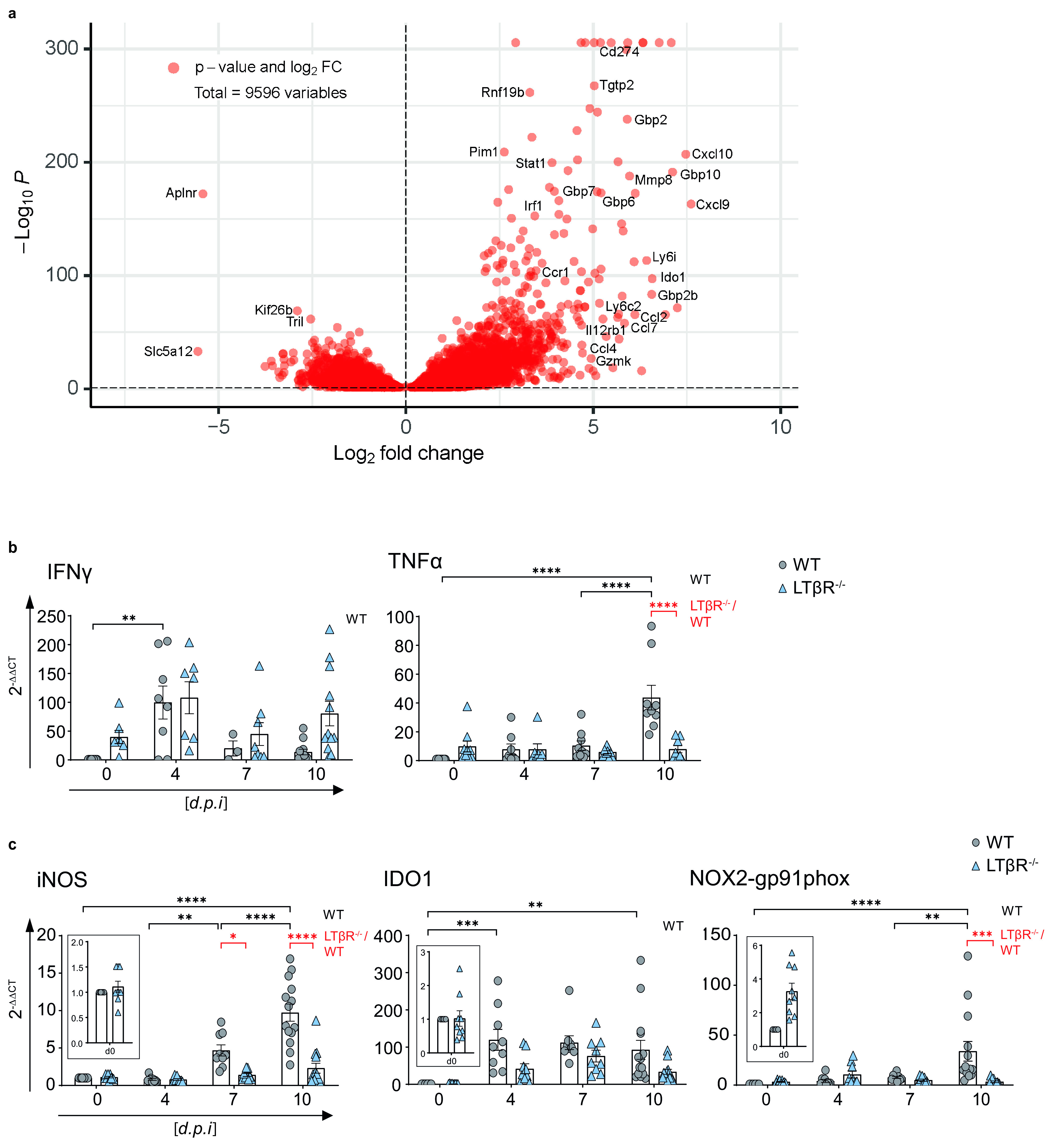
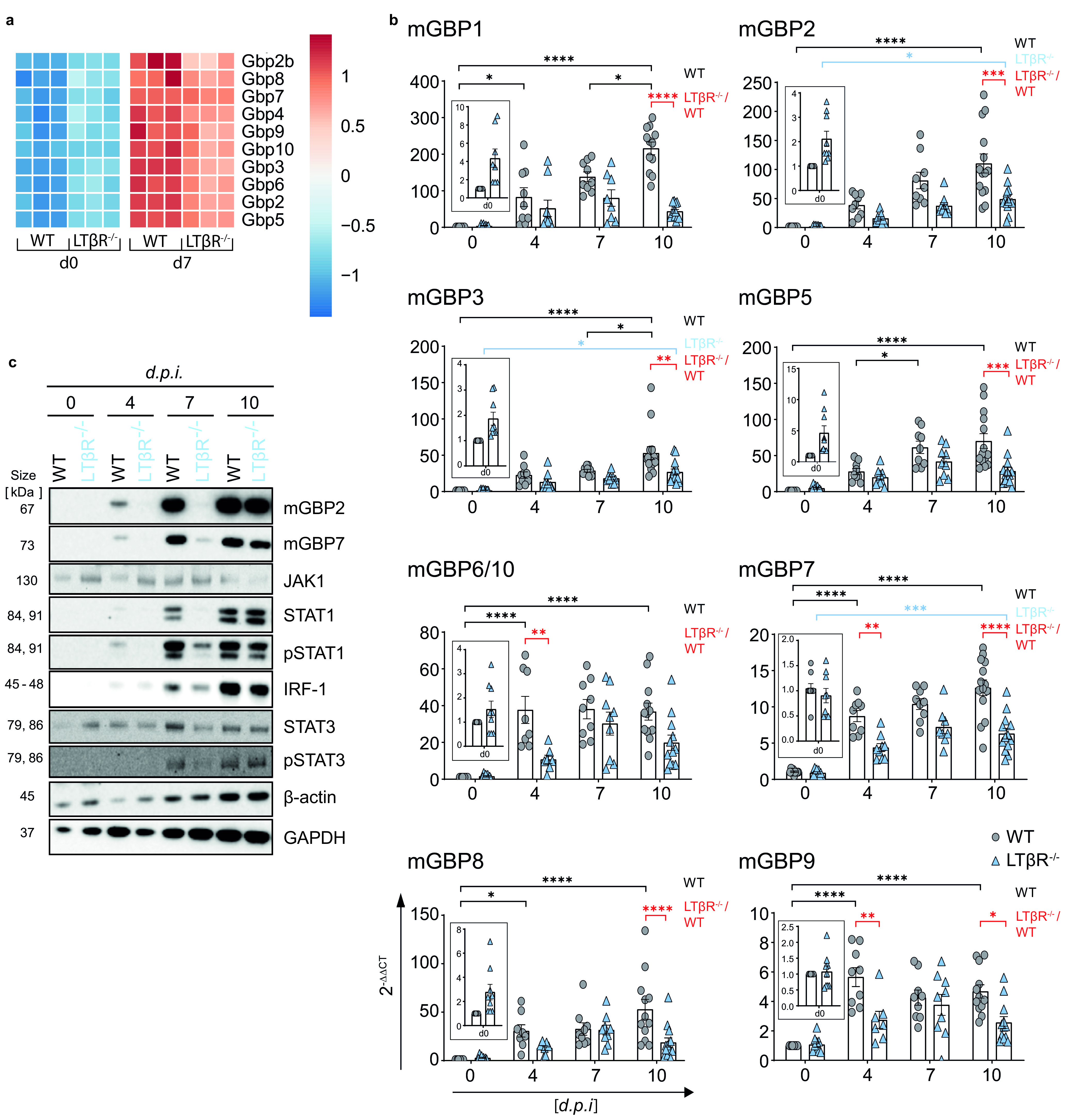
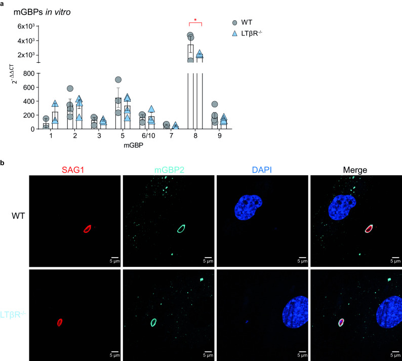
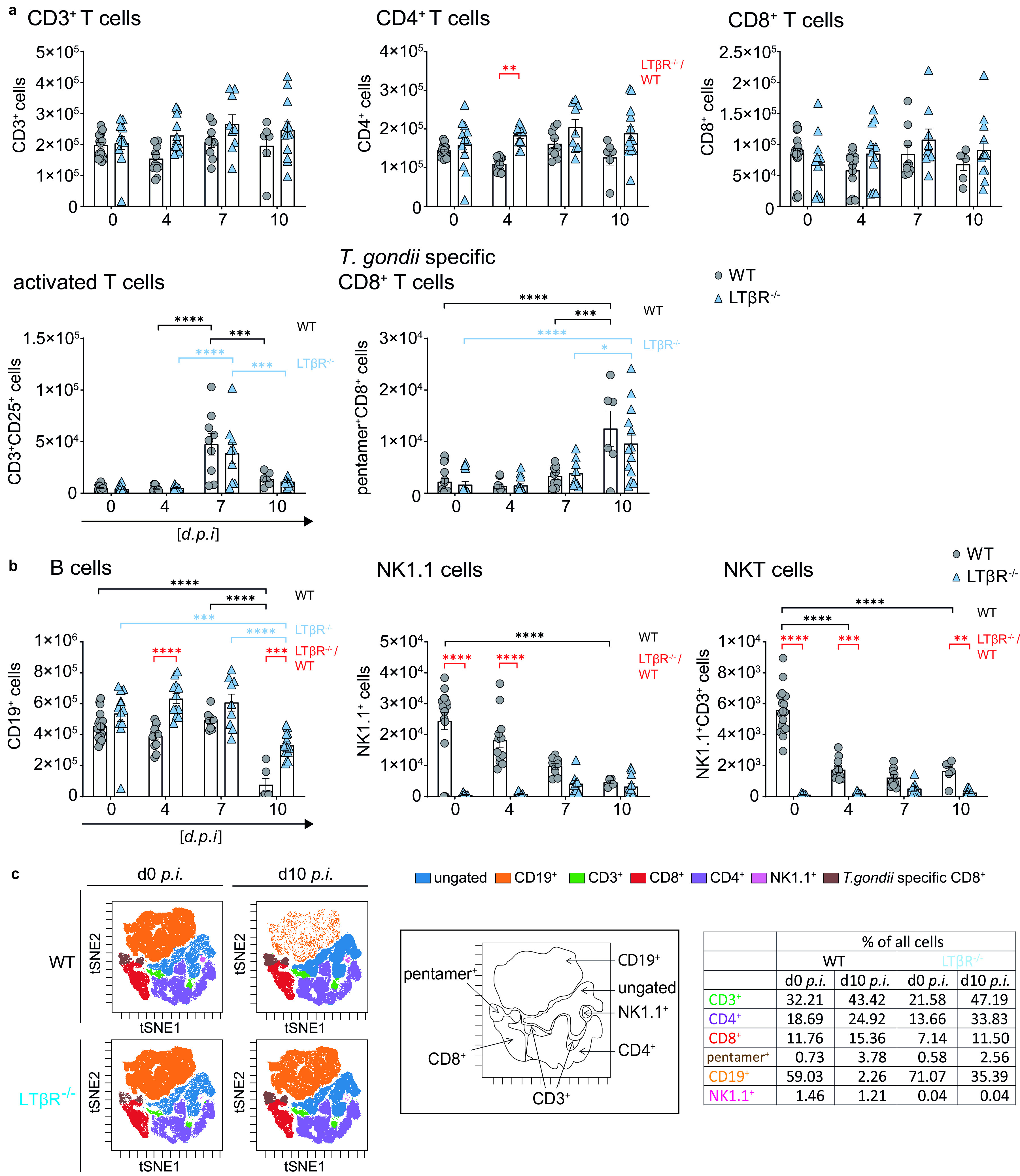
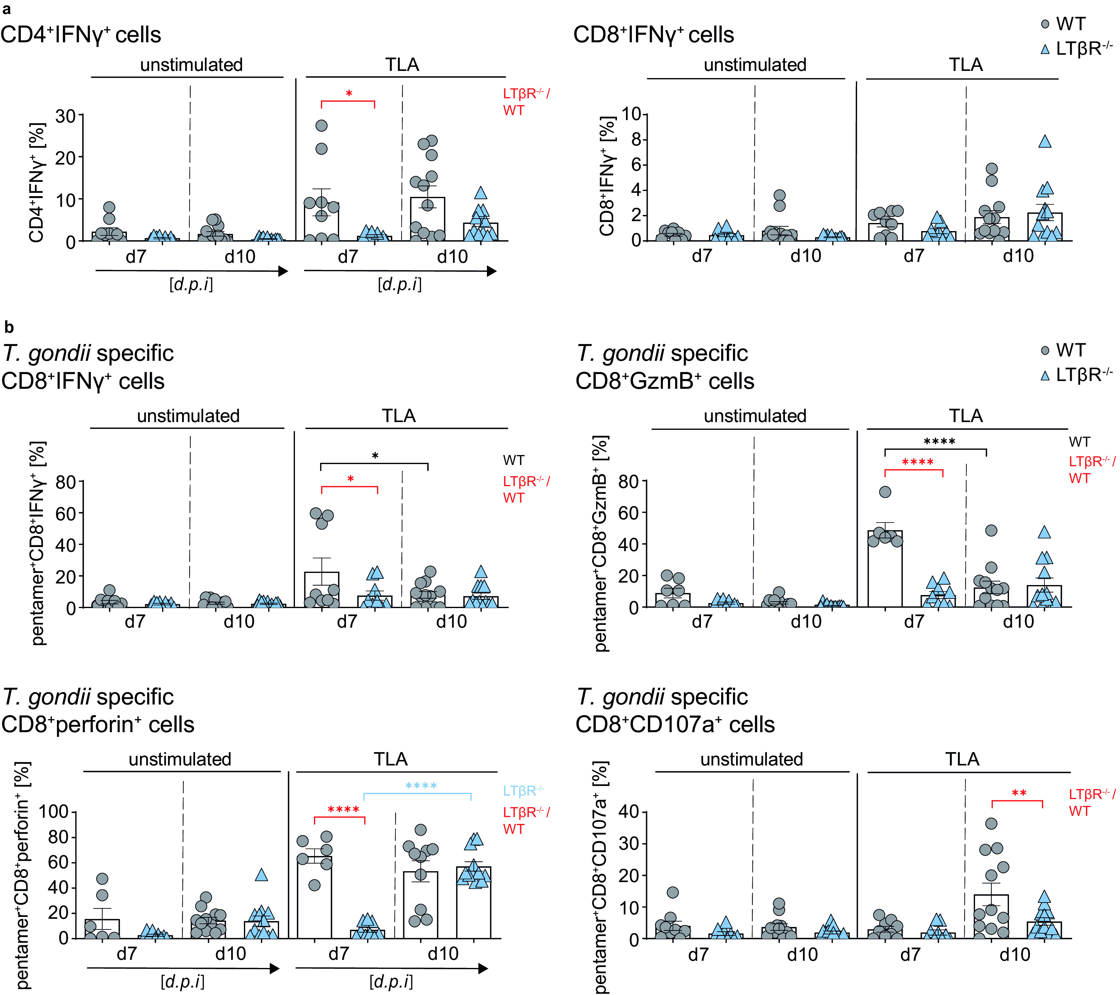
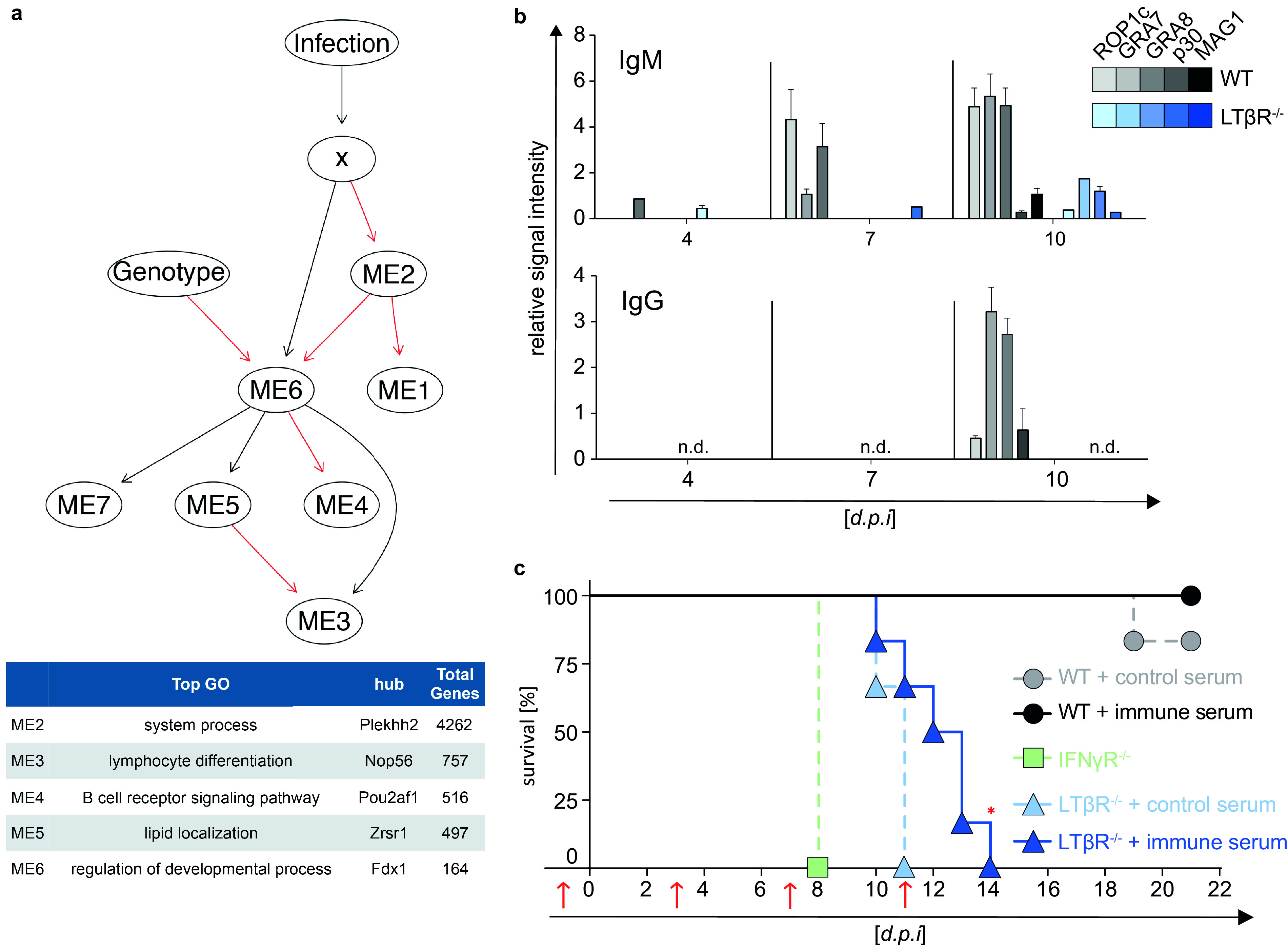
The lymphotoxin β receptor (LTβR) plays an essential role in the initiation of immune responses to intracellular pathogens. In mice, the LTβR is crucial for surviving acute toxoplasmosis; however, until now, a functional analysis was largely incomplete. Here, we demonstrate that the LTβR is a key regulator required for the intricate balance of adaptive immune responses. Toxoplasma gondii-infected LTβR-deficient (LTβR-/-) mice show globally altered interferon-γ (IFN-γ) regulation, reduced IFN-γ-controlled host effector molecule expression, impaired T cell functionality, and an absent anti-parasite-specific IgG response, resulting in a severe loss of immune control of the parasites. Reconstitution of LTβR-/- mice with toxoplasma immune serum significantly prolongs survival following T. gondii infection. Notably, analysis of RNA-seq data clearly indicates a specific effect of T. gondii infection on the B cell response and isotype switching. This study uncovers the decisive role of the LTβR in cytokine regulation and adaptive immune responses to control T. gondii.

Keywords: Toxoplasma gondii; host-pathogen interactions; lymphotoxin.


Figures

FIG 1
FIG 1
LTβR−/− mice show increased parasite load and dysregulated cytokine expression. (a) Survival of T. gondii-infected (ME49, 40 cysts, i.p.) WT (n = 15) and LTβR−/− (n = 13) mice. (b) qRT-PCR analysis of T. gondii DNA (assessing parasite load) in lung, spleen, and muscle tissue of uninfected (d0) and T. gondii-infected WT and LTβR−/− mice (d0 to d7, n ≥ 12; d10, n ≥ 14). (c) Expression of IFN-γ, TNF-α, IL-6, IL-10, and CCL2 in the serum of uninfected and T. gondii-infected WT and LTβR−/− mice (d0 to d7, n ≥ 12; d10, n = 18) analyzed via bead-based immunoassay. The data shown represent at least three independent experiments; symbols represent individual animals, columns represent mean values, and error bars represent the ± SEM. A log rank (Mantel Cox) test was used for statistical analysis represented in panel a. Two-way ANOVA corrected for multiple comparison using Tukey’s post hoc test was used for the statistical analysis represented in panels b and c. *, P < 0.0332; **, P < 0.0021; ***, P < 0.0002; ****, P < 0.0001.
FIG 2
FIG 2
Lungs of LTβR−/− mice show an altered transcriptome after T. gondii infection. (a) Volcano plot showing RNA-seq data of lung tissue of infected WT mice correlated with infected LTβR−/− mice (d7 p.i.; n = 3/group). The dashed horizontal black line represents an adjusted P value of 0.1 (Wald test). (b and c) qRT-PCR analysis of (b) cytokines (IFN-γ and TNF-α) and (c) host effector molecules (iNOS, IDO1, NOX2-gp91phox) in lung tissue from uninfected (d0) and T. gondii-infected (ME49, 40 cysts, i.p.) WT and LTβR−/− mice (d0 to 7, n ≥ 12; d10, n ≥ 14; exception: IFN-γ, n ≥ 3, d0 to 10 p.i.). Data shown in panels b and c represent four independent experiments; symbols represent individual animals, columns represent mean values, and error bars represent the ± SEM. Two-way ANOVA corrected for multiple comparison using Tukey’s post hoc test was used for the statistical analysis. *, P < 0.0332; **, P < 0.0021; ***, P < 0.0002; ****, P < 0.0001.
FIG 3
FIG 3
LTβR deficiency dysregulates IFN-γ signaling in the lung. (a) Heat map of differentially expressed murine guanylate-binding proteins (mGBPs) based on RNA-seq analysis (Wald test and adjusted P value of 0.1) of lung tissue from uninfected (d0) and T. gondii-infected (ME49, 40 cysts i.p., d7 p.i.) WT and LTβR−/− mice (n = 3). (b) qRT-PCR of mGBPs in lung tissue from uninfected and T. gondii-infected WT and LTβR−/− mice (d0 to 7, n ≥ 12; d10, n ≥ 14). The data shown represent four independent experiments; symbols represent individual animals, columns represent mean values, and error bars represent the ± SEM. (c) Immunoblot analysis of proteins involved in or induced via the IFN-γ signaling pathway in lung tissue from uninfected and T. gondii-infected WT and LTβR−/− mice. Two-way ANOVA corrected for multiple comparison using Tukey’s post hoc test was used for the statistical analysis represented in panel b. *, P < 0.0332; **, P < 0.0021; ***, P < 0.0002; ****, P < 0.0001. The data shown in panel c are representative of three independent experiments.
FIG 4
FIG 4
mGBP upregulation and recruitment. (a) qRT-PCR analysis of mGBP mRNA expression of uninfected WT and LTβR−/− MEFs stimulated with IFN-γ (7.5 ng/ml) for 8 h (all n = 3, except for mGBP1, where n = 2). Each symbol represents an individual technical replicate; columns represent mean values, and error bars represent the ± SEM. two-way ANOVA corrected for multiple comparisons by the Sidak post hoc test was used for statistical analysis. *, P < 0.00332. (b) Representative immunofluorescence analysis of T. gondii tachyzoite-infected (multiplicity of infection [MOI], 1:40) WT and LTβR−/− MEFs. Cells were prestimulated with IFN-γ (7.5 ng/ml) for 16 h before being infected with T. gondii tachyzoites for 2 h. T. gondii surface antigen SAG1 was visualized using a Cy3-conjugated secondary antibody, and mGBP2 was visualized using an mGBP2 antiserum (30) followed by an Alexa Fluor 633-conjugated secondary antibody for detection of mGBP2 recruitment toward the T. gondii PV. Cell nuclei were stained using DAPI (4′,6-diamidino-2-phenylindole). The data shown in panels a and b represent at least two independent experiments.
FIG 5
FIG 5
Dysregulated immune cell numbers in LTβR−/− mice. (a) Absolute cell numbers of CD3+, CD4+, CD8+, CD25+CD3+, and pentamer+CD8+ T cells. (b) CD19+, NK1.1+, and NK1.1+CD3+ cells in spleens of uninfected (d0) and T. gondii-infected (ME49, 40 cysts, i.p.) WT and LTβR−/− mice (d0 to d7 p.i., n = 12; d10 p.i., n ≥ 6) determined via flow cytometry. (c) Representative tSNE plots from splenocytes of uninfected and T. gondii-infected (d10 p.i.) WT and LTβR−/− mice. Clustered populations were identified using the indicated markers. The data shown represent at least three independent experiments; symbols represent individual animals, columns represent mean values, and error bars represent the ± SEM. Two-way ANOVA corrected for multiple comparison using Tukey’s post hoc test was used for the statistical analysis represented in panels a and b. *, P < 0.0332; **, P < 0.0021; ***, P < 0.0002; ****, P < 0.0001.
FIG 6
FIG 6
LTβR deficiency impairs T cell effector function in the spleen. (a and b) Intracellular staining of (a) CD4+IFN-γ+ and CD8+IFN-γ+ T cells (%) and (b) cytotoxic granule (GzmB+ or perforin+) containing and degranulating (CD107a+) pentamer+CD8+ T cells of unstimulated and toxoplasma lysate antigen (TLA) ex vivo restimulated splenocytes from T. gondii-infected (d7 and 10 p.i.) WT and LTβR−/− mice (d7, n ≥ 6; d10, n ≥ 10). Representative data of at least two independent experiments; symbols represent individual animals, columns represent mean values, and error bars represent the ± SEM. Two-way ANOVA corrected for multiple comparison using Tukey’s post hoc test was used for statistical analysis. *, P < 0.0332; **, P < 0.0021; ****, P < 0.0001.
FIG 7
FIG 7
Abrogated parasite-specific isotype class switching and reconstitution of mice with T. gondii-specific immune serum. (a) Host-pathogen network prediction model generated based on RNA-seq data of lung tissue of uninfected (d0) and T. gondii-infected (ME49, 40 cysts; d7 p.i.) WT and LTβR−/− mice (n = 3/group). GmicR was used to detect relationships between module eigengenes (ME) and experimental conditions. X represents the total T. gondii gene expression data for each sample; infection and genotype were included as variables. Red lines indicate inverse and black lines positive relationships. Representative gene ontologies and hub genes reported by GmicR for each module are shown in the summary table. (b) T. gondii-specific IgM and IgG antibody response in serum of uninfected (d0) and T. gondii-infected (ME49, 40 cysts, i.p.) WT and LTβR−/− mice (d4 and d7 p.i., n = 15; d10 p.i., n ≥ 20). Shown is a representative result of four independent experiments; bars represent mean values ± SEM. (c) Transfer of serum (red arrows; d1, d3, d7, and d11 p.i.) from uninfected donor WT mice (control serum) or from T. gondii-infected (ME49, 20 cysts, i.p.) donor WT mice (immune serum) into WT and LTβR−/− acceptor mice. On day 0, acceptor mice (n = 6/group) were infected with T. gondii (ME49, 10 cysts, i.p.), and survival was evaluated. IFN-γR−/− mice (n = 3) served as infection controls. The data shown in panel c represent one experiment. A log rank (Mantel Cox) test was used for the statistical analysis represented in panel c. *, P < 0.0332; n.d., not detected.

Similar articles

Cited by

References

    1. Ward-Kavanagh LK, Lin WW, Sedy JR, Ware CF. 2016. The TNF receptor superfamily in co-stimulating and co-inhibitory responses. Immunity 44:1005–1019. 10.1016/j.immuni.2016.04.019. - DOI - PMC - PubMed
    1. Hehlgans T, Pfeffer K. 2005. The intriguing biology of the tumour necrosis factor/tumour necrosis factor receptor superfamily: players, rules and the games. Immunology 115:1–20. 10.1111/j.1365-2567.2005.02143.x. - DOI - PMC - PubMed
    1. Schneider K, Potter KG, Ware CF. 2004. Lymphotoxin and LIGHT signaling pathways and target genes. Immunol Rev 202:49–66. 10.1111/j.0105-2896.2004.00206.x. - DOI - PubMed
    1. Ware CF. 2005. Network communications: lymphotoxins, LIGHT, and TNF. Annu Rev Immunol 23:787–819. 10.1146/annurev.immunol.23.021704.115719. - DOI - PubMed
    1. Ehlers S, Holscher C, Scheu S, Tertilt C, Hehlgans T, Suwinski J, Endres R, Pfeffer K. 2003. The lymphotoxin beta receptor is critically involved in controlling infections with the intracellular pathogens Mycobacterium tuberculosis and Listeria monocytogenes. J Immunol 170:5210–5218. 10.4049/jimmunol.170.10.5210. - DOI - PubMed

Publication types

Substances

LinkOut - more resources