. 2021 Mar;9(3):e001657.
doi: 10.1136/jitc-2020-001657.

Regorafenib enhances antitumor immunity via inhibition of p38 kinase/Creb1/Klf4 axis in tumor-associated macrophages

Affiliations

Regorafenib enhances antitumor immunity via inhibition of p38 kinase/Creb1/Klf4 axis in tumor-associated macrophages

Da-Liang Ou et al. J Immunother Cancer. 2021 Mar.

Abstract

Background: Regorafenib and other multikinase inhibitors may enhance antitumor efficacy of anti-program cell death-1 (anti-PD1) therapy in hepatocellular carcinoma (HCC). Its immunomodulatory effects, besides anti-angiogenesis, were not clearly defined.

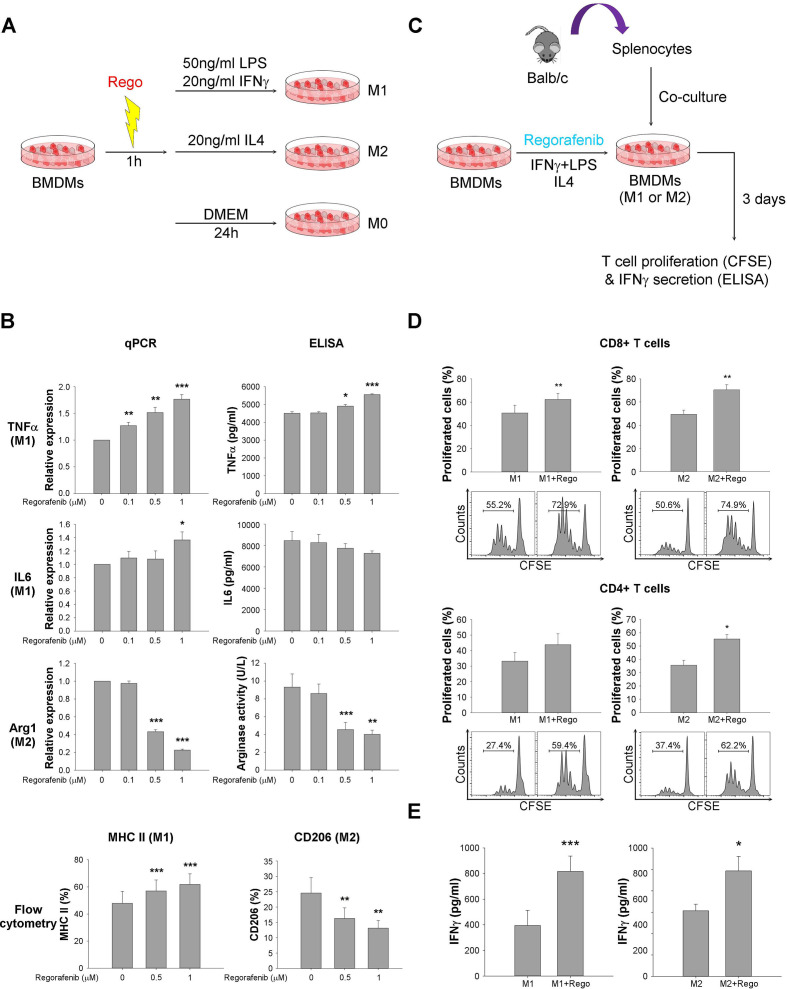
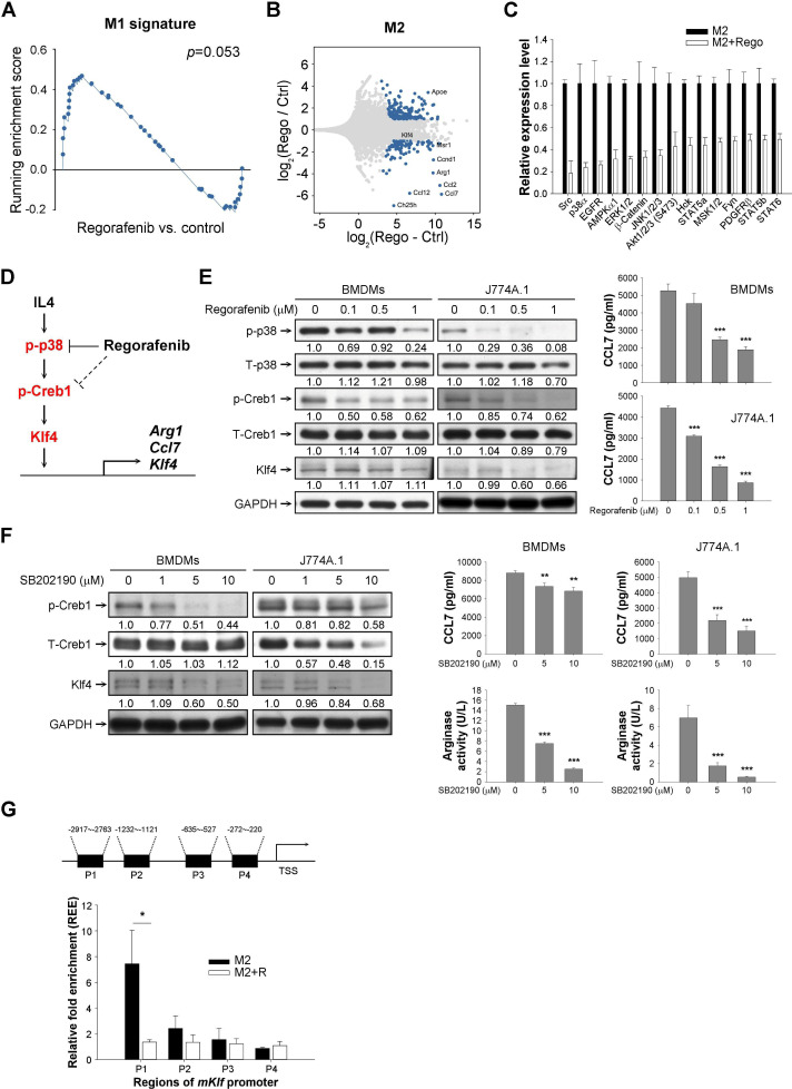
Methods: In vivo antitumor efficacy was tested in multiple syngeneic liver cancer models. Murine bone marrow-derived macrophages (BMDMs) were tested in vitro for modulation of polarization by regorafenib and activation of cocultured T cells. Markers of M1/M2 polarization were measured by quantitative reverse transcription PCR (RT-PCR), arginase activity, flow cytometry, and ELISA. Knockdown of p38 kinase and downstream Creb1/Klf4 signaling on macrophage polarization were confirmed by using knockdown of the upstream MAPK14 kinase, chemical p38 kinase inhibitor, and chromatin immunoprecipitation.

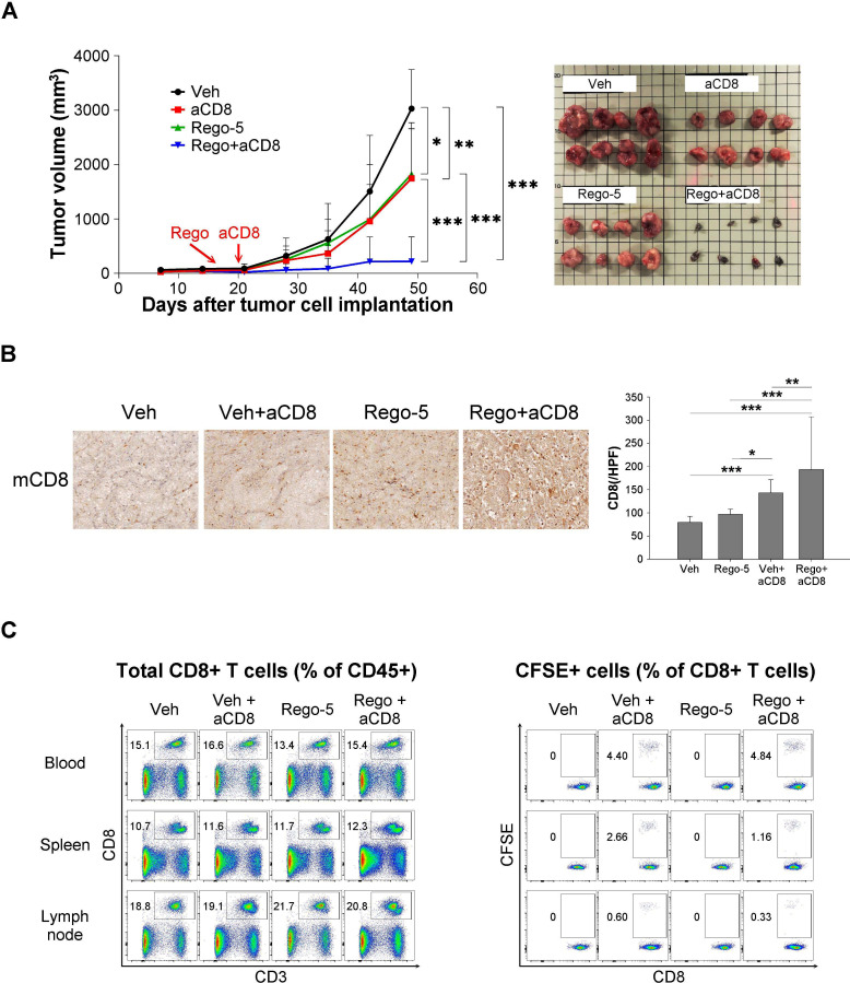
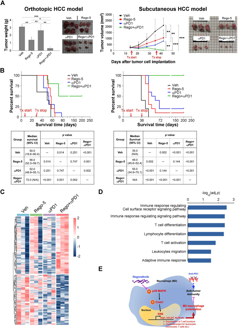
Results: Regorafenib (5 mg/kg/day, corresponding to about half of human clinical dosage) inhibited tumor growth and angiogenesis in vivo similarly to DC-101 (anti-VEGFR2 antibody) but produced higher T cell activation and M1 macrophage polarization, increased the ratio of M1/M2 polarized BMDMs and proliferation/activation of cocultured T cells in vitro, indicating angiogenesis-independent immunomodulatory effects. Suppression of p38 kinase phosphorylation and downstream Creb1/Klf4 activity in BMDMs by regorafenib reversed M2 polarization. Regorafenib enhanced antitumor efficacy of adoptively transferred antigen-specific T cells. Synergistic antitumor efficacy between regorafenib and anti-PD1 was associated with multiple immune-related pathways in the tumor microenvironment.

Conclusion: Regorafenib may enhance antitumor immunity through modulation of macrophage polarization, independent of its anti-angiogenic effects. Optimization of regorafenib dosage for rational design of combination therapy regimen may improve the therapeutic index in the clinic.

Keywords: immunomodulation; immunotherapy; tumor microenvironment.


Conflict of interest statement

Competing interests: Dr A-L Cheng is a consultant for and a member of the speaker’s bureau of Bayer-Schering Pharma. Dr A-L Cheng is a consultant of Novartis, Merck Serono, Eisai, Merck Sharp & Dohme (MSD) Corp., ONXEO, Bayer HealthCare Pharmaceuticals Inc., Bristol-Myers Squibb (BMS) Company, and Ono Pharmaceutical Co., Ltd. Dr C Hsu received research grants from BMS/ONO, Roche, and Ipsen and received honorarium from the following pharmaceutical companies: AstraZeneca, Bayer, BMS/ONO, Eisai, Eli Lilly, Ipsen, Merck Serono, MSD, Novartis, Roche, and TTY Biopharm.

Figures

Figure 1
Figure 1
The immune modulatory effects of regorafenib contribute to its in vivo antitumor efficacy. (A) The in vivo antitumor efficacy of regorafenib in immune-competent (BNL-MEA cells implanted orthotopically into BALB/c mice (N=5) and Hepa1-6 cells implanted subcutaneously into C57BL/6 mice (N=10)) liver cancer models. Mice were treated with vehicle, regorafenib 5 mg/kg/day (Rego-5) for 28 days and the tumor weight and volume were monitored. (B) Tumor-infiltrating T cells (CD4 and CD8), tumor cell proliferation (Ki67), and tumor angiogenesis (CD31) were quantified by immunohistochemical staining. Apoptotic tumor cells were quantified by terminal deoxynucleotidyltransferase dUTP nick end labeling (TUNEL) assay. Tumor samples were collected from BNL-MEA tumor-bearing BALB/c mice treated after 5 days of treatment with vehicle or regorafenib 5 mg/kg/day by gavage. Data were analyzed using 20 images (regions of interest, ROI)/ tumor, 4 tumors from 4 mice in each treatment group. (C) Gene-set enrichment analysis (GSEA) of leukocyte activation and angiogenesis signatures in RNA of tumor bulk from BNL-MEA tumor-bearing BALB/c mice treated with vehicle or regorafenib for 5 days. (D) Murine splenocytes from BALB/c mice were treated regorafenib (0, 0.5, 1, 5, 10 μM) in vitro and the proliferation of CD4+ and CD8+ T cells was detected with carboxyfluorescein succinimidyl ester (CFSE) staining and flow cytometry. Data are presented as the mean±SD from a representative experiment of at least triplicate. *p<0.05; **p<0.01; ***p<0.001, two-tailed Student’s t-test. HCC, hepatocellular carcinoma.
Figure 2
Figure 2
Regorafenib had immunomodulatory effects, which are independent of anti-angiogenesis. (A) Comparison between regorafenib and DC-101, a murine anti-VEGFR2 antibody, in terms of induction of CD8+ T cell infiltration (CD8 staining), angiogenesis inhibition (CD31 staining), and antitumor efficacy. BALB/c mice implanted BNL-MEA cells orthotopically were treated with regorafenib (5 mg/kg/day) or DC-101 (800 μg, intraperitoneal, days 1, 3, 5) for 5 days. (B) Scatter plot comparing the fold changes of genes regulated by regorafenib or DC-101. The blue dots indicated genes whose regulations were considered independent of VEGFR2 inhibition. (C) Enrichment map showing GO terms of regorafenib-regulated, VEGFR2-independent genes. Groups of functionally related gene sets were highlighted. (D) Composition of tumor-infiltrating immune cells analyzed by flow cytometry. Orthotopic liver tumor samples were collected 5 days after treatment start and the percentage of individual immune cell types was measured by flow cytometry. Values are presented as means±SD (n=3 in each group). (E) Regulation of macrophage polarization by regorafenib in vivo, indicated by the change in the ratio of M1 (F4/80+MHCII+CD206-))/M2 (F4/80+MHCII-CD206+) cells measured by multiplex immunofluorescence staining. Multispectral images were acquired to cover the whole area of the specimens; each dot in the left panel represented one acquisition region of interest (ROI). Data were analyzed using 60 ROI images/ tumor, 4 tumors from 4 mice in each treatment group. Right panel, representative spectrally unmixed composite images (×20 magnification) from the multiplex immunofluorescence staining. (F) Gene-set enrichment analysis (GSEA) of signaling pathways related to macrophage activation in tumors treatment with regorafenib or DC-101 compared with vehicle. *, p < 0.05; **, p < 0.01; ***, p < 0.001.
Figure 3
Figure 3
Regorafenib increased proliferation and activation of CD8+ T cells via regulation of macrophage polarization. (A) Design of in vitro study to measure the impact of regorafenib on macrophage polarization. Bone marrow–derived macrophages (BMDMs) were pretreated with regorafenib for 1 hour and then polarized to by interferon-γ (IFNγ)+lipopolysaccharides (LPS) (M1 phenotype) or interleukin-4 (IL4) (M2 phenotype), respectively for 24 hours. M1 (TNFα, IL6, MHC II) and M2 (Arg1, CD206) markers were detected by qPCR, ELISA, flow cytometry, and arginase activity. (B) Expression of M1 markers was enhanced by regorafenib, while expression of M2 markers was suppressed. (C) Design of coculture study to measure the impact of regorafenib-treated BMDMs on T cell function. BMDMs treated with/without regorafenib (1 µM) were cocultured with carboxyfluorescein succinimidyl ester (CFSE)-labeled murine splenocytes at 20:1 ratio for 72 hours. (D) T cell proliferation, measured by CFSE staining and flow cytometry, was enhanced by regorafenib-treated BMDMs. (E) T cell activation, measured by IFN-γ secretion into culture medium using ELISA, was enhanced by regorafenib-treated BMDMs. *, p < 0.05; **, p < 0.01; ***, p <0.001.
Figure 4
Figure 4
Regorafenib may regulate macrophage polarization through suppressing p38MAPK-Creb1-Klf4 pathway. (A) Gene-set enrichment analysis (GSEA) of M1 signature in regorafenib-treated or vehicle-treated M2 macrophages derived from bone marrow–derived macrophages (BMDMs). BMDMs were pretreated with regorafenib 1 µM for 1 hour, polarized to M2 phenotype, and total RNA were harvested for RNA-sequencing. (B) Representative genes regulated by regorafenib in M2 BMDMs. Genes with log (fold changes) (logFC) of >1 or <−1 were listed. (C) Inhibition of representative kinases in BMDMs by regorafenib 1 μM (phospho-kinase array). Kinases with ≥50% suppression of phosphorylation were shown. (D) A proposed mechanism by which regorafenib may prevent the M2 polarization of macrophages. (E) Suppression of p38MAPK and the downstream Creb1 phosphorylation and expression of Klf4 and CCL7 by regorafenib in macrophages. (F) Suppression of Creb1 phosphorylation, Klf4/CCL7 expression, and arginase activity in macrophages by the p38MAPK inhibitor SB202190. The number below each band in the western blot indicated the relative intensity of staining signals measured by ImageJ software (National Institutes of Health, https://imagej.nih.gov/ij/). (G) Binding of the transcription factor Creb1 to the predicted binding sites at the Klf4 promoter. Regorafenib (1 µM) significantly suppressed Creb1 binding to Klf4 promoter, particularly the −2917 to −2763 site, in M2 BMDMs. *, p < 0.05; **, p < 0.01; ***, p <0.001.
Figure 5
Figure 5
Effects of regorafenib on adoptive transfer of antigen-specific cytotoxic T cells. (A) Adoptive transfer of antigen-specific, carboxyfluorescein succinimidyl ester (CFSE)-labeled CD8+ T cells into C57BL/6 mice-bearing gp33-overexpressed Hepa1-6 cells subcutaneously. The antitumor efficacy of antigen-specific CD8+ T cells adoptively transferred into mice-bearing gp33-overexpressed tumors was enhanced by regorafenib (5 mg/kg/day) (N=8). (B) Immunohistochemistry staining and quantification of tumor-infiltrating CD8+ T cells. Data were analyzed using 20 images (regions of interest, ROI)/ tumor, 4 tumors from 4 mice in each treatment group. (C) The transferred T cells in peripheral blood, spleen, and lymph nodes were measured by flow cytometry. *, p < 0.05; **, p < 0.01; ***, p <0.001.
Figure 6
Figure 6
Antitumor synergy between regorafenib and anti-program cell death-1 (anti-PD1) therapy. Synergistic antitumor efficacy between regorafenib (5 mg/kg/day) and anti-PD1 (200 µg/intraperitoneal, ×5) therapy in orthotopic (BNL cell line/BALB/c mice) and subcutaneous (Hepa1-6 cell line/ C57BL/6 mice) syngeneic liver cancer models. (A) The efficacy was measured in terms of tumor weight/volume (orthotopic, N=5; subcutaneous, N=10 in each treatment group). (B) The efficacy was measured in terms of animal survival (N=10 in each treatment group). (C) Differential patterns of gene expression regulated by regorafenib and anti-PD1. Three tumors in each treatment group were subjected to RNA-seq analysis. (D) Over-representative GO terms (adj. p value<0.05) related to genes induced by the combination of regorafenib and anti-PD1. (E) Proposed mechanisms by which regorafenib regulates antitumor immunity through macrophage polarization. HCC, hepatocellular carcinoma. *, p < 0.05; **, p < 0.01; ***, p <0.001.

Similar articles

Cited by

References

    1. Sun C, Mezzadra R, Schumacher TN. Regulation and function of the PD-L1 checkpoint. Immunity 2018;48:434–52. 10.1016/j.immuni.2018.03.014 - DOI - PMC - PubMed
    1. Finn RS, Qin S, Ikeda M, et al. . Atezolizumab plus bevacizumab in unresectable hepatocellular carcinoma. N Engl J Med 2020;382:1894–905. 10.1056/NEJMoa1915745 - DOI - PubMed
    1. Huang Y, Yuan J, Righi E, et al. . Vascular normalizing doses of antiangiogenic treatment reprogram the immunosuppressive tumor microenvironment and enhance immunotherapy. Proc Natl Acad Sci U S A 2012;109:17561–6. 10.1073/pnas.1215397109 - DOI - PMC - PubMed
    1. Huang Y, Goel S, Duda DG, et al. . Vascular normalization as an emerging strategy to enhance cancer immunotherapy. Cancer Res 2013;73:2943–8. 10.1158/0008-5472.CAN-12-4354 - DOI - PMC - PubMed
    1. Hegde PS, Wallin JJ, Mancao C. Predictive markers of anti-VEGF and emerging role of angiogenesis inhibitors as immunotherapeutics. Semin Cancer Biol 2018;52:117–24. 10.1016/j.semcancer.2017.12.002 - DOI - PubMed

Publication types

MeSH terms