. 2021 Mar;9(3):e002022.
doi: 10.1136/jitc-2020-002022.

Effect of cabazitaxel on macrophages improves CD47-targeted immunotherapy for triple-negative breast cancer

Affiliations

Effect of cabazitaxel on macrophages improves CD47-targeted immunotherapy for triple-negative breast cancer

Xu Cao et al. J Immunother Cancer. 2021 Mar.

Abstract

Background: Limited therapeutic options are available for triple-negative breast cancer (TNBC), emphasizing an urgent need for more effective treatment approaches. The development of strategies by targeting tumor-associated macrophages (TAMs) to stimulate their ability of Programmed Cell Removal (PrCR) provides a promising new immunotherapy for TNBC treatment.

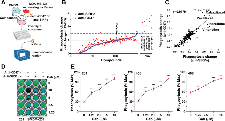
Methods: CD47 is a critical self-protective "don't eat me" signal on multiple human cancers against macrophage immunosurveillance. Using human and mouse TNBC preclinical models, we evaluated the efficacy of PrCR-based immunotherapy by blocking CD47. We performed high-throughput screens on FDA-approved anti-cancer small molecule compounds for agents potentiating PrCR and enhancing the efficacy of CD47-targeted therapy for TNBC treatment.

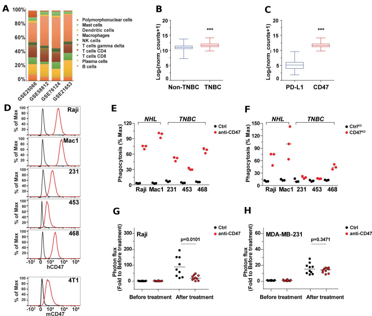
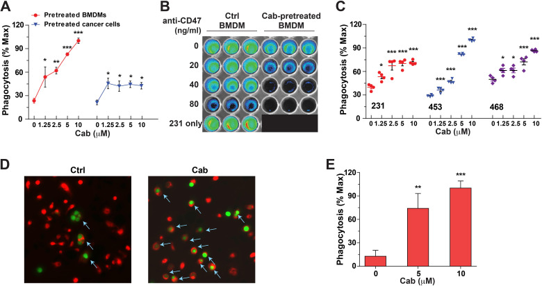
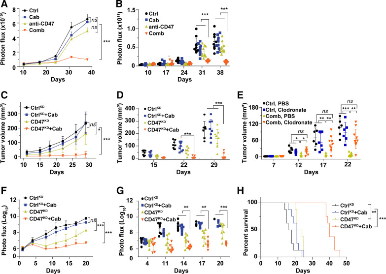
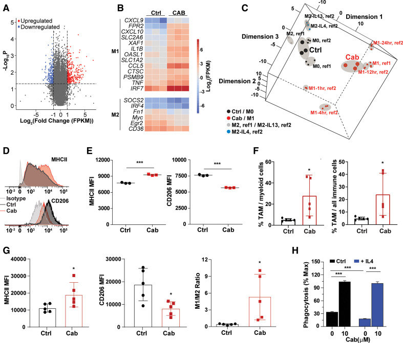
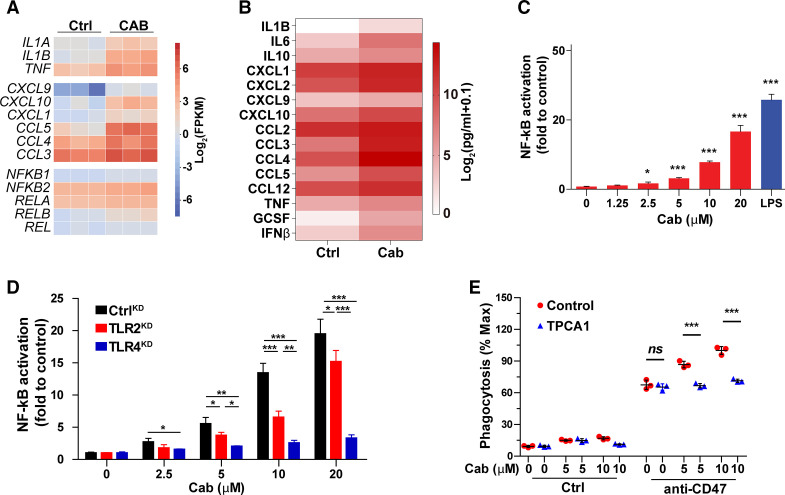
Results: We showed that CD47 was widely expressed on TNBC cells and TAMs represented the most abundant immune cell population in TNBC tumors. Blockade of CD47 enabled PrCR of TNBC cells, but the efficacy was not satisfactory. Our high-throughput screens identified cabazitaxel in enhancing PrCR-based immunotherapy. A combination of CD47 blockade and cabazitaxel treatment yielded a highly effective treatment strategy, promoting PrCR of TNBC cells and inhibiting tumor development and metastasis in preclinical models. We demonstrated that cabazitaxel potentiated PrCR by activating macrophages, independent of its cytotoxicity toward cancer cells. When treated with cabazitaxel, the molecular and phenotypic signatures of macrophages were polarized toward M1 state, and the NF-kB signaling pathway became activated.

Conclusion: The combination of CD47 blockade and macrophage activation by cabazitaxel synergizes to vastly enhance the elimination of TNBC cells. Our results show that targeting macrophages is a promising and effective strategy for TNBC treatment.

Keywords: immunotherapy; macrophages; phagocytosis; triple-negative breast cancer; tumor microenvironment.


Conflict of interest statement

Competing interests: MF declares patent applications pertaining to stimulating TLR/BTK signaling to promote CRT in macrophages assigned to the Stanford University and equity and/or consulting with Forty Seven, Inc.

Figures

Figure 1
Figure 1
CD47 is a therapeutic target in triple-negative breast cancer (TNBC). (A) Inferred composition of 10 immune cell subsets in TNBC biopsies. The gene signature datasets were obtained from Gene Expression Omnibus (GEO) database. The results were analyzed using CIBERSORT. (B) A comparison of CD47 gene expression between non-TNBC (n=974) and TNBC (n=123), basing on TCGA Breast Cancer (BRCA). Whiskers represent min–max values. ***p<0.001 (t-test). (C) A comparison of gene expression of PD-L1 and CD47 on TNBC (n=123), basing on TCGA Breast Cancer (BRCA). Whiskers represent min–max values. ***p<0.001 (t-test). (D) Representative histogram plots showing CD47 expression on NHL cell lines (Raji and Mac-1) and TNBC cell lines (MDA-MB-231, MDA-MB-453, MDA-MB-468 and 4T1). Anti-hCD47 (clone B6H12) or anti-mCD47 (clone miap301) were used for human (Raji, Mac-1, MDA-MB-231, MDA-MB-453 and MDA-MB-468) or mouse (4T1) lines. (E) An in vitro flow cytometry–based phagocytosis assay comparing the efficiency of blocking CD47 with an anti-CD47 antibody between TNBC cell lines (MDA-MB-231, MDA-MB-453 and MDA-MB-468) and NHL cell lines (Raji and Mac-1). Mouse M0 bone marrow–derived macrophages (BMDMs) were used for the assay. Phagocytosis was normalized to the maximal response in the experiments. (F) An in vitro flow cytometry–based phagocytosis assay comparing the efficiency of blocking CD47 by knocking down CD47 gene expression between TNBC cell lines (MDA-MB-231, MDA-MB-453 and MDA-MB-468) and NHL cell lines (Raji and Mac-1). Mouse M0 BMDMs were used for the assay. Phagocytosis was normalized to the maximal response in the experiments. (G and H) In vivo tumor engraftment assay of Raji and MDA-MB-231 cells. Mice engrafted with Raji or MDA-MB-231 cells were treated with vehicle or CD47-blocking antibody. Tumor engraftment and growth were measured by bioluminescence imaging. p=0.0101 (Raji) or 0.3471 (MDA-MB-231) (t-test).
Figure 2
Figure 2
High-throughput screens with FDA-approved anti-cancer small molecule compounds identify cabazitaxel as a PrCR-promoting agent. (A) A schematic showing the experimental design of the high-throughput screen. (B) PrCR-based high-throughput screens of 147 FDA-approved anti-cancer small molecule compounds. Cells were treated with antibodies blocking CD47–Sirpα interaction (anti-CD47 or anti-Sirpα) and subjected to luminescence-based phagocytosis assay. Phagocytosis was normalized to DMSO control. Spots represent individual compounds. (C) The correlation of phagocytosis change between screens with anti-CD47 and anti-Sirpα antibodies. Spots represent individual compounds. (D) Representative bioluminescence images of the luminescence-based phagocytosis assay measuring surviving cancer cells, with MDA-MB-231 cells as the target cells. Mouse M0 bone marrow–derived macrophages (BMDMs) were used for the assay. Cabazitaxel (Cab) was used at doses from 0 to 10 µM. (E) An in vitro luminescence-based phagocytosis assay measuring surviving cancer cells, with MDA-MB-231, MDA-MB-453 or MDA-MB-468 cells as the target cells. Mouse M0 BMDMs were used for the assay. Cells were treated with CD47-blocking antibodies and various concentrations of cabazitaxel (0 µM, 1.25 µM, 2.5 µM, 5 µM, 10 µM). Phagocytosis was normalized to the maximal response in the experiments. Each group was compared with the control group (0 µM cabazitaxel). **p<0.01, ***p<0.001 (one-way ANOVA test). Error bars represent SD.
Figure 3
Figure 3
Cabazitaxel promotes PrCR by directly activating macrophages and independently of its cytotoxic effects toward triple-negative breast cancer (TNBC) cells. (A) An in vitro luminescence-based phagocytosis assay measuring surviving cancer cells with either TNBC cells or mouse M0 bone marrow–derived macrophages (BMDMs) pre-treated with cabazitaxel. TNBC cells or BMDMs were pre-treated with various concentrations of cabazitaxel before cocultured with untreated BMDMs or TNBC cells respectively. Phagocytosis assay was performed in the presence of CD47-blocking antibodies. Phagocytosis was normalized to the maximal response in the experiments. Each group was compared with the control group (0 µM cabazitaxel). *p<0.05, **p<0.01, ***p<0.001 (one-way ANOVA test). Error bars represent SD. (B) Representative bioluminescence images of the luminescence-based phagocytosis assay measuring surviving cancer cells, with MDA-MB-231 cells as the target cells. Mouse M0 BMDMs pre-treated with 10 µM cabazitaxel or DMSO were used for the assay. (C) An in vitro flow cytometry–based phagocytosis assay, with MDA-MB-231, MDA-MB-453 or MDA-MB-468 cells as target cells. Mouse M0 BMDMs pre-treated with DMSO or cabazitaxel were used for the assay. Phagocytosis assay was performed in the presence of CD47-blocking antibodies. Phagocytosis was normalized to the maximal response in the experiments. Each group was compared with the control group (0 µM cabazitaxel). *p<0.05, **p<0.01, ***p<0.001 (one-way ANOVA test). Error bars represent SD. (D) Representative fluorescence microscopic images of a phagocytosis assay, with CD47KD MDA-MB-231 cells as target cells. Mouse M0 BMDMs (red) were stained with CellTrace Far Red, while CD47KD MDA-MB-231 (green) were labeled with CellTrace Calcein Green. Arrows indicated the macrophages (double colors) that phagocytosed cancer cells. (E) An in vitro luminescence-based phagocytosis assay measuring surviving cancer cells, with MDA-MB-231 cells as target cells. Human peripheral blood monocytic cell–derived macrophages were used for the assay. Phagocytosis assay was performed in the presence of CD47-blocking antibodies. Phagocytosis was normalized to the maximal response in the experiments. Each group was compared with the control group (0 µM cabazitaxel). **p<0.01, ***p<0.001 (one-way ANOVA test). Error bars represent SD.
Figure 4
Figure 4
Cabazitaxel significantly enhances the efficacy of blocking CD47 in inhibiting triple-negative breast cancer tumor development and metastasis. (A, B) Growth of tumors developed by MDA-MB-231 cells in RAG2−/− γc−/− mice. Mice orthotopically engrafted with MDA-MB-231 cells were treated with PBS, anti-CD47 antibody, cabazitaxel or a combination of anti-CD47 antibody and cabazitaxel. Tumor growth was measured by bioluminescence imaging. (A) Tumor growth curve, ***p<0.001 (log-linear regression analysis). (B) Tumor burden, ***p<0.001 (one-way ANOVA test). Error bars represent SD. (C, D) Growth of tumors developed by 4T1 cells in BALB/c mice. Mice orthotopically engrafted with CtrlKD or CD47KD 4T1 cells were treated with vehicle or cabazitaxel. Tumor sizes were measured at indicated dates. (C) Tumor growth curve, *0.01<p<0.05, ***p<0.001 (log-linear regression analysis). (D) Tumor burden, *p<0.05, **p<0.01, ***p<0.001 (one-way ANOVA test). Error bars represent SD. (E) Growth of tumors developed by 4T1 cells in BALB/c mice. Mice were orthotopically engrafted with CtrlKD 4T1 and treated with vehicle (Ctrl), or were orthotopically engrafted with CD47KD 4T1 cells and treated with cabazitaxel (Combo). The mice were treated with control liposomes or clodronate liposomes, and tumor sizes were measured at indicated dates. (F, G) Metastasis of 4T1 cells in BALB/c mice. CtrlKD or CD47KD 4T1 cells were intravenously injected to the mice. Colonization and growth of 4T1 cells in the lungs were measured by bioluminescence imaging. (F) Growth curve of lung metastases, ***p<0.001 (log-linear regression analysis). (G) Tumor burden, *p<0.05, **p<0.01, ***p<0.001 (one-way ANOVA test). Error bars represent SD. (H) Survival analysis of metastasis of 4T1 cells in BALB/c mice, **p<0.01, ***p<0.001 (log-rank (Mantel-Cox) test).
Figure 5
Figure 5
Cabazitaxel induces differentiation of macrophages toward M1-like state. (A) Volcano plot showing differentially expressed genes in DMSO-treated and cabazitaxel-treated bone marrow–derived macrophages (BMDMs). Genes with a p value <0.05 and a fold change FC >2 were considered as significantly upregulated or downregulated. BMDMs were generated from three individual BALB/c mice for triplicated samples. (B) A heatmap of gene expression profile by RNA sequencing showing the upregulation of M1-like gene signature and downregulation of M2-like gene signature in cabazitaxel-treated BMDMs, as compared with DMSO-treated BMDMs. (C) Multidimensional scaling (MDS) analysis of RNAseq results from DMSO-treated and cabazitaxel-treated BMDMs. Two references (ref1 and ref2) were used for MDS analysis in which M1 and M2 polarization were induced. In ref2, M1 was induced by LPS+IFNɤ for different time points (1, 4, 12, 24 hours) and M2 was induced by IL4 or IL13. The dots representing DMSO-treated and cabazitaxel-treated BMDMs were presented with bigger size than the dots representing the references. Replicates of the same sample are circled in a shade. (D) Representative FACS plots showing the expression of MHCII (top) and CD206 (bottom) on BMDMs stimulated with cabazitaxel or DMSO (control). (E) Mean fluorescent intensity (MFI) of MHCII (left) and CD206 (right) expression on human peripheral blood monocytic cell–derived macrophages stimulated with cabazitaxel or DMSO (control). ***p<0.001 (t-test). (F) The proportion of F4/80+tumor-associated macrophages (TAMs) in CD11b+ myeloid cells, and the proportion of F4/80+CD11b+tumor-associated macrophages in CD45+ immune cells, in tumors developed by 4T1 cells in BALB/c mice. Mice were treated with vehicle or cabazitaxel. *p<0.05 (t-test). Error bars represent SD. (G) MFI of MHCII (left) and CD206 (middle) expression, and the ratio of M1:M2 TAMs (right) in tumors developed by 4T1 cells in BALB/c mice. Mice were treated with vehicle or cabazitaxel. *p<0.05 (t-test). (H) An in vitro luminescence-based phagocytosis assay, with MDA-MB-231 cells as the target cells. Mouse M0 BMDMs were stimulated with control medium or IL4 (10 ng/mL, to induced M2 polarization) for 48 hours and then treated with DMSO or cabazitaxel. Phagocytosis assay was performed in the presence of CD47-blocking antibodies. Phagocytosis was normalized to the maximal response in the experiments. ***p<0.001 (one-way ANOVA test). Error bars represent SD.
Figure 6
Figure 6
Activation of NF-kB signaling is critical for cabazitaxel-induced enhancement of PrCR. (A) A heatmap of gene expression prolife by RNA sequencing showing the upregulation of genes regulated or relevant to NF-kB signaling in cabazitaxel-treated bone marrow–derived macrophages (BMDMs), as compared with DMSO-treated BMDMs. (B) Measurement of cytokine and chemokine secretion profiling of BMDMs treated with DMSO (control) or cabazitaxel by a Luminex assay. Scale bars indicate log2(pg/mL+0.1). (C) Activation of NF-kB signaling pathway, as measured by a NF-kB reporter system. RAW 264.7 macrophages were stimulated with various concentrations of cabazitaxel or 1.25 ng/mL LPS (positive control). Unstimulated RAW 264.7 cells acted as a negative control. Each group was compared with the control group (0 µM cabazitaxel). *p<0.05, ***p<0.001 (one-way ANOVA test). Error bars represent SD. (D) Activation of NF-kB signaling pathway, as measured by a NF-kB reporter system. CtrlKD, TLR2KD or TLR4KD RAW 264.7 macrophages were stimulated with various concentrations of cabazitaxel. Unstimulated RAW 264.7 cells acted as a negative control. Each group was compared with the control group (0 µM cabazitaxel). *p<0.05, **p<0.01, ***p<0.001 (one-way ANOVA test). Error bars represent SD. (E) An in vitro flow cytometry–based phagocytosis assay, with MDA-MB-231 cells as target cells. Mouse M0 BMDMs were pre-treated with various concentrations of cabazitaxel in absence or presence of TPCA-1 or CD47-blocking antibodies. Phagocytosis was normalized to the maximal response in the experiments. ***p<0.001 (one-way ANOVA test). Error bars represent SD.

Similar articles

Cited by

References

    1. Bianchini G, Balko JM, Mayer IA, et al. . Triple-negative breast cancer: challenges and opportunities of a heterogeneous disease. Nat Rev Clin Oncol 2016;13:674–90. 10.1038/nrclinonc.2016.66 - DOI - PMC - PubMed
    1. Garrido-Castro AC, Lin NU, Polyak K. Insights into molecular classifications of triple-negative breast cancer: improving patient selection for treatment. Cancer Discov 2019;9:176–98. 10.1158/2159-8290.CD-18-1177 - DOI - PMC - PubMed
    1. Schmid P, Adams S, Rugo HS, et al. . Atezolizumab and nab-paclitaxel in advanced triple-negative breast cancer. N Engl J Med 2018;379:2108–21. 10.1056/NEJMoa1809615 - DOI - PubMed
    1. Coulie PG, Van den Eynde BJ, van der Bruggen P, et al. . Tumour antigens recognized by T lymphocytes: at the core of cancer immunotherapy. Nat Rev Cancer 2014;14:135–46. 10.1038/nrc3670 - DOI - PubMed
    1. Ruella M, Kalos M. Adoptive immunotherapy for cancer. Immunol Rev 2014;257:14–38. 10.1111/imr.12136 - DOI - PubMed

Publication types

MeSH terms