. 2021 Mar 22;12(4):307.
doi: 10.1038/s41419-021-03593-z.

The adipokine Retnla deficiency increases responsiveness to cardiac repair through adiponectin-rich bone marrow cells

Affiliations

The adipokine Retnla deficiency increases responsiveness to cardiac repair through adiponectin-rich bone marrow cells

Yong Sook Kim et al. Cell Death Dis. .

Abstract

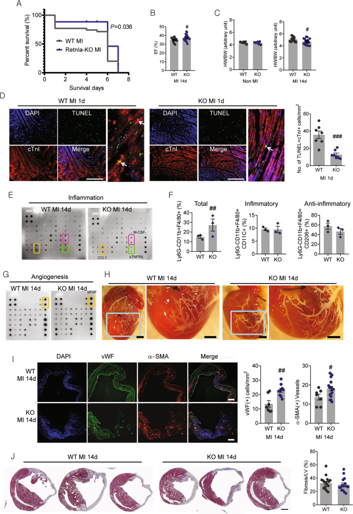
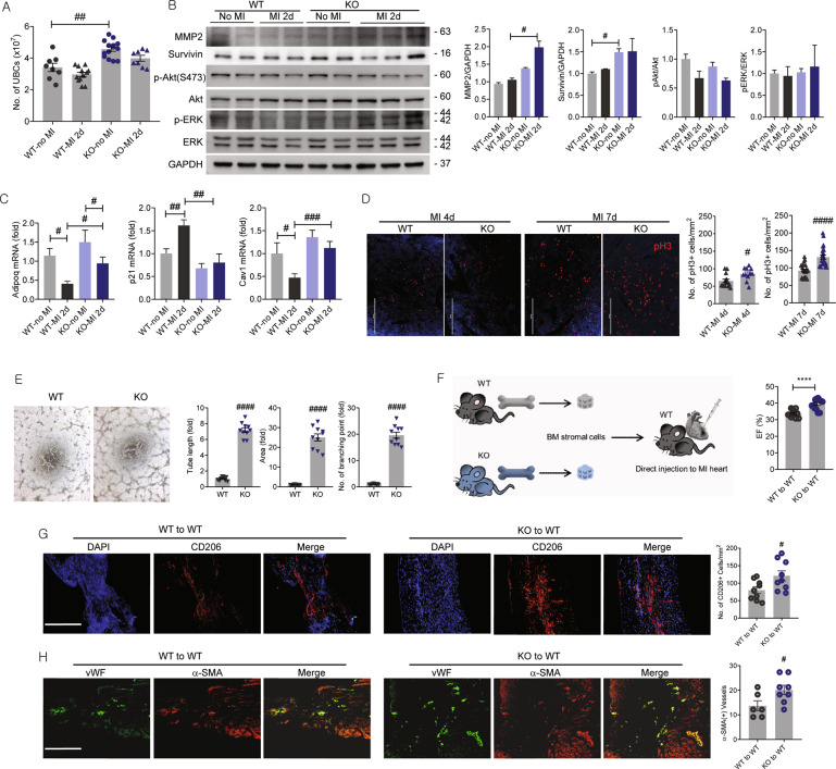
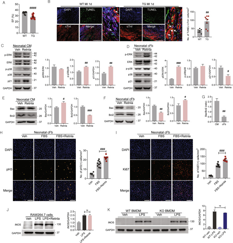
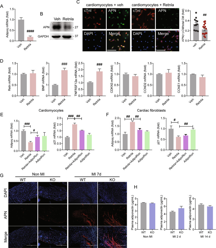
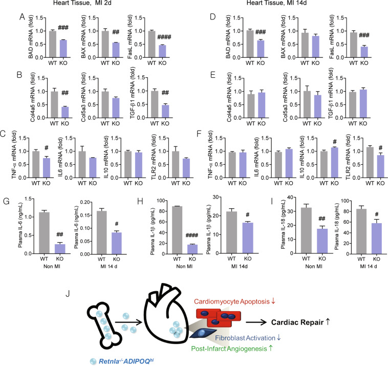
Resistin-like alpha (Retnla) is a member of the resistin family and known to modulate fibrosis and inflammation. Here, we investigated the role of Retnla in the cardiac injury model. Myocardial infarction (MI) was induced in wild type (WT), Retnla knockout (KO), and Retnla transgenic (TG) mice. Cardiac function was assessed by echocardiography and was significantly preserved in the KO mice, while worsened in the TG group. Angiogenesis was substantially increased in the KO mice, and cardiomyocyte apoptosis was markedly suppressed in the KO mice. By Retnla treatment, the expression of p21 and the ratio of Bax to Bcl2 were increased in cardiomyocytes, while decreased in cardiac fibroblasts. Interestingly, the numbers of cardiac macrophages and unsorted bone marrow cells (UBCs) were higher in the KO mice than in the WT mice. Besides, phosphorylated histone H3(+) cells were more frequent in bone marrow of KO mice. Moreover, adiponectin in UBCs was notably higher in the KO mice compared with WT mice. In an adoptive transfer study, UBCs were isolated from KO mice to transplant to the WT infarcted heart. Cardiac function was better in the KO-UBCs transplanted group in the WT-UBCs transplanted group. Taken together, proliferative and adiponectin-rich bone marrow niche was associated with substantial cardiac recovery by suppression of cardiac apoptosis and proliferation of cardiac fibroblast.


Conflict of interest statement

The authors declare no competing interests.

Figures

Fig. 1
Fig. 1. Retnla knockout protects mouse heart from myocardial infarction injury.
A Survival rates of the experimental groups; wild type (WT, n = 15) mice and Retnla knockout (KO, n = 17) mice with or without myocardial infarction (MI). WT MI n = 47, KO MI n = 36. B Left ventricular ejection fraction (LVEF) was determined by echocardiography in Retnla KO (n = 19) and WT littermates (n = 18) 14 days after MI. C Heart weight/body weight (HW/BW) ratios of mice were calculated before and at 14 days after MI. (WT; n = 7, KO; n = 7, WT MI; n = 18, KO MI; n = 16). D Cardiomyocyte apoptosis (white arrows) in the peri-infarct area was determined by TUNEL staining. cTnI cardiac troponin I. Scale bar, 200 μm. (WT; n = 7, KO; n = 7). E Inflammation-related mediators were analyzed by using antibody array membranes. F Three days after MI, cardiac macrophages were isolated to compare the cell number and phenotype. Ly6G(−)CD11b(+)F4/80(+)CD11C(−) cells are pro-inflammatory macrophages and Ly6G(−)CD11b(+)F4/80(+)CD206(+) cells are anti-inflammatory macrophages. The number of Ly6G(−)CD11b9(+)F4/80(+) cardiac macrophages was higher in the KO mice (n = 3, three mice per sample each) than in the WT mice (n = 3, three mice per sample each). G Angiogenesis-related mediators were analyzed by using antibody array membranes. H Microfil perfusion of hearts from WT and Retnla KO mice with MI for 14 days. I Left ventricular vascularity was assessed by immunostaining of von Willebrand factor (vWF) and α-smooth muscle actin (α-SMA). Representative images of vWF stained capillaries and α-SMA(+)-vessels in the infarcted heart. (WT; n = 7, KO; n = 10). Scale bar, 100 μm. J Representative Masson’s trichrome staining images of WT (n = 17) and Retnla KO mice (n = 15) after 14 days of MI. Quantitative analysis of ventricular fibrosis (expressed as a percentage of total left ventricular area) in WT and KO. Scale bar, 1000 μm. EF ejection fraction, FS fractional shortening. Data are represented as mean ± SEM. #P < 0.05, ##P < 0.01, ###P < 0.001 (by Student’s t test).
Fig. 2
Fig. 2. Characterization of Retnla knockout in unsorted bone marrow cells exhibiting cardiac protective effect.
A The number of unsorted bone marrow cells (UBCs) was higher in Retnla knockout (KO) mice regardless of myocardial infarction (MI). B Cell survival and proliferation-related mediators were analyzed in UBCs isolated from WT and Retnla KO mice by western blot. C UBCs were isolated from WT and KO mice 2 days after MI, and mRNA levels of adiponectin (Adipoq), p21, and caveolin-1 (Cav1) were quantified by real-time PCR. D The number of phosphorylated histone H3 (pH3)-positive cells in bone marrow was quantified, and the representative images showed pH3-positive cells (red fluorescence) in the bone marrow. (WT MI 4d; n = 14, KO MI 4d; n = 11, WT MI 7d; n = 23, KO MI 7d; n = 23). Scale bar, 200 μm. E To compare the endogenous angiogenic potential of WT and KO mice, mesenchymal stem cells (MSCs) were used. MSCs were isolated from bone marrow and tube formation assay was performed. Tube length, tubular area, and the number of branching points were analyzed in MSCs from wild type and Retnla KO mice. Scale bar, 500 μm. F UBCs were isolated from WT (n = 12) or KO mice (n = 13), and injected into infarcted heart of wild-type mice (n = 12 in the WT to WT mice, n = 13 in the KO to WT mice). After 2 weeks, cardiac function was analyzed, and the representative echocardiograms were shown. G, H Immunofluorescence staining showed larger distributions of anti-inflammatory CD206(+) macrophages and vascular density in the KO mice than in the WT mice. EF ejection fraction, FS fractional shortening. Scale bars, 200 μm. Data are represented as mean ± SEM. #P < 0.05, ##P < 0.01, ###P < 0.001 (by Student’s t test or two-way ANOVA). The image of the mouse (http://clipart-library.com/clipart/724544.htm) is obtained from clipart library and used under the Creative Commons Attribution-Share Alike 4.0 International license (http://creativecommons.org/licenses/by-sa/4.0/).
Fig. 3
Fig. 3. Effects of Retnla protein treatment on cardiomyocytes, cardiac fibroblasts, and macrophages.
A Cardiac function was assessed by echocardiography in wild type (WT, n = 13) and Retnla transgenic (TG) mice (n = 20) after myocardial infarction (MI). B Cardiomyocyte apoptosis (white arrows) in the peri-infarct area was determined by TUNEL staining. cTnI cardiac troponin I. (WT; n = 7, KO; n = 8). Scale bar, 200 μm. C, D Phosphorylation of stress-activated ERK and p38 and cell cycle arrest-related proteins p21, and were assessed in neonatal rat cardiomyocytes (CM) or neonatal rat cardiac fibroblasts (cFb) by western blotting. Cells were treated with veh or Retnla (500 ng/mL) for 24 h. E, F Expression of pro-apoptotic Bax and antiapoptotic Bcl2 was determined by western blot analysis in cardiomyocytes and cardiac fibroblasts treated with vehicle (veh) or Retnla (500 ng/mL) protein for 24 h. G The ratios of Bax to Bcl2 were calculated in cardiomyocytes and cardiac fibroblasts and showed significant difference. H, I Proliferation of cardiac fibroblasts were induced with FBS with or without Retnla treatment for 24 h. Proliferating cell markers, phosphorylated histone H3 (pH3) and Ki67, were used and quantified by counting pH3(+) cells (veh; n = 6, FBS; n = 9, FBS + Retnla; n = 8) and Ki67(+) cells (veh; n = 6, FBS; n = 10, FBS + Retnla; n = 7). J Induction of inducible nitric oxide synthase (iNOS), a pro-inflammatory marker, was assessed in RAW264.7 cells stimulated with lipopolysaccharide (LPS, 100 μg/mL) for 24 h in the presence of Retnla (500 ng/mL). K Bone marrow-derived macrophages (BMDMs) were derived from WT mice and Retnla KO, and stimulated with LPS (100 μg/mL) for 24 h. Induction of iNOS was compared by western blot. Data are represented as mean ± SEM. #P < 0.05, ##P < 0.01, ###P < 0.001 (by Student’s t test or one-way ANOVA with Bonferroni’s multiple comparisons test).
Fig. 4
Fig. 4. Involvement of adiponectin in cardiac protection mediated by unsorted bone marrow cells.
A The mRNA level of adiponectin (Adipoq) was assessed in neonatal rat cardiomyocytes treated with vehicle (veh) or Retnla (500 ng/mL) for 24 h. B Adiponectin (APN) protein expression was determined by western blot in neonatal rat cardiomyocytes. C Endogenous APN expression was assessed by immunofluorescence staining in neonatal rat cardiomyocytes treated with veh (n = 12) or Retnla (500 ng/mL, n = 15). Adiponectin-expressing cardiomyocytes were counted and quantified. Scale bars, 200 μm. D Relative gene expression analysis of resistin (Retn), brain natriuretic peptide (BNP), tumor necrosis factor receptor superfamily member 12a (TNFRSF12A), cyclin-dependent kinase inhibitor 3 (CDKN3), cyclin D2 (CCND2), and cyclin E1 (CCNE1) in cardiomyocytes treated with veh or Retnla (500 ng/mL) for 24 h. E, F AdipoRon, adiponectin receptor agonist that binds adiponectin receptor 1 and 2, partially restored the transcriptional levels of adiponectin and p21 in Retnla-treated cardiomyocytes and cardiac fibroblasts. G APN protein expression was determined in the myocardium. Scale bar, 400 μm. H Circulating level of APN was measured in WT mice (n = 8) and KO mice (n = 8) at 0 day, 2 days, and 14 days after MI. Data are represented as mean ± SEM. #P < 0.05, ##P < 0.01, ###P < 0.001 (by Student’s t test or one-way ANOVA with Bonferroni’s multiple comparisons test).
Fig. 5
Fig. 5. Expressional changes of signaling molecules of apoptosis, inflammation, fibrosis, and tissue repair in the heart tissues.
Heart tissues were collected at day 2 (AC) and 14 (DF) after MI to analyzed signaling molecules. BCL2-associated agonist of cell death (BAD), BCL2-associated X, apoptosis regulator (BAX), and Fas ligand (FasL) are cell death-related molecules. Collagen type IV, α5 (Col4α5), collagen type V, α3 (Col5α3), and transforming growth factor-β1 (TGF-β1) are fibrosis-related molecules. Tumor necrosis factor-α (TNF-α), interleukin-6 (IL-6), IL-10, and toll-like receptor 2 (TLR2) are inflammation-related molecules. GI Circulating levels of interleukin-6 (IL-6), IL-1β, and IL-18 in WT mice (n = 5) and KO mice (n = 5) with or without MI were measured. Data are represented as mean ± SEM. #P < 0.05, ##P < 0.01, ###P < 0.001 (by Student’s t test). J Schematic representation showing the proliferative bone marrow milieu with high adiponectin is associated with improved cardiac repair in infarcted myocardium. The images of bone (https://www.onlinewebfonts.com/icon/559079) and heart (https://www.onlinewebfonts.com/icon/493852) is obtained from clipart library, and used under the Creative Commons Attribution-Share Alike 4.0 International license (http://creativecommons.org/licenses/by-sa/4.0/).

Similar articles

Cited by

References

    1. Ziaeian B, Fonarow GC. Epidemiology and aetiology of heart failure. Nat. Rev. Cardiol. 2016;13:368–378. doi: 10.1038/nrcardio.2016.25. - DOI - PMC - PubMed
    1. Fan D, Takawale A, Lee J, Kassiri Z. Cardiac fibroblasts, fibrosis and extracellular matrix remodeling in heart disease. Fibrogenes. Tissue Repair. 2012;5:15. doi: 10.1186/1755-1536-5-15. - DOI - PMC - PubMed
    1. Fadini GP, et al. Circulating stem cells and cardiovascular outcomes: from basic science to the clinic. Eur. Heart J. 2020;41:4271–4282. doi: 10.1093/eurheartj/ehz923. - DOI - PMC - PubMed
    1. Mathur A, et al. The effect of intracoronary infusion of bone marrow-derived mononuclear cells on all-cause mortality in acute myocardial infarction: the BAMI trial. Eur. Heart J. 2020;41:3702–3710. doi: 10.1093/eurheartj/ehaa651. - DOI - PMC - PubMed
    1. Steppan CM, et al. A family of tissue-specific resistin-like molecules. Proc. Natl Acad. Sci. USA. 2001;98:502–506. doi: 10.1073/pnas.98.2.502. - DOI - PMC - PubMed

Publication types

Substances