. 2021 Mar 22;11(1):6585.
doi: 10.1038/s41598-021-85927-7.

Genome-wide identification of MIKC-type genes related to stamen and gynoecium development in Liriodendron

Affiliations

Genome-wide identification of MIKC-type genes related to stamen and gynoecium development in Liriodendron

Huanhuan Liu et al. Sci Rep. .

Abstract

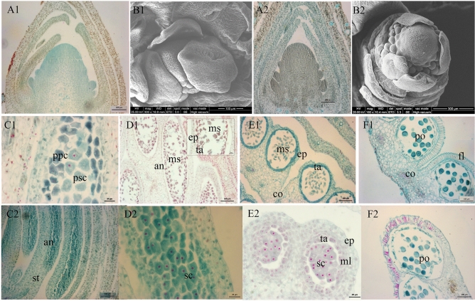
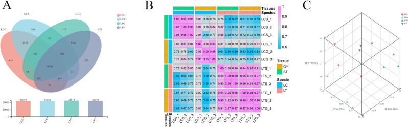
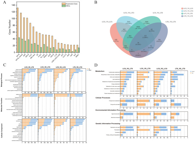
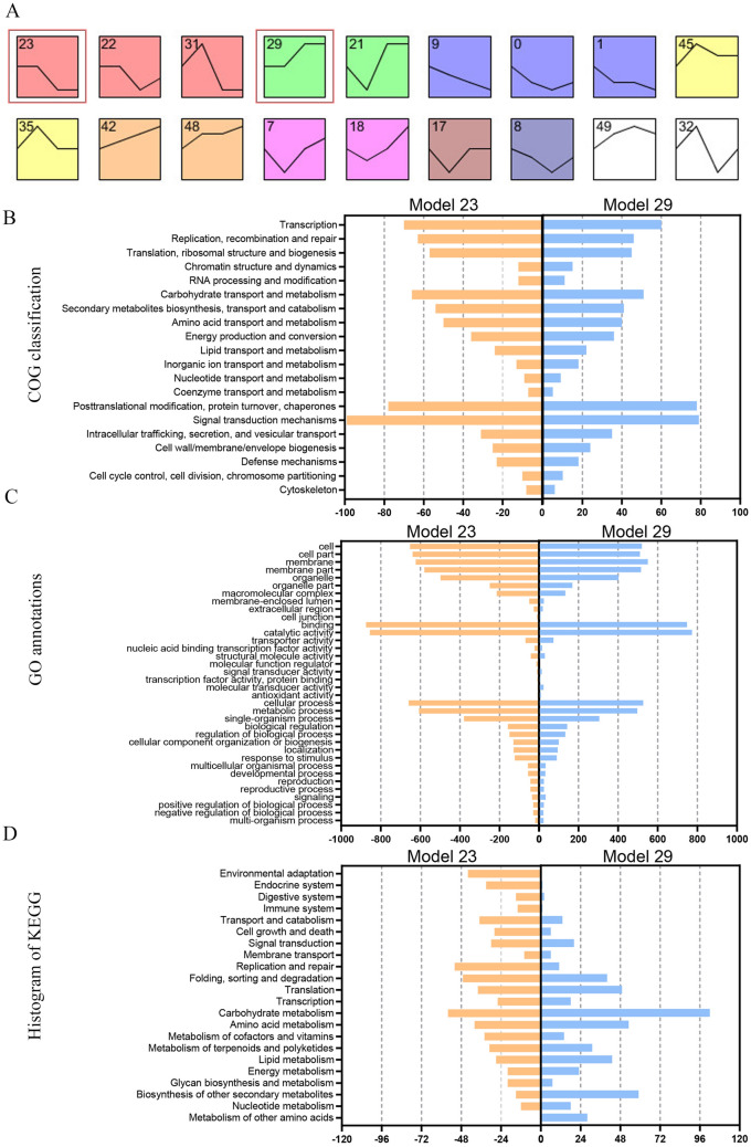
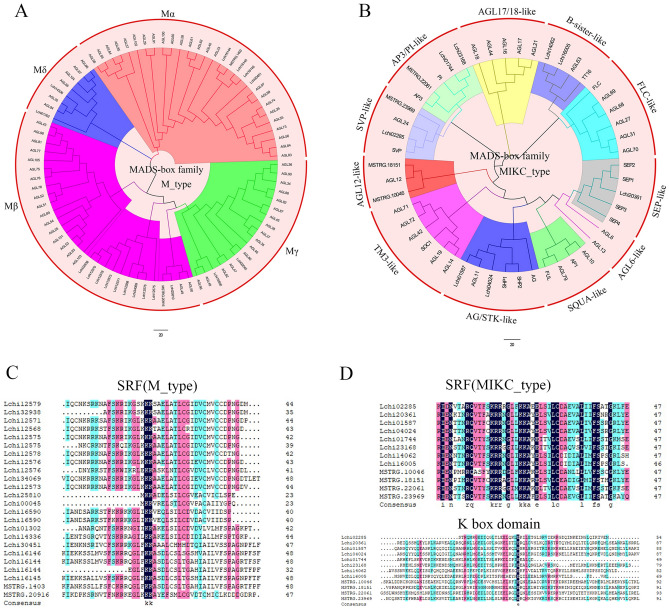
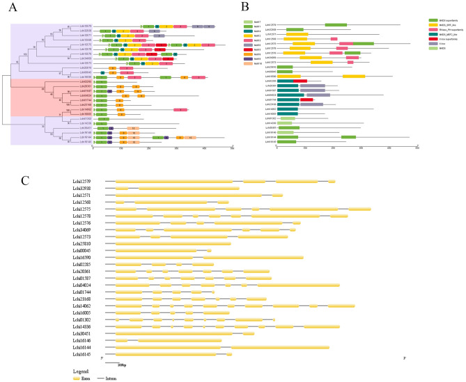
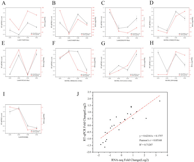
The organogenesis and development of reproductive organs, i.e., stamen and gynoecium, are important floral characteristics that are closely related to pollinators and reproductive fitness. As a genus from Magnoliaceae, Liriodendron has only two relict species: L. chinense and L. tulipifera. Despite the similar flower shapes of these species, their natural seed-setting rates differ significantly, implying interspecies difference in floral organogenesis and development. MADS-box genes, which participate in floral organogenesis and development, remain unexplored in Liriodendron. Here, to explore the interspecies difference in floral organogenesis and development and identify MADS-box genes in Liriodendron, we examined the stamen and gynoecium primordia of the two Liriodendron species by scanning electron microscopy combined with paraffin sectioning, and then collected two types of primordia for RNA-seq. A total of 12 libraries were constructed and 42,268 genes were identified, including 35,269 reference genes and 6,999 new genes. Monoterpenoid biosynthesis was enriched in L. tulipifera. Genome-wide analysis of 32 MADS-box genes was conducted, including phylogenetic trees, exon/intron structures, and conserved motif distributions. Twenty-six genes were anchored on 17 scaffolds, and six new genes had no location information. The expression profiles of MIKC-type genes via RT-qPCR acrossing six stamen and gynoecium developmental stages indicates that the PI-like, AG/STK-like, SEP-like, and SVP-like genes may contribute to the species-specific differentiation of the organogenesis and development of reproductive organs in Liriodendron. Our findings laid the groundwork for the future exploration of the mechanism underlying on the interspecific differences in reproductive organ development and fitness in Liriodendron.


Conflict of interest statement

The authors declare no competing interests.

Figures

Figure 1
Figure 1
Morphological and cytological observations of the stamen. (A1–B1) Stamen primordia of L. chinense; (A2–B2) Stamen primordia of L. tulipifera; (C1–F1) Ddevelopment process of anther primordium of L. chinense; (C2–F2) Development process of anther primordium of L. tulipifera; A1, B1, A2, D1, E1, F1, C2, and F2: bar, 100 μm; B2: bar, 300 μm; C1, D2, and E3: bar, 20 μm; an anther, co connectivum, ep epidermis, fl fibrous layer, ml middle layer, ms microsporocyte, nu nucellus, po pollen, ppc primary parietal cell, psc primary sporogenous cell, sc sporogenous cell; st stamen, ta tapetum.
Figure 2
Figure 2
Morphological and cytological observations of the gynoecium. (A1-B1) Gynoecium primordia of L. chinense; (A2-B2) Gynoecium primordia of L. tulipifera; (C1-F1) Ovule primordia; C2–H2: The two ovules in an ovary; A1, A2, B2, E1, C2, D2, E2, and F2: bar, 100 μm; B1: bar, 300 μm; D1, F1, G2, and H2: bar, 20 μm; C2: bar, 200 μm; ca carpel, ii inner integument, in integument, me megasporocyte, nu nucellus, oi outer integument, ov ovule.
Figure 3
Figure 3
Sample relationship analysis based on all expressed genes. (A) Venn diagram among all samples; (B) Correlation between two samples analysis; (C) Principal component analysis.
Figure 4
Figure 4
TF family prediction and GO functional annotation and KEGG pathway category analysis of DEGs. (A) TF family prediction of all expressed genes and all DEGs; (B) Number of DEGs identified in four comparisons; (C) GO functional annotation analysis of DEGs identified in four comparisons; (D) KEGG pathway category analysis of DEGs identified in four comparisons. Note: Fig. 4A was analyzed by Origin software (2017, https://www.originlab.com/).
Figure 5
Figure 5
Expression clustering analysis between L. chinense and L. tulipifera. (A) Expression clustering of all samples; (B) COG classification analysis of Models 23 and 29; (C) GO functional annotation analysis of Models 23 and 29; (D) KEGG pathway category analysis of Models 23 and 29. (A) was conducted using STEM software (v1.3.8, http://www.cs.cmu.edu/~jernst/stem/) according to the criterion TPM ≥ 0.5 by the STEM clustering method and 16 model profiles were significant (P < 0.01).
Figure 6
Figure 6
Construction of phylogenetic trees of the MADS-box proteins from L. chinense and A. thaliana and alignment of the SRF and K-box conserved domains. (A) Phylogenetic tree of type-I (M-type) MADS-box proteins from L. chinense (12) and A. thaliana (65) classified into 4 subgroups; (B) Phylogenetic tree of type-II (MIKC-type) MADS-box proteins from L. chinense (20) and A. thaliana (62) classified into 11 subgroups; (C) SRF conserved domain of M-type proteins; (D) SRF and K-box conserved domains of MIKC-type proteins. Notes: Phylogenetic trees were constructed by Clustal X (v2.1, http://www.clustal.org/clustal2/) and MEGA 7 software (https://www.megasoftware.net/) with the neighbor-joining (NJ) method with a bootstrap value of 1,000 and with the ML method, and they were edited using FigTree software (v1.4.3, http://tree.bio.ed.ac.uk/software/figtree/). The conserved domains of SRF and K-box were aligned using DNAMAN software (v10, https://www.lynnon.com/dnaman.html).
Figure 7
Figure 7
Distribution of conserved motifs (A), domains (B) and gene structures (C) of MADS-box proteins in L. chinense. All figures were analyzed by TB Tools software (v1.068, http://www.tbtools.com/).
Figure 8
Figure 8
Validation of RNA-seq data by RT-qPCR assay. (A–I) Expression profiling of 9 MADS-box family genes; (J) Scatter plot showing the correlation between RNA-seq and RT-qPCR data. Note: All data and figures were analyzed by Origin software (2017, https://www.originlab.com/).
Figure 9
Figure 9
Heat map of highly expressed MADS-box genes in the stamen primordia and gynoecium primordia of L. chinense and L. tulipifera. Note: The heat map was analyzed using TB Tools software (v1.068, http://www.tbtools.com/).
Figure 10
Figure 10
Heat map of the MIKC-type MADS-box genes in different stages of the stamens of L. chinense and L. tulipifera. (A1–F1) Stages 1–6 of the stamens of L. chinense; (A2–F2) Stages 1–6 of the stamens of L. tulipifera; (G) Heat map of the MIKC-type MADS-box genes of the stamens; (A1,B1,A2,B2) bar, 1 mm; C1-F1, C2-F2: bar, 5 mm. (G) was drawn using TB Tools software (v1.068, http://www.tbtools.com/).
Figure 11
Figure 11
Heat map of the MIKC-type MADS-box genes in different stages of the gynoecia of L. chinense and L. tulipifera. (A1–F1) Stage 1–6 of the gynoecia of L. chinense; (A2–F2) Stage 1–6 of the gynoecia of L. tulipifera; (G) Heat map of the MIKC-type MADS-box genes of the gynoecia; (A1,B1,A2,B2) bar, 1 mm; (C1-F1,C2-F2) bar, 5 mm. Note: Fig. 11G was analyzed using TB Tools software (v1.068, http://www.tbtools.com/).

Similar articles

Cited by

References

    1. Li HG, Chen L, Liang CY, Huang MR. A case study on provenance testing of tulip tree (Liriodendron spp) Chin. For. Sci. Technol. 2005;19(5):6–13. doi: 10.3969/j.issn.1000-8101.2005.05.005. - DOI
    1. Massoni J, Couvreur TL, Sauquet H. Five major shifts of diversification through the long evolutionary history of Magnoliidae (angiosperms) BMC Evol. Biol. 2015;15(1):49. doi: 10.1186/s12862-015-0320-6. - DOI - PMC - PubMed
    1. Xu F, Rudall PJ. Comparative floral anatomy and ontogeny in Magnoliaceae. Plant Syst. Evol. 2006;258:1–15. doi: 10.1007/s00606-005-0361-1. - DOI
    1. Wang S, Xie Y. Red List of Endangered Plants in China. Beijing; 2004.
    1. Fan RW, Ye JG, Yin ZF, Gao HG, You LX. Studies on seed development and embryogenesis in Liriodendron chinense (Hemsl) Sary. Acta Bot. Sin. 1992;34(6):437–442.

Publication types

Substances