. 2021 Mar 4;11(9):4467-4482.
doi: 10.7150/thno.54245. eCollection 2021.

Pancreatic ductal deletion of S100A9 alleviates acute pancreatitis by targeting VNN1-mediated ROS release to inhibit NLRP3 activation

Affiliations

Pancreatic ductal deletion of S100A9 alleviates acute pancreatitis by targeting VNN1-mediated ROS release to inhibit NLRP3 activation

Hong Xiang et al. Theranostics. .

Abstract

Recent studies have proven that the overall pathophysiology of pancreatitis involves not only the pancreatic acinar cells but also duct cells, however, pancreatic duct contribution in acinar cells homeostasis is poorly known and the molecular mechanisms leading to acinar insult and acute pancreatitis (AP) are unclear. Our previous work also showed that S100A9 protein level was notably increased in AP rat pancreas through iTRAQ-based quantitative proteomic analysis. Therefore, we investigated the actions of injured duct cells on acinar cells and the S100A9-related effects and mechanisms underlying AP pathology in the present paper. Methods: In this study, we constructed S100A9 knockout (s100a9-/-) mice and an in vitro coculture system for pancreatic duct cells and acinar cells. Moreover, a variety of small molecular inhibitors of S100A9 were screened from ChemDiv through molecular docking and virtual screening methods. Results: We found that the upregulation of S100A9 induces cell injury and inflammatory response via NLRP3 activation by targeting VNN1-mediated ROS release; and loss of S100A9 decreases AP injury in vitro and in vivo. Moreover, molecular docking and mutant plasmid experiments proved that S100A9 has a direct interaction with VNN1 through the salt bridges formation of Lys57 and Glu92 residues in S100A9 protein. We further found that compounds C42H60N4O6 and C28H29F3N4O5S can significantly improve AP injury in vitro and in vivo through inhibiting S100A9-VNN1 interaction. Conclusions: Our study showed the important regulatory effect of S100A9 on pancreatic duct injury during AP and revealed that inhibition of the S100A9-VNN1 interaction may be a key therapeutic target for this disease.

Keywords: S100A9; VNN1; acinar cells; acute pancreatitis; duct cells.


Conflict of interest statement

Competing Interests: The authors have declared that no competing interest exists.

Figures

Figure 1
Figure 1
Loss of S100A9 decreases pancreatic injury in AP mice. (A) Overview of the targeting strategy for constructing s100a9-/- mice. (B) PCR identification of homozygote, heterozygote and WT of s100a9-/- mice. (C) Gross images of the pancreas indicated that the s100a9-/- pancreas showed milder symptoms, including edema, hemorrhage and cholestasis. (D) s100a9-/- mice showed lower serum enzymes (n = 8) and decreased inflammatory factor levels (n = 3). (E) HE staining images proved that both pancreatic duct and acinar injuries were obviously alleviated in the s100a9-/- pancreas. (F) Double staining experiments showed that S100A9 protein levels were upregulated around the pancreatic duct (CK19-positive area). Data are presented as the mean ± SEM; *P < 0.05, **P < 0.01 vs. SO mice; #P < 0.05, ##P < 0.01 vs. AP mice.
Figure 2
Figure 2
Establishment of a coculture system for STC-injured ductal cells and primary acinar cells. (A) Bright field images and flow cytometry results indicated that a large number of damaged and apoptotic cells occurred among H6C7 cells and primary acinar cells. (B) MTT results showed that the cell viability of primary acinar cells in the coculture system was significantly decreased compared with that of the ctrl group (n = 6). (C) AMS-CK19 double staining results showed that CK19 (green fluorescence, marker of ductal cells) was markedly upregulated in primary acinar cells. (D) Flow cytometry and laser confocal microscopy results showed that Ca2+ release and oscillation were all increased in STC-injured H6C7 cells. (E) ELISA results showed that single S100A8 and S100A9 levels were all significantly increased, but the S100A8/9 dimer level has no obvious change in STC-injured H6C7 cells (n = 5). (F) S100A8 and S100A9 mRNA and protein expression levels were notably increased in STC-injured H6C7 cells (n = 3). (G) Levels of inflammatory factors were markedly elevated in STC-injured H6C7 cells (n = 3). Data are presented as the mean ± SEM; *P < 0.05 and **P < 0.01 vs. ctrl group.
Figure 3
Figure 3
S100A8 and S100A9 are important elements in STC-induced ductal cell injury. (A) Knockdown of S100A8 or S100A9 significantly increased the viability of H6C7 cells after STC pretreatment (n = 6). (B) Knockdown of S100A8 or S100A9 markedly inhibited inflammatory factor releases (IL-1β, IL-6, IL-8 and IL-18) in STC-pretreated H6C7 cells (n = 3). (C) Knockdown of S100A8 or S100A9 notably decreased apoptosis in STC-pretreated H6C7 cells. (D) Inflammatory factors (IL-1β, IL-6, IL-8 and IL-18) levels were significantly elevated in S100A8 or S100A9-overexpressing H6C7 cells (n = 3). (E) Cell viability of H6C7 cells were notably decreased in S100A8 or S100A9-overexpressing H6C7 cells (n = 6). (F) Apoptosis in H6C7 cells was increased in S100A8 or S100A9-overexpressing H6C7 cells. Data are presented as the mean ± SEM, *P < 0.05 and **P < 0.01 vs. shRNA NC or NC group; #P < 0.05 and ##P < 0.01 vs. shRNA NC+STC group.
Figure 4
Figure 4
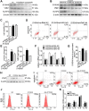
STC promotes NLRP3 activation through increasing S100A9/ VNN1 mediated ROS release. (A) Inflammatory factors (IL-1β, IL-6, IL-8 and IL-18) levels were remarkedly up-regulated in S100A9 group after pre-treating with Paq and FPS-ZM1 compared to NC group (n = 3). (B) Silver staining results of S100A9 IP experiment. (C) Co-IP experiment results further indicated that the S100A9 protein can pull down the VNN1 protein. (D) VNN1 protein level was notably upregulated in STC-injured H6C7 cells, and its expression wasn't notably changed after inhibiting TLR4 and RAGE signalings. (E) STC induced the increases of VNN1 expression and cysteamine release, and the decreases of γ-GCS and GSH contents (n = 5). (F) RR6 inhibited VNN1 enzyme activity, but STC had on obvious effect on it (n = 5). (G-I) ROS release, and downstream NLRP3 expression, caspase-1 activation, gasdermin D cleavage and IL-1β level were all upregulated in STC-injured H6C7 cells (n = 5). Data are presented as the mean ± SEM; *P < 0.05 and **P < 0.01 vs. NC or ctrl group; #P < 0.05 and ##P < 0.01 vs. STC group.
Figure 5
Figure 5
Extracellular S100A9 increases apoptosis and inflammatory response by targeting VNN1. (A) Protein expression levels of S100A9, VNN1 and NLRP3 were all downregulated after knocking down S100A9 in H6C7 cells. (B) Knockdown of VNN1 reduced VNN1 and NLRP3 protein levels in S100A9 plasmid pretreated-H6C7 cells but had no obvious effects on S100A9 protein expression. (C-D) Knockdown of VNN1 significantly increased cell viability (n = 6), and decreased apoptosis in S100A9-overexpressing H6C7 cells. (E) Knockdown of VNN1 improved oxidative stress by upregulating γ-GCS and GSH release in S100A9-overexpressing H6C7 cells (n = 6). (F) Knockdown of VNN1 notably downregulated inflammatory factor levels in S100A9-overexpressing H6C7 cells (n = 3). (G) ELISA result showed that S100A9 release in dep-S100A9 group is significantly downregulated compared with S100A9 group (n = 5). (H-K) VNN1 expression, cell apoptosis, ROS and inflammatory factors (n = 3) were notably decreased in dep-S100A9 group compared with S100A9 group. Data are presented as the mean ± SEM; *P < 0.05 and **P < 0.01 vs. NC or S100A9+shRNA NC group; ##P < 0.01 vs. S100A9 group.
Figure 6
Figure 6
Loss of S100A9 inhibits NLRP3 activation by decreasing VNN1-mediated ROS release. (A) Double staining experiments showed that both S100A9 and VNN1 were all upregulated in the pancreatic tissue of AP mice. (B) s100a9-/- mice showed decreased ROS (red fluorescence) release. (C) s100a9-/- mice showed increased γ-GCS and GSH levels (n = 8). (D) s100a9-/- mice showed downregulated NLRP3 expression. Data are presented as the mean ± SEM; **P < 0.01 vs. SO mice; ##P < 0.01 vs. AP mice.
Figure 7
Figure 7
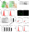
The S100A9 protein has an interaction with the VNN1 protein. (A) Binding model between S100A9 protein and VNN1 protein; the surfaces of S100A9 and VNN1 are colored orange and green, respectively. (B) Co-IP results indicated that S100A9 antibody pulled down less VNN1 protein in S100A9-MT group than in S100A9-WT group. (C-D) Ca2+ release and oscillation were all remarkedly downregulated in the MT variant of S100A9 compared with S100A9-WT. (E) S100A9-MT plasmid had no obvious effect on S100A9 protein expression but could downregulate VNN1 and NLRP3 protein levels compared with S100A9-WT. (F-I) S100A9-MT significantly increased cell viability (n = 6), and decreased cell apoptosis, inflammatory factor releases (n = 3) and ROS level of H6C7 cells compared with S100A9-WT. Data are presented as the mean ± SEM; *P < 0.05 and **P < 0.01 vs. S100A9-WT group.
Figure 8
Figure 8
Effects of small molecular inhibitors of S100A9-VNN1 interaction on STC-induced H6C7 cells injury. (A) The binding models of compounds 2, 5 and 8 with S100A9 protein. (B-D) Effects of compounds 2, 5 and 8 against STC-induced apoptosis, ROS release and inflammatory response (n = 3). Data are presented as the mean ± SEM; *P < 0.05 and **P < 0.01 vs. ctrl group; #P < 0.05 and ##P < 0.01 vs. STC group.
Figure 9
Figure 9
Effects of small molecular inhibitors of S100A9-VNN1 interaction on AP injury. (A) Compounds 2 and 5 at the dose of 10 mg/kg/day for 2 days have no obvious pancreatic, hepatic and renal toxicities (n = 6). (B-E) Compounds 2 and 5 at the doses of 10 mg/kg/day for 2 days can significantly inhibit α-AMS and LPS activities (n = 6) and improve AP damage through increasing γ-GCS and GSH (n = 6), and decreasing inflammatory response (n = 3). Data are presented as the mean ± SEM; **P < 0.01 vs. SO mice; ##P < 0.01 vs. AP mice.

References

    1. van Dijk SM, Hallensleben NDL, van Santvoort HC, Fockens P, van Goor H, Bruno MJ. et al. Acute pancreatitis: recent advances through randomised trials. Gut. 2017;66:2024–32. - PubMed
    1. Tenner S, Baillie J, DeWitt J, Vege SS, American College of Gastroenterology. American College of Gastroenterology guideline: management of acute pancreatitis. Am J Gastroenterol. 2013;108:1400–15. 1416. - PubMed
    1. Lankisch PG, Ape M, Banks PA, Acute pancreatitis. Lancet. 2015; 386: 85-96. - PubMed
    1. Trikudanathan G, Wolbrink DRJ, van Santvoort HC, Mallery S, Freeman M, Besselink MG. Current concepts in severe acute and necrotizing pancreatitis: an evidence-based approach. Gastroenterology. 2019;156:1994–2007.e3. - PubMed
    1. Hines QJ, Pandol SJ. Management of severe acute pancreatitis. BMJ. 2019;367:l6227. - PubMed

Publication types

MeSH terms