. 2021 Jun;97(6):1194-1209.
doi: 10.1111/cbdd.13841. Epub 2021 Apr 4.

Association of sigma-1 receptor with dopamine transporter attenuates the binding of methamphetamine via distinct helix-helix interactions

Affiliations

Association of sigma-1 receptor with dopamine transporter attenuates the binding of methamphetamine via distinct helix-helix interactions

Liang Xu et al. Chem Biol Drug Des. 2021 Jun.

Abstract

Dopamine transporter (DAT) and sigma-1 receptor (σ1R) are potential therapeutic targets to reduce the psychostimulant effects induced by methamphetamine (METH). Interaction of σ1R with DAT could modulate the binding of METH, but the molecular basis of the association of the two transmembrane proteins and how their interactions mediate the binding of METH to DAT or σ1R remain unclear. Here, we characterize the protein-ligand and protein-protein interactions at a molecular level by various theoretical approaches. The present results show that METH adopts a different binding pose in the binding pocket of σ1R and is more likely to act as an agonist. The relatively lower binding affinity of METH to σ1R supports the role of antagonists as inhibitors that protect against METH-induced effects. We demonstrate that σ1R could bind to Drosophila melanogaster DAT (dDAT) through interactions with either the transmembrane helix α12 or α5 of dDAT. Our results showed that the truncated σ1R displays stronger association with dDAT than the full-length σ1R. Although different helix-helix interactions between σ1R and dDAT lead to distinct effects on the dynamics of individual protein, both associations attenuate the binding affinity of METH to dDAT, particularly in the interactions with the helix α5 of dDAT. Together, the present study provides the first computational investigation on the molecular mechanism of coupling METH binding and the association of σ1R with dDAT.

Keywords: association; binding affinity; dopamine transporter; methamphetamine; sigma-1 receptor.


Conflict of interest statement

CONFLICT OF INTEREST

The authors declare no competing financial interest.

Figures
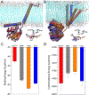
FIGURE 1.
FIGURE 1.
(A) Initial conformations of σ1R bound to METH (red), GM4 (agonist, gray), GMJ (antagonist, orange), and COC (blue), aligned with respect to the conformation of σ1R bound METH; (B) Representative conformations of σ1R bound to different ligands after 500-ns MD simulations; (C) The calculated binding energy for the binding of different ligand to σ1R; (D) The calculated conformational energy of σ1R bound to different ligands.
FIGURE 2.
FIGURE 2.
Interactions of METH (A), GM4 (B), GMJ (C), and COC (D) with σ1R. The ligands are shown in CPK representations, and the interacting residues are shown in Licorice representations. Residues are colored based on their types: blue for basic, red for acidic, green for polar, and white for nonpolar residues. Non-interacting residues D126 and E172 are shown in thin Licorice representations. A contact occurs if any atom of the ligand is within 3 Å of any atom of protein residues, and only residues with a contact frequency > 50% over the last-50 ns MD simulations are shown.
FIGURE 3.
FIGURE 3.
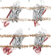
Representative binding conformations of dDAT with the truncated σ1R (residues 1–102) (A, B), and the full-length σ1R (C, D). The structure of σ1R is shown in red, and the molecule of METH in the binding pocket is shown in blue. The bound Na+ and Cl ions in dDAT are shown in yellow and cyan, respectively. The phosphate atoms of the lipid heads are shown as orange spheres to indicate the boundary of the membrane.
FIGURE 4.
FIGURE 4.
Interaction energies calculated for the association of dDAT with the truncated σ1R (A, B), and the full-length σ1R (C, D). (truncated, 1) corresponds to the truncated σ1R bound to the helix α12 of dDAT (Figure 3A); (truncated, 2) corresponds to the truncated σ1R bound to the helix α5 of dDAT (Figure 3B); (full-length, 1) corresponds to the full-length σ1R bound to the helix α12 of dDAT (Figure 3C); and (full-length, 2) corresponds to the full-length σ1R bound to the helix α5 of dDAT (Figure 3D).
FIGURE 5.
FIGURE 5.
The binding energy (A) and conformational energy (B) calculated for the binding of METH to dDAT in different systems. σ1R(1) corresponds to σ1R bound to the helix α12 of dDAT (Figure 3A and 3C); and σ1R(2) corresponds to σ1R bound to the helix α5 of dDAT (Figure 3B and 3D).
FIGURE 6.
FIGURE 6.
Helix-helix interactions between dDAT and the full-length σ1R. (A) Interactions of σ1R with the helix α12 of dDAT; and (B) Interactions of σ1R with the helix α5 of dDAT. The interacting parts of σ1R and dDAT are shown in red and white cartoon, respectively. Residues are colored based on their types: blue for basic, red for acidic, green for polar, and white for nonpolar residues. Two residues are considered in contact if a pair of (any) atoms belonging to two residues is closer than 3.0 Å, and only residues with a contact frequency > 50% over the last-50 ns MD simulations are shown.
FIGURE 7.
FIGURE 7.
Dynamic cross-correlation maps (DCCMs) constructed for dDAT bound to the truncated σ1R (A, B), and the full-length σ1R (C, D). The DCCMs for the truncated σ1R are not shown in A and B.

Similar articles

Cited by

References

    1. Abraham MJ, Murtola T, Schulz R, Páll S, Smith JC, Hess B, & Lindahl E (2015). Gromacs: High Performance Molecular Simulations through Multi-Level Parallelism from Laptops to Supercomputers. SoftwareX, 1–2, 19–25. doi:10.1016/j.softx.2015.06.001 - DOI
    1. Abramyan AM, Yano H, Xu M, Liu L, Naing S, Fant AD, & Shi L (2020). The Glu102 Mutation Disrupts Higher-Order Oligomerization of the Sigma 1 Receptor. Computational and Structural Biotechnology Journal, 18, 199–206. doi:10.1016/j.csbj.2019.12.012 - DOI - PMC - PubMed
    1. Amadei A, Ceruso MA, & Di Nola A (1999). On the Convergence of the Conformational Coordinates Basis Set Obtained by the Essential Dynamics Analysis of Proteins’ Molecular Dynamics Simulations. Proteins: Structure, Function, and Genetics, 36, 419–424. doi:10.1002/(sici)1097-0134(19990901)36:4<419::aid-prot5>3.0.co;2-u - DOI - PubMed
    1. Berendsen HJC, Postma JPM, van Gunsteren WF, DiNola A, & Haak JR (1984). Molecular Dynamics with Coupling to an External Bath. The Journal of Chemical Physics, 81, 3684–3690. doi:10.1063/1.448118 - DOI
    1. Berendsen HJC, van der Spoel D, & van Drunen R (1995). Gromacs: A Message-Passing Parallel Molecular Dynamics Implementation. Computer Physics Communications, 91, 43–56. doi:10.1016/0010-4655(95)00042-e - DOI

Publication types

MeSH terms

LinkOut - more resources