. 2021 Oct;236(10):7045-7057.
doi: 10.1002/jcp.30367. Epub 2021 Mar 23.

Higher infectivity of the SARS-CoV-2 new variants is associated with K417N/T, E484K, and N501Y mutants: An insight from structural data

Affiliations

Higher infectivity of the SARS-CoV-2 new variants is associated with K417N/T, E484K, and N501Y mutants: An insight from structural data

Abbas Khan et al. J Cell Physiol. 2021 Oct.

Abstract

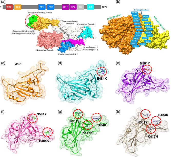
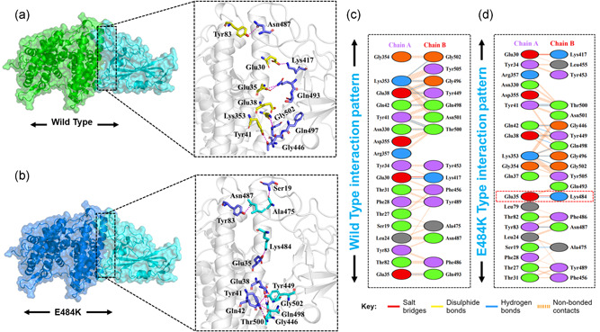
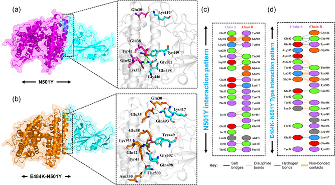
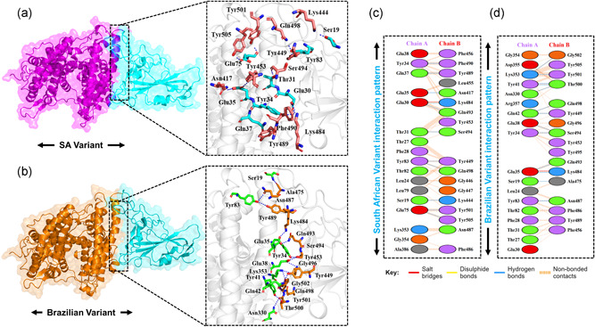
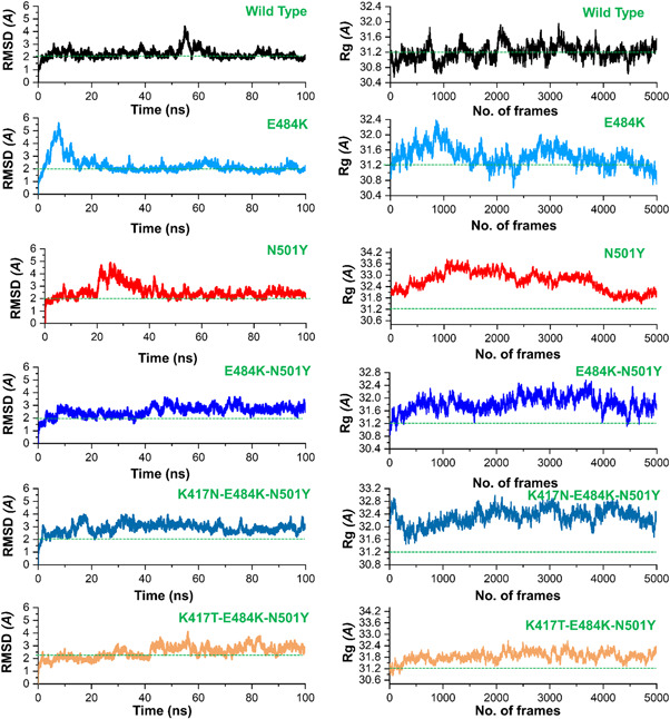
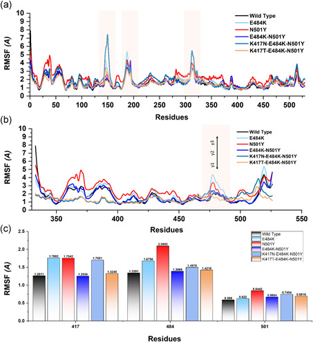
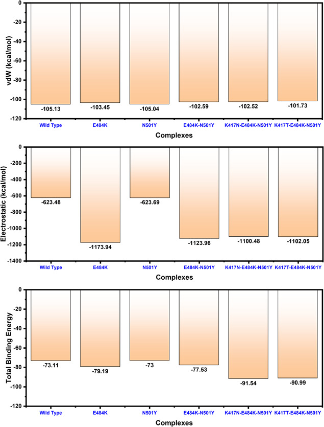
The evolution of the SARS-CoV-2 new variants reported to be 70% more contagious than the earlier one is now spreading fast worldwide. There is an instant need to discover how the new variants interact with the host receptor (ACE2). Among the reported mutations in the Spike glycoprotein of the new variants, three are specific to the receptor-binding domain (RBD) and required insightful scrutiny for new therapeutic options. These structural evolutions in the RBD domain may impart a critical role to the unique pathogenicity of the SARS-CoV-2 new variants. Herein, using structural and biophysical approaches, we explored that the specific mutations in the UK (N501Y), South African (K417N-E484K-N501Y), Brazilian (K417T-E484K-N501Y), and hypothetical (N501Y-E484K) variants alter the binding affinity, create new inter-protein contacts and changes the internal structural dynamics thereby increases the binding and eventually the infectivity. Our investigation highlighted that the South African (K417N-E484K-N501Y), Brazilian (K417T-E484K-N501Y) variants are more lethal than the UK variant (N501Y). The behavior of the wild type and N501Y is comparable. Free energy calculations further confirmed that increased binding of the spike RBD to the ACE2 is mainly due to the electrostatic contribution. Further, we find that the unusual virulence of this virus is potentially the consequence of Darwinian selection-driven epistasis in protein evolution. The triple mutants (South African and Brazilian) may pose a serious threat to the efficacy of the already developed vaccine. Our analysis would help to understand the binding and structural dynamics of the new mutations in the RBD domain of the Spike protein and demand further investigation in in vitro and in vivo models to design potential therapeutics against the new variants.

Keywords: KD (dissociation constant); MD simulation; SARS-CoV-2; new variants; protein-protein docking.


Conflict of interest statement

The authors declare that there are no conflict of interests.

Figures

Figure 1
Figure 1
Structural representation of the spike glycoprotein (PDB ID:6M0J) and the receptor‐binding domain of the SARS‐CoV‐2. (a) shows the distribution of different domains differentiated with different colors. The RBD domain is shown as yellow specifically. (B) shows the binding interface of the ACE2 and spike RBD. (c) wild type, (d) E484K, (e) N501Y, and (f) E484K‐N501Y, (g) K417N‐E484K‐N501Y, and (h) shows the K417T‐E484K‐N501Y structure of the spike RBD
Figure 2
Figure 2
Docking representation of the Wild type and E484K mutant complexes. (a) represent the binding interface of the wild‐type complex along with its stick representation of the key hydrogen interactions. (b) shows the binding interface and stick representation of the key hydrogen bonding interactions of the E484K mutant. (c, d) represent the 2D interactions representation including hydrogen, salt bridges, and nonbonded interactions in wild type and E484K complex
Figure 3
Figure 3
Docking representation of the N501Y and E484K‐ N501Y mutant complexes. (a) represent the binding interface of the N501Y complex along with its stick representation of the key hydrogen interactions. (b) shows the binding interface and stick representation of the key hydrogen bonding interactions of the E484K‐ N501Y mutant. (c, d) represent the 2D interactions representation including hydrogen, salt bridges and nonbonded interactions in N501Y and E484K‐ N501Y complex
Figure 4
Figure 4
Docking representation of the South African and Brazilian mutant complexes. (a) represent the binding interface of the South African complex along with its stick representation of the key hydrogen interactions. (b) shows the binding interface and stick representation of the key hydrogen bonding interactions of the Brazilian mutant. (c, d) represent the 2D interactions representation including hydrogen, salt bridges, and nonbonded interactions in South African and Brazilian complex
Figure 5
Figure 5
The figure represents the RMSDs and Rg(s) of all the complexes. The RMSD and Rg of the wild type is shown in black color while the other mutants are given in different colors. RMSDs, root mean square deviations
Figure 6
Figure 6
This figure represents the residual flexibility index of the wild‐type and mutant complexes. (a) shows the RMSFs of the complexes while the fluctuated regions are highlighted in light orange color. (b) represent the residual flexibility index of the wild and mutant spike RBD only. The three important loops required for interaction with ACE2 are represented with γ1, γ2, and γ3. These regions γ1 (474–485), γ2 (488–490), and γ3 (494–505) are crucial for binding. (c) show the individual residue flexibility, that is, K417N, K417T, E48K, and N501Y. RMSFs, root mean square fluctuations
Figure 7
Figure 7
Free energy calculation results obtained from MD simulation trajectory of the wild type and mutant complexes. The top bar graph shows the vdW contribution by each complex, the 2nd bar graph shows the electrostatic energy while the bottom bar graph shows the total binding energy. All the energies given here are calculated in kcal/mol

References

    1. Abraham, S. , Kienzle, T. E. , Lapps, W. , & Brian, D. A. (1990). Deduced sequence of the bovine coronavirus spike protein and identification of the internal proteolytic cleavage site. Virology, 176(1), 296–301. - PMC - PubMed
    1. Ali, A. , Khan, A. , Kaushik, A. C. , Wang, Y. , Ali, S. S. , Junaid, M. , Saleem, S. , Cho, W. C. , Mao, X. , & Wei, D.‐Q. (2019). Immunoinformatic and systems biology approaches to predict and validate peptide vaccines against Epstein–Barr virus (EBV). Scientific Reports, 9(1), 1–12. - PMC - PubMed
    1. Ali, A. , & Vijayan, R. (2020). Dynamics of the ACE2–SARS‐CoV‐2/SARS‐CoV spike protein interface reveal unique mechanisms. Scientific Reports, 10(1), 1–12. - PMC - PubMed
    1. Davies, N. G. , Barnard, R. C. , Jarvis, C. I. , Kucharski, A. J. , Munday, J. , Pearson, C. A. , Russell, T. W. , Tully, D. C. , Abbott, S. , & Gimma, A. (2020). Estimated transmissibility and severity of novel SARS‐CoV‐2 Variant of Concern 202012/01 in England. medRxiv.
    1. Dominguez, C. , Boelens, R. , & Bonvin, A. M. (2003). HADDOCK: A protein−protein docking approach based on biochemical or biophysical information. Journal of the American Chemical Society, 125(7), 1731–1737. - PubMed

Publication types

Substances