. 2021 Mar 23;11(1):30.
doi: 10.1186/s13550-021-00773-y.

Carotid plaque inflammatory activity assessed by 2-[18F]FDG-PET imaging decrease after a neurological thromboembolic event

Affiliations

Carotid plaque inflammatory activity assessed by 2-[18F]FDG-PET imaging decrease after a neurological thromboembolic event

Laerke Urbak et al. EJNMMI Res. .

Abstract

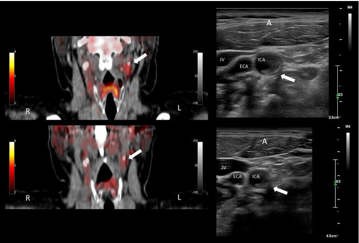
Background: Atherosclerotic plaque vulnerability is comprised by plaque composition driven by inflammatory activity and these features can be depicted with 3D ultrasound and 2-[18F]FDG-PET, respectively. The study investigated timely changes in carotid artery plaque inflammation and morphology after a thromboembolic event with PET/CT and novel ultrasound volumetric grayscale median (GSM) readings. Patients with a single hemisphere-specific neurological symptom and the presence of an ipsilateral carotid artery atherosclerotic plaque were prospectively included to both 2-[18F]FDG PET/CT and 3D ultrasound scans of the plaque immediately after their event and again three months later. On PET/CT images the maximum standardized uptake value (SUVmax) was measured and the volumetric ultrasound acquisitions were analyzed using a semiautomated software measuring GSM values.

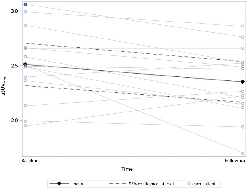
Results: Baseline scans were performed by a mean of 7 days (range 2-14) after the symptom and again after 98 days (range 91-176). For the entire group (n = 14), we found a decrease in average SUVmax from baseline to follow-up of - 0.18 (95% confidence interval: - 0.34 to - 0.02, P = 0.034). GSM did not increase significantly over time (mean change: + 2.21, 95% confidence interval: - 17.02 to 21.44, P = 0.808).

Conclusion: A decrease in culprit lesion 2-[18F]FDG-uptake 3 months after an event indicates a decrease in inflammatory activity, suggesting that carotid plaque stabilization over time. 3D ultrasound morphological quantitative differences in GSM were not detectable after 3 months.

Keywords: 3D ultrasound; Carotid artery plaque; FDG PET; Inflammation; Vulnerable plaque.


Conflict of interest statement

Henrik Sillesen has received research grants and speakers honoraria from Philips Ultrasound.

Figures

Fig. 1
Fig. 1
Schematic example of the calculated average max standardized uptake value (aSUVmax) and grayscale median (GSM). ECA = external carotid artery. ICA = internal carotid artery. CCA = common carotid artery. The 3D ultrasound acquisition of the plaque was sliced in 2-mm segments from the flow divider, and the slice containing the maximum plaque thickness was identified. The slice with the maximum plaque thickness and the two slices on each side were used in analyses. For the PET/CT, the flow divider was used as landmark for alignment of SUVmax readings to the ultrasound readings. An average SUVmax was calculated for this 1 cm long plaque volume. GSM was automatically calculated by the software for the volume of the 10-mm-long plaque segment
Fig. 2
Fig. 2
Flowchart of the included patients. Of the 19 included four were excluded. Two were excluded after withdrawal of consent, one died because of an, at the time of inclusion, undiagnosed cancer and one scan was lost
Fig. 3
Fig. 3
Change in average maximum standardized uptake values (aSUVmax) from baseline to follow-up. The black diamonds represent the mean aSUVmax at baseline and follow-up with the 95% confidence interval as the dotted line. Mean difference in aSUVmax of − 0.18, CI95%: − 0.34 to − 0.02, P = 0.034. The light blue lines represent each patient with the circles marking the aSUVmax at baseline and follow-up. The patient with recurrent symptoms undergoing carotid endarterectomy before the follow-up scan is marked as a filled circle
Fig. 4
Fig. 4
Baseline and follow-up patient example of 2-[18F]FDG-PET / CT-angiography and ultrasound images. Images from one patient. To the left 2-[18F]FDG-PET/CT angiography fusion, frontal MPR and to the right ultrasound images, axial plane. The top images are from baseline and the bottom follow-up. The white arrow points to the plaque on the symptomatic side

Similar articles

Cited by

References

    1. Ois A, Gomis M, Rodriguez-Campello A, Cuadrado-Godia E, Jimenez-Conde J, Pont-Sunyer C, et al. Factors associated with a high risk of recurrence in patients with transient ischemic attack or minor stroke. Stroke. 2008;39(6):1717–1721. doi: 10.1161/strokeaha.107.505438. - DOI - PubMed
    1. Coull AJ, Lovett JK, Rothwell PM. Population based study of early risk of stroke after transient ischaemic attack or minor stroke: implications for public education and organisation of services. BMJ. 2004;328(7435):326. doi: 10.1136/bmj.37991.635266.44. - DOI - PMC - PubMed
    1. Naylor AR, Ricco JB, de Borst GJ, Debus S, de Haro J, Halliday A, et al. Editor's choice—management of atherosclerotic carotid and vertebral artery disease: 2017 clinical practice guidelines of the European Society for Vascular Surgery (ESVS) Eur J Vasc Endovasc Surg. 2018;55(1):3–81. doi: 10.1016/j.ejvs.2017.06.021. - DOI - PubMed
    1. Falk E, Shah PK, Fuster V. Coronary plaque disruption. Circulation. 1995;92(3):657–671. doi: 10.1161/01.CIR.92.3.657. - DOI - PubMed
    1. Giroud D, Li JM, Urban P, Meier B, Rutishauer W. Relation of the site of acute myocardial infarction to the most severe coronary arterial stenosis at prior angiography. Am J Cardiol. 1992;69(8):729–732. doi: 10.1016/0002-9149(92)90495-k. - DOI - PubMed

LinkOut - more resources