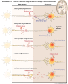
Deep grey matter injury in multiple sclerosis: a NAIMS consensus statement
- PMID: 33757115
- PMCID: PMC8370433
- DOI: 10.1093/brain/awab132
Deep grey matter injury in multiple sclerosis: a NAIMS consensus statement
Abstract
Although multiple sclerosis has traditionally been considered a white matter disease, extensive research documents the presence and importance of grey matter injury including cortical and deep regions. The deep grey matter exhibits a broad range of pathology and is uniquely suited to study the mechanisms and clinical relevance of tissue injury in multiple sclerosis using magnetic resonance techniques. Deep grey matter injury has been associated with clinical and cognitive disability. Recently, MRI characterization of deep grey matter properties, such as thalamic volume, have been tested as potential clinical trial end points associated with neurodegenerative aspects of multiple sclerosis. Given this emerging area of interest and its potential clinical trial relevance, the North American Imaging in Multiple Sclerosis (NAIMS) Cooperative held a workshop and reached consensus on imaging topics related to deep grey matter. Herein, we review current knowledge regarding deep grey matter injury in multiple sclerosis from an imaging perspective, including insights from histopathology, image acquisition and post-processing for deep grey matter. We discuss the clinical relevance of deep grey matter injury and specific regions of interest within the deep grey matter. We highlight unanswered questions and propose future directions, with the aim of focusing research priorities towards better methods, analysis, and interpretation of results.
Keywords: MRI; atrophy; deep grey matter; multiple sclerosis; thalamus.
© The Author(s) (2021). Published by Oxford University Press on behalf of the Guarantors of Brain. All rights reserved. For permissions, please email: journals.permissions@oup.com.
Figures


References
-
- Trapp BD, Peterson J, Ransohoff RM, Rudick R, Mörk S, Bö L.. Axonal transection in the lesions of multiple sclerosis. N Engl J Med. 1998;338(5):278–285. - PubMed
-
- Kutzelnigg A, Lassmann H.. Cortical lesions and brain atrophy in MS. J Neurologic Sci. 2005;233(1-2):55–59. - PubMed
-
- Bakshi R, Czarnecki D, Shaikh ZA.. Brain MRI lesions and atrophy are related to depression in multiple sclerosis. Neuroreport. 2000;11(6):1153–1158. - PubMed
-
- Cifelli A, Arridge M, Jezzard P, Esiri MM, Palace J, Matthews PM.. Thalamic neurodegeneration in multiple sclerosis. Ann Neurol. 2002;52(5):650–653. - PubMed
Publication types
MeSH terms
Grants and funding
LinkOut - more resources
Full Text Sources
Other Literature Sources
Medical

