. 2021 Mar 23;18(1):81.
doi: 10.1186/s12974-021-02127-w.

Specific depletion of resident microglia in the early stage of stroke reduces cerebral ischemic damage

Affiliations

Specific depletion of resident microglia in the early stage of stroke reduces cerebral ischemic damage

Ting Li et al. J Neuroinflammation. .

Abstract

Background: Ischemia can induce rapid activation of microglia in the brain. As key immunocompetent cells, reactive microglia play an important role in pathological development of ischemic stroke. However, the role of activated microglia during the development of ischemia remains controversial. Thus, we aimed to investigate the function of reactive microglia in the early stage of ischemic stroke.

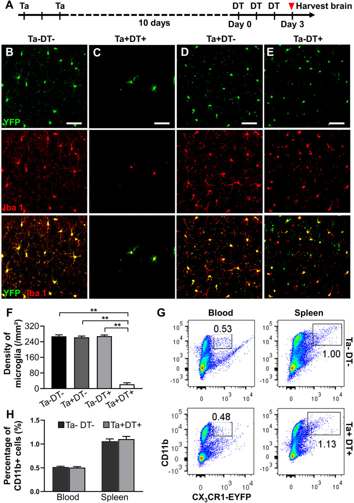
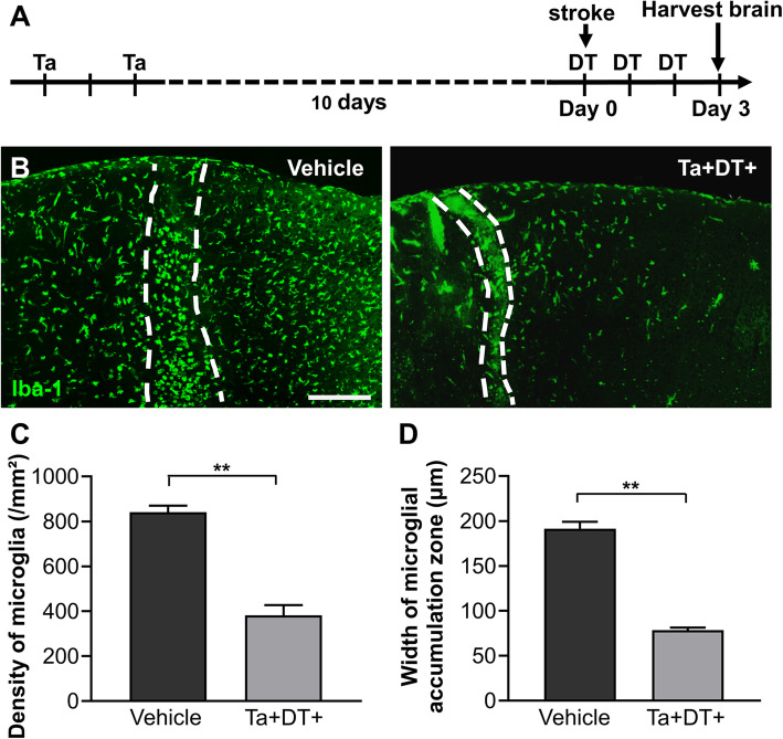
Methods: A Rose Bengal photothrombosis model was applied to induce targeted ischemic stroke in mice. CX3CR1CreER:R26iDTR mice were used to specifically deplete resident microglia through intragastric administration of tamoxifen (Ta) and intraperitoneal injection of diphtheria toxin (DT). At day 3 after ischemic stroke, behavioral tests were performed. After that, mouse brains were collected for further histological analysis and detection of mRNA expression of inflammatory factors.

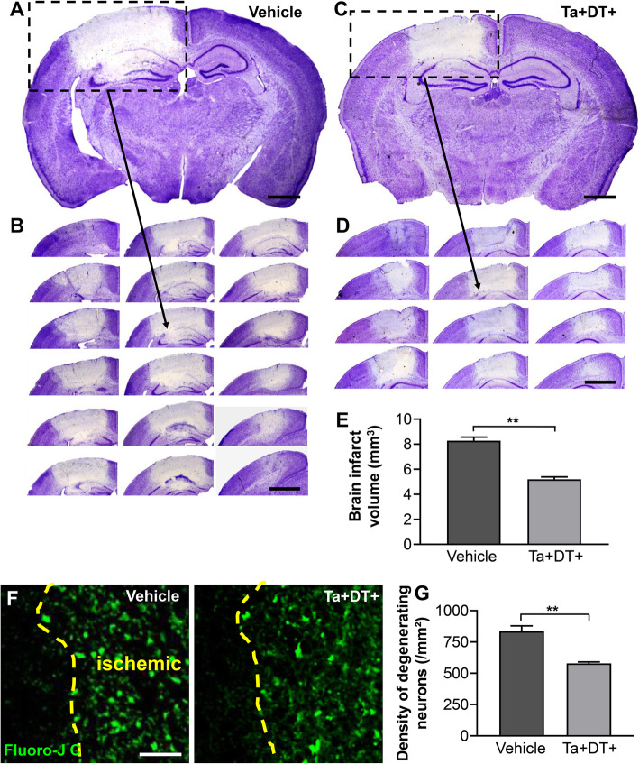
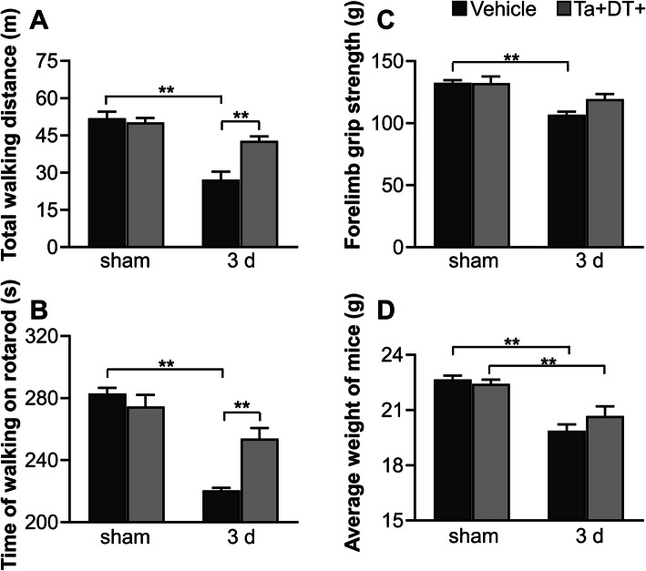
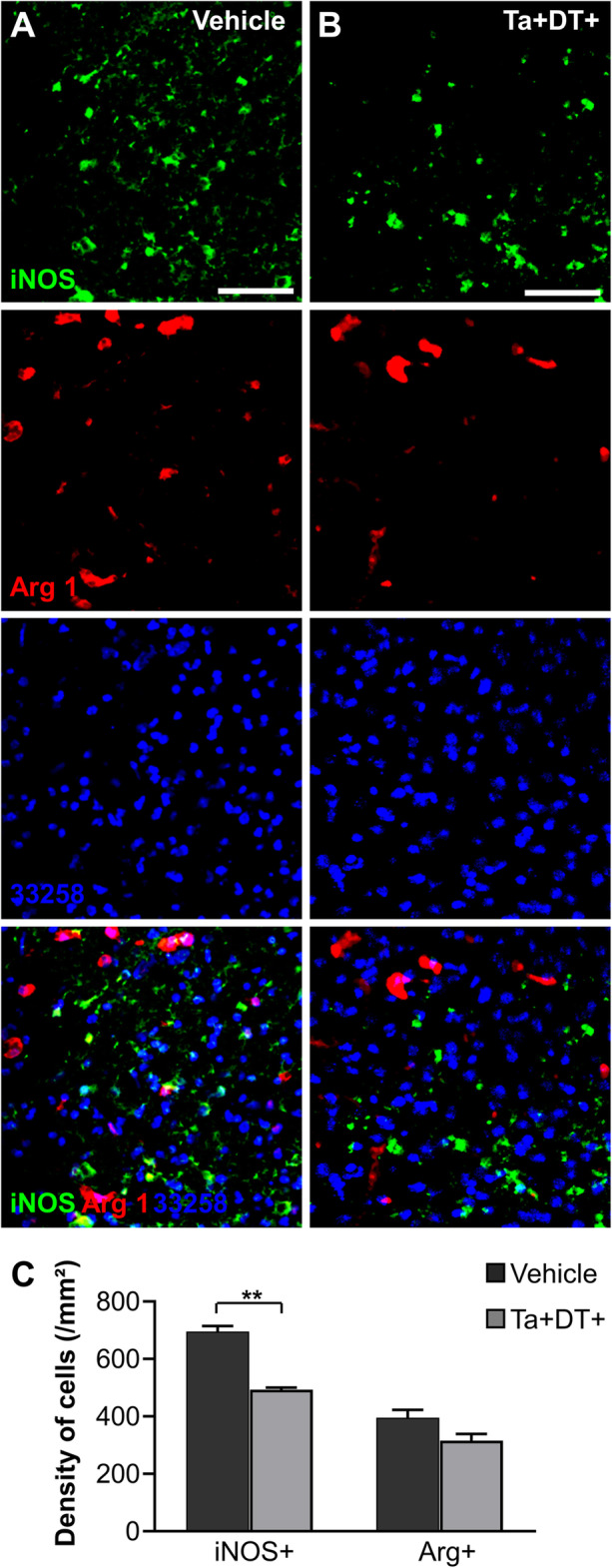
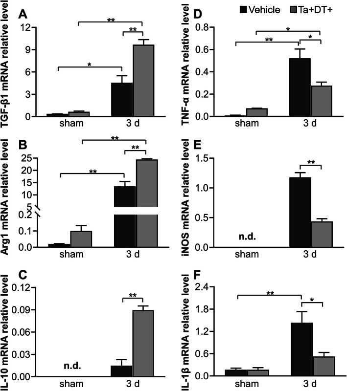
Results: The results showed that specific depletion of microglia resulted in a significant decrease in ischemic infarct volume and improved performance in motor ability 3 days after stroke. Microglial depletion caused a remarkable reduction in the densities of degenerating neurons and inducible nitric oxide synthase positive (iNOS+) cells. Importantly, depleting microglia induced a significant increase in the mRNA expression level of anti-inflammatory factors TGF-β1, Arg1, IL-10, IL-4, and Ym1 as well as a significant decline of pro-inflammatory factors TNF-α, iNOS, and IL-1β 3 days after stroke.

Conclusions: These results suggest that activated microglia is an important modulator of the brain's inflammatory response in stroke, contributing to neurological deficit and infarct expansion. Modulation of the inflammatory response through the elimination of microglia at a precise time point may be a promising therapeutic approach for the treatment of cerebral ischemia.

Keywords: Depletion; Function; Inflammation; Ischemia; Microglia.


Conflict of interest statement

All authors claim that there are no conflict of interest.

Figures

Fig. 1
Fig. 1
Microglia depletion efficiency of CX3CR1CreER/+:R26iDTR/+ transgenic mice. (a) Timeline of Ta and DT administration in the selective microglial depletion model. (b) Represent images of microglia after vehicle treatment. Microglia evenly distributed throughout the cerebral cortex in mice brain without microglial depletion. (c) Represent images of microglia after Ta and DT treatment. After drug treatment, most native microglia were depleted and the remnants exhibited appearance similar to activated state and distributed randomly. (d) Represent images of microglia only with intragastric administration of Ta but not DT. (e) Represent images of microglia only with intraperitoneal injection of DT but not Ta. No significant effect was found on the phenotype and density of microglia in cortex between single- and no-drug groups (b-e scale bar = 50 μm). (f) Densities of microglia in the cerebral cortex with drug treatment or not (n ≥5, **p < 0.01). (g) FACS analysis showing percentage of CD11b+ cells in the blood and spleen of mice after vehicle or Ta+DT+ treatment. (h) Quantification of the FACS analysis results shown in (g) (n = 4)
Fig. 2
Fig. 2
Microgliosis in CX3CR1CreER/+:R26iDTR/+ transgenic mice 3 days after ischemic stroke. (a) Timeline of drug administration and tissue processing 3 days post stroke. (b) Distribution of microglia at the lesion site in vehicle-treated mice and microglia-depleted mice 3 days post stroke. Iba-1 was used to highlight microglia in CX3CR1CreER/+:R26iDTR/+ mice. Microglial accumulation zones were delineated by the dashed lines (scale bar = 200 μm). (c) Density of microglia in the accumulation zone with and without microglia depletion. (d) Width of the accumulation zone of reactive microglia with and without microglial depletion (n ≥ 3, **p < 0.01)
Fig. 3
Fig. 3
Depletion of microglia decreased the infarct volume and neuronal degeneration 3 days after ischemic stroke. (a) Representative Nissl stained coronal brain slice of vehicle-treated mouse after stroke (scale bar = 1 mm). (b) Coronal sections in accordance to the boxed region in (a) illustrating the whole infarct (scale bar = 2 mm). (c) Representative Nissl stained coronal brain slice of microglia-depleted mouse after stroke (scale bar = 1 mm). (d) Coronal sections in accordance to the boxed region in (c) illustrating the whole infarct (scale bar = 2 mm). (e) Calculated brain infarct volumes 3 days post stroke, in mice with Ta and DT treatment or not (n ≥ 6, **p < 0.01). (f) Representative confocal images of FJC-labeled degenerating neurons in the ischemic areas of strokeTa−DT− and strokeTa+DT+ mice. The boundaries between ischemic area and normal tissue were delineated by the dashed lines (scale bar = 50 μm). (g) Densities of degenerating neurons in the border area underwent microglia depletion or not. Note that neurodegeneration greatly decreased in microglia-devoid mice 3 days after stroke (n ≥ 3, **p < 0.01)
Fig. 4
Fig. 4
Effects of microglial depletion on mice behavioral performance and body weight. (a-c) Mice behavioral performance in spontaneous activity test, rotarod test, and grip strength test with or without ischemic stroke. (d) Body weight of mice with or without ischemic stroke. Microglial depletion did not show obvious effect on body weight of mice. Note that ischemia itself greatly impacted on behavioral performance and body weight (n ≥ 5, *p <0.05, **p <0.01)
Fig. 5
Fig. 5
Depleting microglia reduced inflammatory cells at the lesion site. (a) Representative images showing iNOS+ cells and Arg1+ cells at the lesion site without microglial depletion. (b) Representative images showing iNOS+ cells and Arg1+ cells after microglia depletion (a-b Scale bars = 50 μm). (c) Densities of iNOS+ cells and Arg1+ cells in the periphery area of lesion site with microglia depletion or not (n = 4, *p <0.05, **p <0.01)
Fig. 6
Fig. 6
qRT-PCR analysis of mRNA expression of inflammatory factors in the presence and absence of microglia. (a-c) Relative mRNA levels of anti-inflammatory factors TGF-β1, Arg1, IL-10 with and without microglial depletion. (d-f) Relative mRNA levels of pro-inflammatory factors TNF-α, iNOS, and IL-1β with and without microglial depletion (n ≥ 3, *p <0.05, **p < 0.01. n.d. = not detectable)

References

    1. Huang X, Cheripelli BK, Lloyd SM, Kalladka D, Moreton FC, Siddiqui A, Ford I, Muir KW. Alteplase versus tenecteplase for thrombolysis after ischaemic stroke (ATTEST): a phase 2, randomised, open-label, blinded endpoint study. Lancet Neurol. 2015;14(4):368–376. doi: 10.1016/S1474-4422(15)70017-7. - DOI - PubMed
    1. Bivard A, Levi C, Krishnamurthy V, McElduff P, Miteff F, Spratt NJ, Bateman G, Donnan G, Davis S, Parsons M. Perfusion computed tomography to assist decision making for stroke thrombolysis. Brain. 2015;138(7):1919–1931. doi: 10.1093/brain/awv071. - DOI - PMC - PubMed
    1. Donnan GA, Fisher M, Macleod M, Davis SM. Stroke. Lancet. 2008;371(9624):1612–1623. doi: 10.1016/S0140-6736(08)60694-7. - DOI - PubMed
    1. Xiong XY, Liu L, Yang QW. Functions and mechanisms of microglia/macrophages in neuroinflammation and neurogenesis after stroke. Prog Neurobiol. 2016;142:23–44. doi: 10.1016/j.pneurobio.2016.05.001. - DOI - PubMed
    1. Wake H, Moorhouse AJ, Jinno S, Kohsaka S, Nabekura J. Resting microglia directly monitor the functional state of synapses in vivo and determine the fate of ischemic terminals. J Neurosci. 2009;29(13):3974–3980. doi: 10.1523/JNEUROSCI.4363-08.2009. - DOI - PMC - PubMed

Substances