Review
. 2021 Mar 23;9(1):49.
doi: 10.1186/s40478-021-01152-3.

Space-occupying brain lesions, trauma-related tau astrogliopathy, and ARTAG: a report of two cases and a literature review

Affiliations
Review

Space-occupying brain lesions, trauma-related tau astrogliopathy, and ARTAG: a report of two cases and a literature review

Adam D Bachstetter et al. Acta Neuropathol Commun. .

Abstract

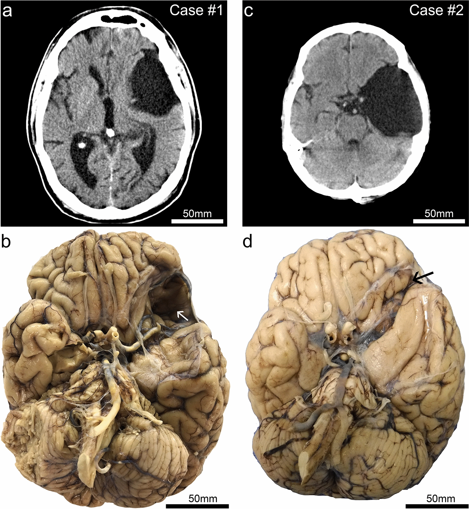
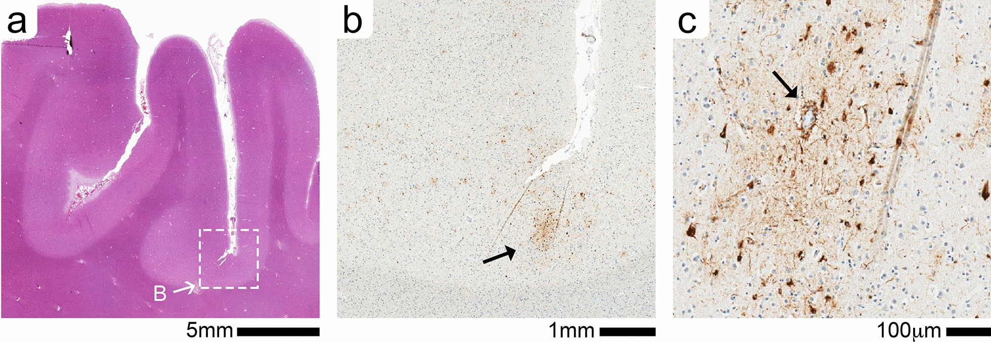
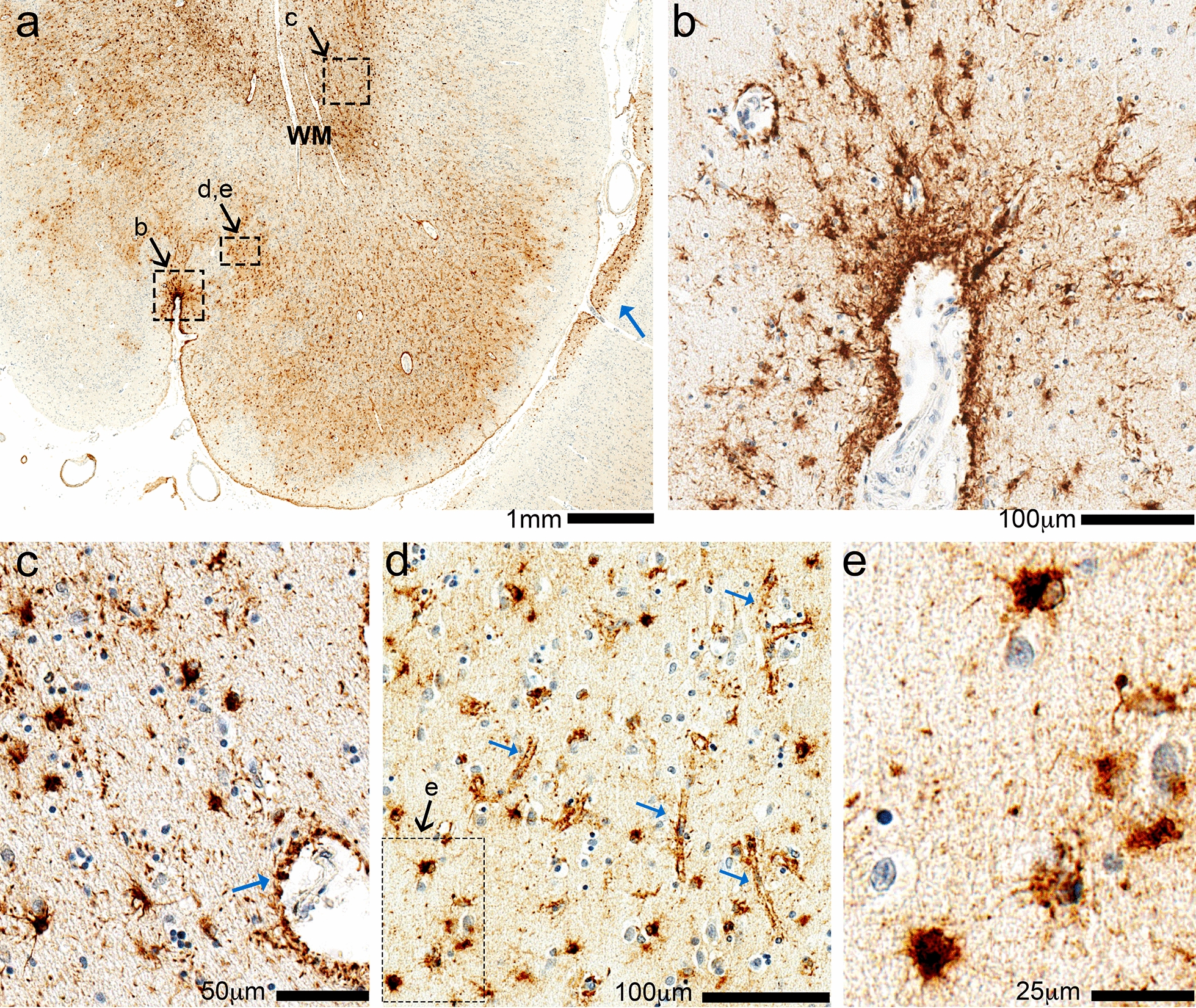
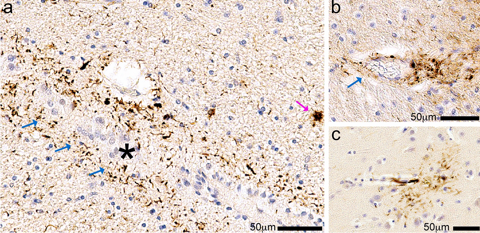
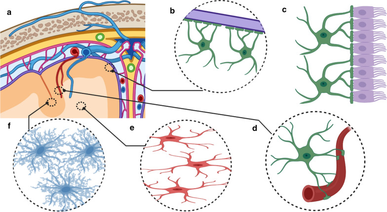
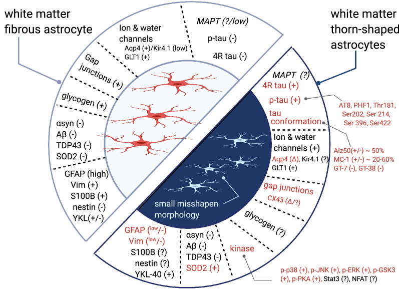
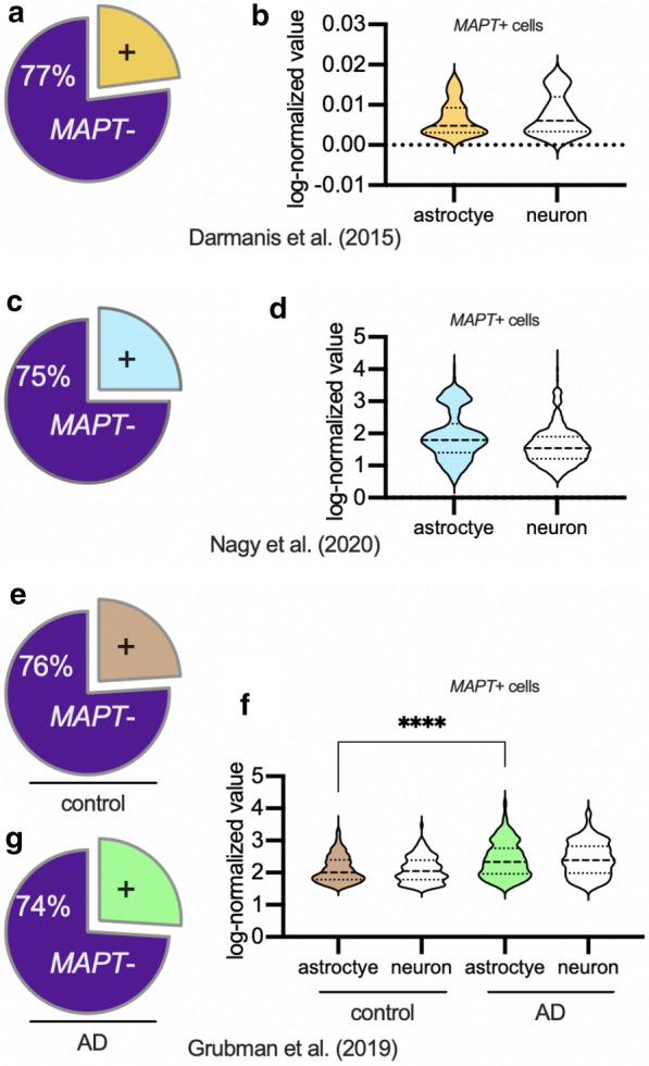
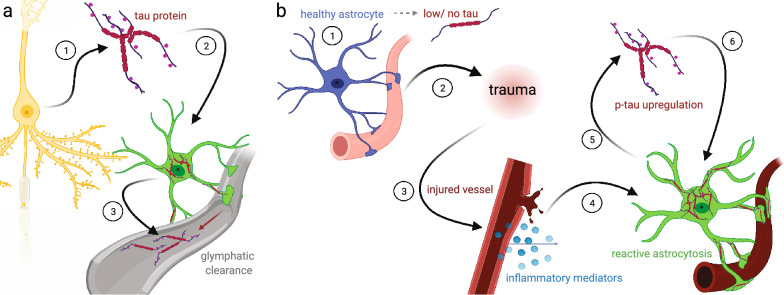
Astrocytes with intracellular accumulations of misfolded phosphorylated tau protein have been observed in advanced-stage chronic traumatic encephalopathy (CTE) and in other neurodegenerative conditions. There is a growing awareness that astrocytic tau inclusions are also relatively common in the brains of persons over 70 years of age-affecting approximately one-third of autopsied individuals. The pathologic hallmarks of aging-related tau astrogliopathy (ARTAG) include phosphorylated tau protein within thorn-shaped astrocytes (TSA) in subpial, subependymal, perivascular, and white matter regions, whereas granular-fuzzy astrocytes are often seen in gray matter. CTE and ARTAG share molecular and histopathologic characteristics, suggesting that trauma-related mechanism(s) may predispose to the development of tau astrogliopathy. There are presently few experimental systems to study the pathobiology of astrocytic-tau aggregation, but human studies have made recent progress. For example, leucotomy (also referred to as lobotomy) is associated with a localized ARTAG-like neuropathology decades after the surgical brain injury, suggesting that chronic brain injury of any type may predispose to later life ARTAG. To examine this idea in a different context, we report clinical and pathologic features of two middle-aged men who came to autopsy with large (> 6 cm in greatest dimension) arachnoid cysts that had physically displaced and injured the subjects' left temporal lobes through chronic mechanical stress. Despite the similarity of the size and location of the arachnoid cysts, these individuals had dissimilar neurologic outcomes and neuropathologic findings. We review the evidence for ARTAG in response to brain injury, and discuss how the location and molecular properties of astroglial tau inclusions might alter the physiology of resident astrocytes. These cases and literature review point toward possible mechanism(s) of tau aggregation in astrocytes in response to chronic brain trauma.

Keywords: FTLD; Meninges; NFTs; PSP; TBI; Tangles; Tauopathy.


Conflict of interest statement

The authors declare that they have no competing interests.

Figures

Fig. 1
Fig. 1
Clinical (premortem) computerized tomography (CT) scans, and photographs of brain after autopsy and fixation, in two cases with large arachnoid cysts. Case #1 is a male, 72 years old at death. This CT scan (a) was acquired at age 68. (b) The brain at autopsy following fixation from Case #1 shows the large left-sided arachnoid cyst cavity. Both the anterior temporal lobe and the orbitofrontal frontal lobe are affected by the lesion. Note that on the surface of the frontal cortex within the cyst cavity there is discoloration (white arrow). By contrast, the right side lacks evidence of indentation or contusion in the orbitofrontal cortex. Case #2 is a male, 64 years old at death. The CT scan (c) was acquired at age 56. As seen in the brain at autopsy following fixation (d), the arachnoid cyst is larger in the rostral-caudal axis in Case #2 in comparison to that of Case #1. However, the lesion is shallower—the affected region is smaller in the dorsal–ventral axis, and there was less impact on the orbitofrontal cortex (black arrow) in Case #2 compared to Case #1
Fig. 2
Fig. 2
CTE-like neuronal pathology in frontal cortex (middle frontal gyrus) of Case #1. (a) Low magnification of H&E-stained section shows the location (white box) of the CTE-like perivascular cluster of phosph-tau (p-tau) illustrated in adjacent PHF1 + stained section (b). A single cluster of PHF-1 + staining seen in the depth of the cortical sulci (B, black arrow). (c) At a higher magnification the large vessel (black arrow) is surrounded by p-tau + neuronal and astrocyte staining
Fig. 3
Fig. 3
Phospho-tau (p-tau) immunohistochemistry in the left fronto-parietal region of Case 1. In the low-magnification photomicrograph (a), the pia mater is near the bottom and white matter (WM) near the top. This is a section of the left fronto-parietal cortex, which is caudal to the region most directly impacted by the arachnoid cyst, and shows widespread p-tau (PHF-1) immunoreactivity in the subpial, gray matter, and white matter regions. In each of these compartments there is prominent staining of p-tau surrounding small blood vessels, and also in cells with morphologic features of astrocytes. Subpial staining is demonstrated in a small sulcus (b) and additional subpial staining is shown at low power in the adjacent gyrus (blue arrow in a). (c) White matter shows p-tau cells with astrocyte morphology, as well as staining around blood vessels resembling astrocyte foot processes (blue arrow). (d) The astrocytic p-tau in gray matter highlight pericapillary staining (blue arrows); the inset shows compact p-tau + cells with astrocyte features (e)
Fig. 4
Fig. 4
Sparse glial phospho-tau (p-tau) immunohistochemistry in Case 2. In this case, p-tau (PHF-1) immunoreactive structures were much more sparse, mostly non-neuronal, and were apparent in the medial temporal lobe. (a) In the hippocampus, p-tau immunostaining was found surrounding the rostral fringe of the inferior horn of the lateral ventricle (blue arrows; the ependymal lining denoted with an asterisk). Note that a few TSAs are stained (e.g., magenta arrow). (b) Shows p-tau immunoreactivity surrounding Virchow–Robin spaces of arterioles (blue arrow). Scattered p-tau + cells with delicate glial processes were also observed, such as that shown in (c) from the left-sided temporal cortex
Fig. 5
Fig. 5
Illustration of morphology and location of aging-related tau astrogliopathy neuropathological changes (ARTAG–NC). (a) Phosphorylated tau protein (p-tau) immunopositive astrocytes can broadly be found in five parts of the brain. (The illustration is not meant to be specific for any neuroanatomical region of the brain). TSAs are seen in the border-associated astrocytes of the subpial (b) and subependymal borders (c) (location of C not shown on A). (d) Perivascular astrocytes are the third border associated TSA. (It is not yet defined if these are exclusively arterial or are also venous associated astrocytes). (e) TSAs are also seen in the white matter. (f) In the gray matter, GFAs are the second disease-defining astrocyte morphology
Fig. 6
Fig. 6
Comparison of white matter fibrous versus tau + thorn-shaped astrocytes. A summary of the molecular characteristics of healthy white matter fibrous astrocytes is shown in comparison to reported changes in white matter thorn-shaped astrocytes. The text highlighted in red are known changes in the thorn-shaped astrocytes in ARTAG
Fig. 7
Fig. 7
MAPT (Tau mRNA transcript) expression in astrocytes from human adult brain. Shown are results from three different single-cell and single-nucleus RNAseq studies (from outside laboratories) which analyzed mRNA transcripts in human brain samples. Pie charts (a, c, e, g) show that the proportion of astrocytes that express MAPT was remarkably similar (~ 1/4th of astrocytes evaluated express MAPT transcript) in all three studies. Among the astrocytes that expressed any MAPT (b, d, f), the amount of MAPT transcripts detected in astrocytes was approximately the same as in neurons in the same brains. In AD astrocytes that express MAPT (f), the level of MAPT transcripts detected was higher than in MAPT expressing control astrocytes
Fig. 8
Fig. 8
Two hypothesis of ARTAG development. (a) In the neuronal centric model, a neuron expresses the majority of the tau protein (1). The neuron releases this tau protein (2) which (after trauma or axonal breakage) may be taken up by an astrocyte (3) during the glymphatic removal of the tau protein. This leads to the accumulation of exogenous tau, and the seeding of aggregated astrocyte tau. (b) In the astrocyte centric model, most healthy astrocytes express no or very low levels of MAPT. Changes in the brain environment—from an injury, aging, disease, or a combination of factors—leads to a reactive astrocyte response (1), which may be centered around small blood vessels (2, 3). The reactive astrocytes then upregulate the expression of MAPT and increase kinase activity (4), which leads to the hyperphosphorylation and aggregation of tau (5), causing p-tau + aggregates in astrocyte cell bodies and perivascular foot processes (6)

Similar articles

Cited by

References

    1. Arena JD, Johnson VE, Lee EB, Gibbons GS, Smith DH, Trojanowski JQ, Stewart W. Astroglial tau pathology alone preferentially concentrates at sulcal depths in chronic traumatic encephalopathy neuropathologic change. Brain Commun. 2020;2(2):210. doi: 10.1093/braincomms/fcaa210. - DOI - PMC - PubMed
    1. McKee AC. The neuropathology of chronic traumatic encephalopathy: the status of the literature. Semin Neurol. 2020;40(04):359–369. doi: 10.1055/s-0040-1713632. - DOI - PubMed
    1. Shively SB, Edgerton SL, Iacono D, Purohit DP, Qu BX, Haroutunian V, Davis KL, Diaz-Arrastia R, Perl DP. Localized cortical chronic traumatic encephalopathy pathology after single, severe axonal injury in human brain. Acta Neuropathol. 2017;133(3):353–366. doi: 10.1007/s00401-016-1649-7. - DOI - PMC - PubMed
    1. Rabiei K, Tisell M, Wikkelso C, Johansson BR. Diverse arachnoid cyst morphology indicates different pathophysiological origins. Fluids Barriers CNS. 2014;11(1):5. doi: 10.1186/2045-8118-11-5. - DOI - PMC - PubMed
    1. Choi JU, Kim DS. Pathogenesis of arachnoid cyst: congenital or traumatic? Pediatr Neurosurg. 1998;29(5):260–266. doi: 10.1159/000028733. - DOI - PubMed

Publication types