. 2021 Mar 23;12(1):203.
doi: 10.1186/s13287-021-02273-1.

Mesenchymal stem cells cultured in serum-free medium ameliorate experimental peritoneal fibrosis

Affiliations

Mesenchymal stem cells cultured in serum-free medium ameliorate experimental peritoneal fibrosis

Kohei Nagasaki et al. Stem Cell Res Ther. .

Abstract

Background: Mesenchymal stem cells (MSCs) provide potential treatments for peritoneal fibrosis. However, MSCs cultured in media containing serum bring risks of infection and other problems. In this study, we compared the effect of human MSCs in serum-free medium (SF-MSCs) on peritoneal fibrosis with that of MSCs cultured in medium containing 10% fetal bovine serum (10%MSCs).

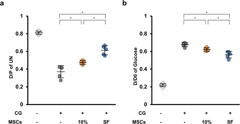
Methods: Peritoneal fibrosis was induced by intraperitoneally injecting 0.1% chlorhexidine gluconate (CG). SF-MSCs or 10%MSCs were intraperitoneally administered 30 min after the CG injection. Ten days after the CG and MSC injections, we performed histological analyses and peritoneal equilibrium testing. In the in vitro experiments, we used transforming growth factor (TGF)-β1-stimulated human peritoneal mesothelial cells incubated in conditioned medium from MSCs to examine whether the SF-MSCs showed enhanced ability to produce antifibrotic humoral factors.

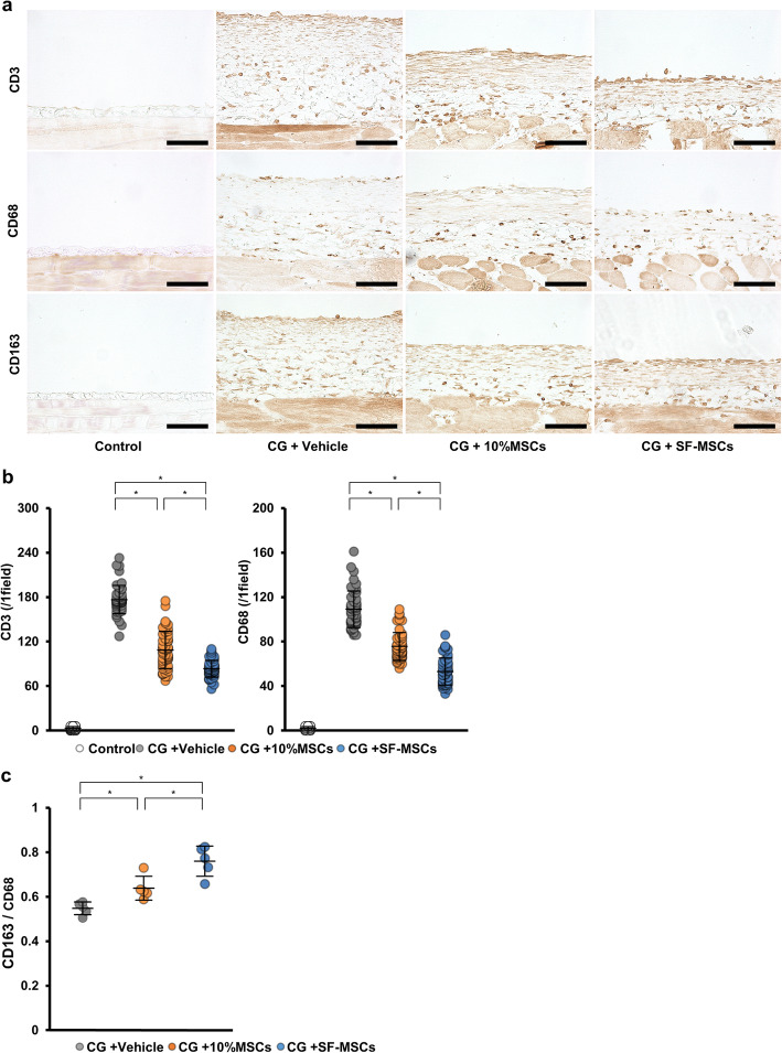
Results: Histological staining showed that the SF-MSCs significantly suppressed CG-induced cell accumulation and thickening compared with that of the 10%MSCs. Additionally, the SF-MSCs significantly inhibited mesenchymal cell expression, extracellular matrix protein deposition and inflammatory cell infiltration. Peritoneal equilibration testing showed that compared with administering 10%MSCs, administering SF-MSCs significantly reduced the functional impairments of the peritoneal membrane. The in vitro experiments showed that although the conditioned medium from MSCs suppressed TGF-β1 signaling, the suppression did not significantly differ between the SF-MSCs and 10%MSCs.

Conclusions: Serum-free culture conditions can enhance the antifibrotic abilities of MSCs by suppressing inflammation. Administering ex vivo expanded SF-MSCs may be a potential therapy for preventing peritoneal fibrotic progression.

Keywords: Immunosuppression; Mesenchymal stem cells; Peritoneal dialysis; Peritoneal fibrosis; Serum-free culture condition.


Conflict of interest statement

The Department of Stem Cell Biology and Medicine, Graduate School of Biomedical & Health Sciences, Hiroshima University, is a collaborative research laboratory funded by TWOCELLS Company, Limited. Emeritus Prof. Kato is the vice president of TWOCELLS Company, Limited. All remaining authors have declared that no conflicts of interest exist.

Figures

Fig. 1
Fig. 1
Effects of mesenchymal stem cell (MSC) injection on chlorhexidine gluconate (CG)-induced peritoneal cell density and thickness. Thirty minutes after injecting 0.1% CG, rats were intraperitoneally injected with MSCs cultured in DMEM containing 10% FBS (10%MSCs; 5 × 106 cells) or MSCs cultured in serum-free medium (SF-MSCs; 5 × 106 cells). After 10 days, the parietal peritoneum was examined via (a) hematoxylin-eosin staining and Masson’s trichrome staining to assess the peritoneal cell density and thickness, respectively. b Graph showing the cell density or thickness in each group. Control, control rats without CG injection; CG + vehicle, CG-injected rats treated with the vehicle; CG + 10%MSCs, CG-injected rats treated with 10%MSCs; CG + SF-MSCs, CG-injected rats treated with SF-MSCs. We examined 50 randomly chosen fields from 5 rats per group as described in the “Materials and Methods” section. *P < 0.05 (Kruskal–Wallis test)
Fig. 2
Fig. 2
Effects of MSC injection on fibrosis markers and collagen expression in CG-induced peritoneal fibrosis. a Immunohistochemical analyses of TGF-β1, a profibrotic marker, α-smooth muscle actin (α-SMA), a marker for myofibroblasts, collagen I and collagen III expressions in peritoneal tissues were performed on day 10 after MSC injection. b Graph showing the percentage of TGF-β1-positive areas or α-SMA-positive areas, and the area of collagen-I-positive staining or collagen-III-positive staining, per group. We measure 50 fields from 5 rats per group. Abbreviations are as in Fig. 1. *P < 0.05 (Kruskal–Wallis test)
Fig. 3
Fig. 3
Effects of MSC injection on CD3+ T-lymphocyte and macrophage infiltration and on M2 macrophage polarization in CG-induced peritoneal fibrosis. a Immunohistochemical analyses for CD3+ T lymphocytes, CD68+ macrophages, and CD163+ M2 macrophages. We measured 50 fields from 5 rats per group. b Graph showing the number of CD3- and CD68-positive cells per group. c For assessing the polarization into CD163+ M2 macrophages, the ratio of CD163+ cells (M2 macrophages) to CD68+ cells (total macrophages) were calculated for each rat (n = 5 rats per group). Abbreviations are as in Fig. 1. *P < 0.05 (Kruskal–Wallis test)
Fig. 4
Fig. 4
Effects of MSC injection on CG-induced peritoneal membrane dysfunction. Rats were instilled with PD solution (4.25% dialysis solution) at 100 mL/kg body weight. After 30 min, PD solution and plasma were collected. a Dialysate-to-plasma concentration ratio for urea nitrogen (D/P of UN); b dialysate-to-baseline dialysate concentration ratio for glucose (D/D0 of glucose). Abbreviations are as in Fig. 1. n = 5 rats per group. *P < 0.05 (Kruskal–Wallis test)
Fig. 5
Fig. 5
Effects of conditioned medium (CM) from MSCs on the TGF-β1/Smad signaling pathway and the proliferative ability of MSCs cultured in STK2 serum-free medium or 10% FBS-containing medium. HPMCs were grown, then the medium was replaced with CM from 10%MSCs, CM from SF-MSCs, or control medium. After 12 h, HPMCs were treated with TGF-β1 for 30 min (pSmad2, pSmad3) or 24 h (α-SMA), and whole-cell lysates were prepared and subjected to western blot analysis. Western blot analysis of a pSmad2, b pSmad3, and c α-SMA expression. Graph showing the densitometric analysis of pSmad2, pSmad3, and α-SMA expression normalized to Smad2, Smad3, and GAPDH expression. n = 5 per group. *P < 0.05 (Kruskal–Wallis test). d MSCs were cultured in DMEM containing 10% FBS or STK2 for 0, 12, 24, and 48 h. Surviving cells were subsequently assessed via WST-1 assay. Graph showing the absorbance value at each time point. n = 5 per group. *P < 0.05 (Mann–Whitney U test)
Fig. 6
Fig. 6
Effects of culture medium on TSG-6 expression in MSCs. MSCs were cultured in the following media: DMEM containing 10% FBS (Sigma), DMEM containing 10% FBS (HyClone), DMEM containing exosome-depleted 10% FBS (System Biosciences), 2% serum-supplemented media dedicated to MSC culture (Promo Cell), and STK2 serum free medium. Total RNA was extracted using TRIzol Reagent. Graph showing the TSG-6 mRNA level normalized to β-actin mRNA level. n = 5 per group. *P < 0.05 (Kruskal–Wallis test)

Similar articles

Cited by

References

    1. Liyanage T, Ninomiya T, Jha V, Neal B, Patrice HM, Okpechi I, Zhao MH, Lv J, Garg AX, Knight J, Rodgers A, Gallagher M, Kotwal S, Cass A, Perkovic V. Worldwide access to treatment for end-stage kidney disease: a systematic review. Lancet. 2015;385(9981):1975–1982. doi: 10.1016/S0140-6736(14)61601-9. - DOI - PubMed
    1. Mehrotra R, Chiu YW, Kalantar-Zadeh K, Bargman J, Vonesh E. Similar outcomes with hemodialysis and peritoneal dialysis in patients with end-stage renal disease. Arch Intern Med. 2011;171(2):110–118. doi: 10.1001/archinternmed.2010.352. - DOI - PubMed
    1. Elshahat S, Cockwell P, Maxwell AP, Griffin M, O'Brien T, O'Neill C. The impact of chronic kidney disease on developed countries from a health economics perspective: a systematic scoping review. PLoS One. 2020;15(3):e0230512. doi: 10.1371/journal.pone.0230512. - DOI - PMC - PubMed
    1. Williams JD, Craig KJ, Topley N, Von Ruhland C, Fallon M, Newman GR, et al. Morphologic changes in the peritoneal membrane of patients with renal disease. J Am Soc Nephrol. 2002;13(2):470–479. - PubMed
    1. Margetts PJ, Churchill DN. Acquired ultrafiltration dysfunction in peritoneal dialysis patients. J Am Soc Nephrol. 2002;13(11):2787–2794. - PubMed

Publication types

Substances

LinkOut - more resources