. 2021:62:100069.
doi: 10.1016/j.jlr.2021.100069. Epub 2021 Mar 20.

Octanoate is differentially metabolized in liver and muscle and fails to rescue cardiomyopathy in CPT2 deficiency

Affiliations

Octanoate is differentially metabolized in liver and muscle and fails to rescue cardiomyopathy in CPT2 deficiency

Andrea S Pereyra et al. J Lipid Res. 2021.

Abstract

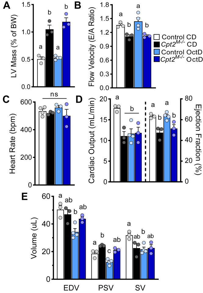
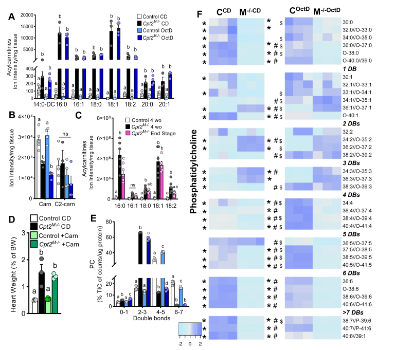
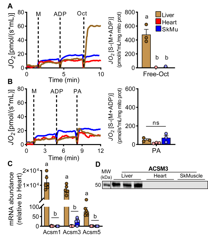
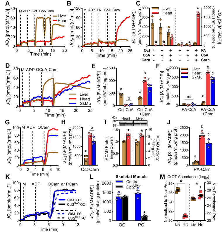
Long-chain fatty acid oxidation is frequently impaired in primary and systemic metabolic diseases affecting the heart; thus, therapeutically increasing reliance on normally minor energetic substrates, such as ketones and medium-chain fatty acids, could benefit cardiac health. However, the molecular fundamentals of this therapy are not fully known. Here, we explored the ability of octanoate, an eight-carbon medium-chain fatty acid known as an unregulated mitochondrial energetic substrate, to ameliorate cardiac hypertrophy in long-chain fatty acid oxidation-deficient hearts because of carnitine palmitoyltransferase 2 deletion (Cpt2M-/-). CPT2 converts acylcarnitines to acyl-CoAs in the mitochondrial matrix for oxidative bioenergetic metabolism. In Cpt2M-/- mice, high octanoate-ketogenic diet failed to alleviate myocardial hypertrophy, dysfunction, and acylcarnitine accumulation suggesting that this alternative substrate is not sufficiently compensatory for energy provision. Aligning this outcome, we identified a major metabolic distinction between muscles and liver, wherein heart and skeletal muscle mitochondria were unable to oxidize free octanoate, but liver was able to oxidize free octanoate. Liver mitochondria, but not heart or muscle, highly expressed medium-chain acyl-CoA synthetases, potentially enabling octanoate activation for oxidation and circumventing acylcarnitine shuttling. Conversely, octanoylcarnitine was oxidized by liver, skeletal muscle, and heart, with rates in heart 4-fold greater than liver and, in muscles, was not dependent upon CPT2. Together, these data suggest that dietary octanoate cannot rescue CPT2-deficient cardiac disease. These data also suggest the existence of tissue-specific mechanisms for octanoate oxidative metabolism, with liver being independent of free carnitine availability, whereas cardiac and skeletal muscles depend on carnitine but not on CPT2.

Keywords: carnitine palmitoyltransferase; carnitine shuttle; fatty acid oxidation; medium-chain fatty acids; mitochondria.


Conflict of interest statement

Conflict of interest The authors declare that they have no conflicts of interest with the contents of this article.

Figures

Fig. 1
Fig. 1
Dietary octanoate failed to attenuate cardiac hypertrophy in Cpt2M/ mice. A: Transverse, H&E-stained sections of control and Cpt2M/ hearts (left) and photography of half heart post fixative (right). B: Heart weight of control and Cpt2M/ mice in response to 20% medium-chain supplemented chow diet. C: Diet composition of formulated control (CD) and octanoate (OctD) diets. D: Plasma ketones in response to 4 weeks on diets, n = 6–9. E: Body, (F) organ, and (G) heart weight of male and female control and Cpt2M/ mice fed control or OctD, n = 4–6. Data are presented as mean ± SEM. Statistical analysis by 2-way ANOVA. Means depicting a different letter indicate significant differences between groups (P ≤ 0.05). The scale bar represents 1,000 μm. B-OHB, beta-hydroxybutyrate; BW, body weight; Cpt2, carnitine palmitoyltransferase 2.
Fig. 2
Fig. 2
Dietary octanoate did not improve heart function in Cpt2M/ mice and increased cardiac output in control mice. A: Left ventricular mass relative to body weight, (B) flow velocity, and (C–E) cardiac functional parameters derived from 2D echocardiogram and Doppler analysis of control and Cpt2M/ mice on control or OctD, females, n = 3–4. Data are presented as mean ± SEM. Statistical analysis by two-way ANOVA. Means depicting a different letter indicate significant differences between groups (P ≤ 0.05). Cpt2, carnitine palmitoyltransferase 2; EDV, end-diastolic volume; PSV, peak-systolic volume; SV, stroke volume.
Fig. 3
Fig. 3
Effects of long- and medium-chain ketogenic diets on cardiac remodeling and metabolic programs. A–F: Cardiac mRNA abundance normalized to control group on control diet (CD) of pathological hypertrophy markers, mTOR pathway, and myokines from control and Cpt2M−/− mice fed either CD, OctD, or LCKD, females, n = 4–6. G, H: Phosphorylation levels of PDH enzyme at Serine293 and mRNA abundance of PDH kinase (Pdk4) as regulators of pyruvate metabolism in hearts from control and Cpt2M−/− mice fed either CD, OctD, or LCKD, females, n = 4–6. Statistical analysis by 2-way ANOVA. Means depicting a different letter indicate significant differences between groups (P ≤ 0.05). Cpt2, carnitine palmitoyltransferase 2; LCKD, long-chain fatty acid ketogenic diet; mTOR, mechanistic target of rapamycin; OctD, octanoate diet; PDH, pyruvate dehydrogenase; Pdk4, pyruvate dehydrogenase kinase 4.
Fig. 4
Fig. 4
CPT2-deficient hearts are resistant to diet-induced alterations in cardiac acylcarnitines and phospholipid acyl-chain composition. A, B: Cardiac acylcarnitines in control and Cpt2M/ mice fed control and OctD, n = 3–5 or (C) at 4 weeks of age and end-stage heart failure, n = 4–6. D: Heart mass in control and Cpt2M/ with and without dietary carnitine supplementation, n = 4–5. E: Sum of and (F) heat map for normalized abundance of phosphatidylcholine species arranged by unsaturation degree, in control and Cpt2M/ mice fed control or OctD, n = 3. Data are presented as mean ± SEM, all from female mice. Statistical analysis by 2-way ANOVA. Means depicting a different letter indicate significant differences between groups (P ≤ 0.05). ∗by genotype, #by diet among controls, and $by diet among Cpt2M/. Cpt2, carnitine palmitoyltransferase 2; DBs, double bonds; wo, weeks of.
Fig. 5
Fig. 5
Differential octanoate oxidation between liver, heart, and skeletal muscle. A, B: Rates of oxygen consumption in isolated mitochondria of liver, heart, and skeletal muscle as representative traces over time and quantification of maximum rates during administration of malate (M) and ADP along with free octanoate (Oct) or free palmitate (PA) as substrate, n = 3. Relative abundance of ACSM isoforms by (C) mRNA and (D) expression of ACSM3 protein in liver, heart, and skeletal muscle, n = 3–6. Data are presented as mean ± SEM, all from male mice. Statistical analysis by 1-way ANOVA. Means depicting a different letter indicate significant differences between groups (P ≤ 0.05). ACSM, medium-chain acyl-CoA synthetase.
Fig. 6
Fig. 6
Octanoylcarnitine is oxidized by liver, heart, and muscle in a CPT2-independent manner. A, B: Rates of oxygen consumption in isolated mitochondria of liver and heart as representative trace over time and (C) quantitation of maximum rates during administration of malate (M) and ADP with octanoate (Oct) or palmitate (PA) as substrates, followed by CoA and free l-carnitine (Carn), n = 3. D: Rates of oxygen consumption in isolated mitochondria from liver, heart, and skeletal muscle as representative trace and (E, F) quantitation of maximum rates when given M and ADP with octanoyl-CoA (OCoA) or palmitoyl-CoA followed by carnitine, n = 3. G: Rates of oxygen consumption in isolated mitochondria of liver, heart, and skeletal muscle as representative trace and (H) quantitation of maximum rates given octanoylcarnitine (OCarn) as substrate, n = 3. I:MCAD protein and activity in heart and liver, n = 3. J: Rates of palmitoylcarnitine oxidation in isolated mitochondria of liver, heart, and skeletal muscle. K: Representative trace of oxygen consumption and (L) quantification of maximum rates in isolated mitochondria of skeletal muscle from control or Cpt2Sk−/− mice given octanoylcarnitine and palmitoylcarnitine (PCarn), n = 3. M: Relative carnitine O-octanoyltransferase (CrOT) abundance in isolated mitochondria from liver and heart as detected by discovery proteomics, n = 5. Data are presented as mean ± SEM, all from male mice. ∗P ≤ 0.05 by one-way ANOVA (E, F, H, J, and M) or Student's t-test (I and J). Means depicting a different letter indicate significant differences between groups (P ≤ 0.05). MCAD, medium-chain acyl-CoA dehydrogenase.
Fig. 7
Fig. 7
Schematic of octanoate oxidation in liver and muscles. Speculative metabolic pathway for octanoate oxidation in liver and muscles (cardiac and skeletal). Dietary-free octanoate reaches the liver through the portal circulation where liver, but not muscle, can activate free octanoate within the mitochondria via medium-chain acyl-CoA synthetases (ACSM). Muscles and liver mitochondria metabolize octanoylcarnitine as a product of peroxisomal oxidation. The enzyme that converts octanoylcarnitine back to octanoyl-CoA in the mitochondria is not CPT2 but could be carnitine O-octanoyltransferase (CrOT). Octanoyl-CoA enters mitochondrial oxidation through medium-chain acyl-CoA dehydrogenase (MCAD) and can be oxidized for energy production in liver and muscles or can be used for ketone body production in liver. β-Oxi, beta-oxidation; Carn, free carnitine; CoA, coenzyme A; TCA, tricarboxylic acid cycle; VLCFA, very long chain fatty acid. Created with BioRender.com.

References

    1. Ormerod J.O.M., Frenneaux M.P., Sherrid M.V. Myocardial energy depletion and dynamic systolic dysfunction in hypertrophic cardiomyopathy. Nat. Rev. Cardiol. 2016;13:677–687. - PubMed
    1. Bianco H.T., Izar M.C., Póvoa R.M., Bombig M.T., Fonseca H.A., Helfenstein T., Ferreira C.E., Nicolau J.C., Neto A.A., Feio C.M., Cerci M.S., Fonseca F.A. Left ventricular hypertrophy and QTc dispersion are predictors of long-term mortality in subjects with type 2 diabetes. Int. J. Cardiol. 2014;176:1170–1172. - PubMed
    1. Struthers A.D., Morris A.D. Screening for and treating left-ventricular abnormalities in diabetes mellitus: a new way of reducing cardiac deaths. Lancet. 2002;359:1430–1432. - PubMed
    1. Brady T.M. The role of obesity in the development of left ventricular hypertrophy among children and adolescents. Curr. Hypertens. Rep. 2015;18:1–7. - PMC - PubMed
    1. Murdolo G., Angeli F., Reboldi G., Di Giacomo L., Aita A., Bartolini C., Vedecchia P. Left ventricular hypertrophy and obesity: only a matter of fat? High Blood Press. Cardiovasc. Prev. 2015;22:29–41. - PubMed

MeSH terms

Supplementary concepts

LinkOut - more resources