. 2021 Jan-Jun:296:100575.
doi: 10.1016/j.jbc.2021.100575. Epub 2021 Mar 21.

Coordinate regulation of systemic and kidney tryptophan metabolism by the drug transporters OAT1 and OAT3

Affiliations

Coordinate regulation of systemic and kidney tryptophan metabolism by the drug transporters OAT1 and OAT3

Jeffry C Granados et al. J Biol Chem. 2021 Jan-Jun.

Abstract

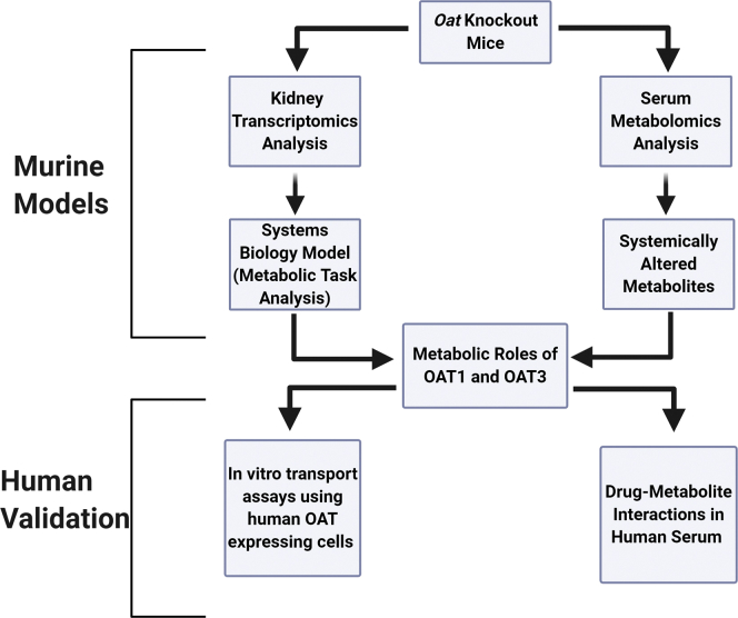
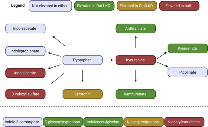
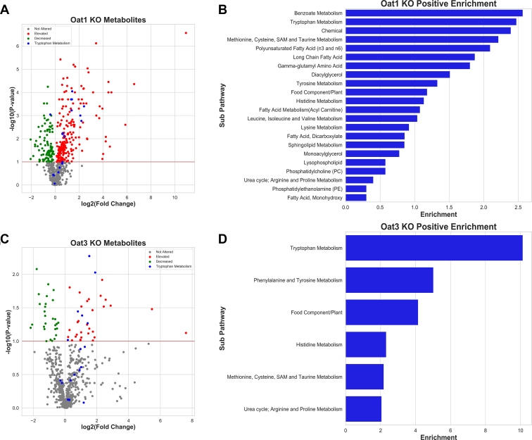
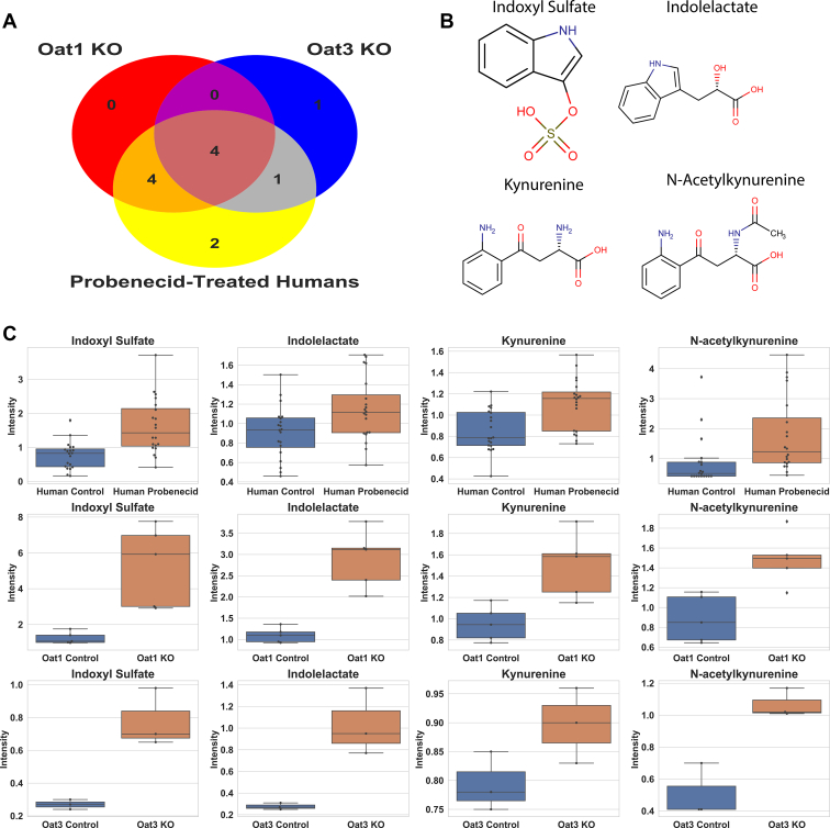
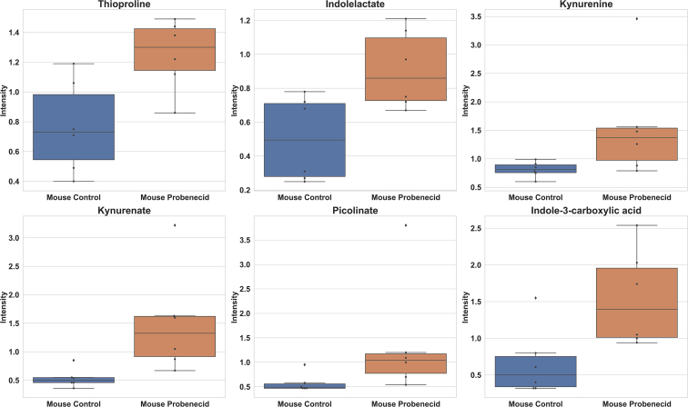
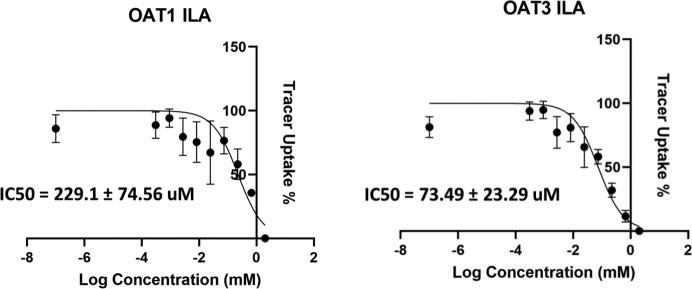
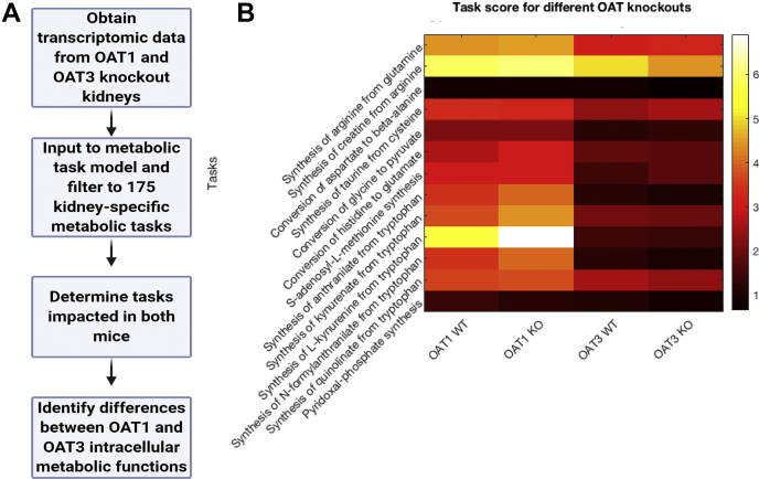
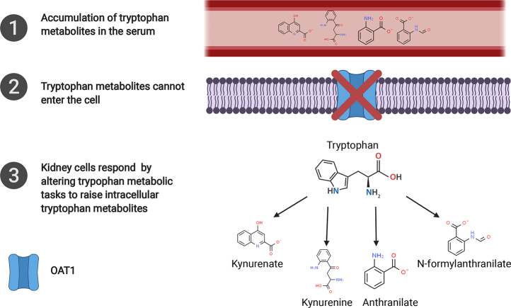
How organs sense circulating metabolites is a key question. Here, we show that the multispecific organic anion transporters of drugs, OAT1 (SLC22A6 or NKT) and OAT3 (SLC22A8), play a role in organ sensing. Metabolomics analyses of the serum of Oat1 and Oat3 knockout mice revealed changes in tryptophan derivatives involved in metabolism and signaling. Several of these metabolites are derived from the gut microbiome and are implicated as uremic toxins in chronic kidney disease. Direct interaction with the transporters was supported with cell-based transport assays. To assess the impact of the loss of OAT1 or OAT3 function on the kidney, an organ where these uptake transporters are highly expressed, knockout transcriptomic data were mapped onto a "metabolic task"-based computational model that evaluates over 150 cellular functions. Despite the changes of tryptophan metabolites in both knockouts, only in the Oat1 knockout were multiple tryptophan-related cellular functions increased. Thus, deprived of the ability to take up kynurenine, kynurenate, anthranilate, and N-formylanthranilate through OAT1, the kidney responds by activating its own tryptophan-related biosynthetic pathways. The results support the Remote Sensing and Signaling Theory, which describes how "drug" transporters help optimize levels of metabolites and signaling molecules by facilitating organ cross talk. Since OAT1 and OAT3 are inhibited by many drugs, the data implies potential for drug-metabolite interactions. Indeed, treatment of humans with probenecid, an OAT-inhibitor used to treat gout, elevated circulating tryptophan metabolites. Furthermore, given that regulatory agencies have recommended drugs be tested for OAT1 and OAT3 binding or transport, it follows that these metabolites can be used as endogenous biomarkers to determine if drug candidates interact with OAT1 and/or OAT3.

Keywords: chronic kidney disease; drug transport; drug transporters; gut microbiome; kidney; kidney metabolism; organ crosstalk; tryptophan; uremic toxins; xenobiotic.


Conflict of interest statement

Conflicts of interest The authors declare that they have no conflicts of interest with the contents of this article.

Figures

Figure 1
Figure 1
Strategy for determining metabolic role of drug transporters. By combining tissue-specific transcriptomic data with serum metabolomic data, the local and systemic roles of the OAT1 and OAT3 transporters can be determined. Using this framework, we investigated the role of each transporter in cellular metabolism of the relevant tissue (kidney), as well as the role the transporters play in controlling metabolite concentrations in the serum. These studies were supported with clinically relevant human data as described in Results.
Figure 2
Figure 2
Systemic tryptophan metabolites regulated by OAT1, OAT3, or both transporters. The metabolites measured in both knockout animals are shown in the figure with an edge between two nodes representing one or several reactions that lead to production of the metabolite. Note that, apart from the four common metabolites depicted, different tryptophan metabolites accumulate in the Oat1 KO and the Oat3 KO. The five metabolites at the bottom have yet to be placed in the biochemical map but are known to be tryptophan derivatives. Statistical significance was determined by Welch's t-test. Individual fold changes for the altered metabolites are as follows: Indolelactate (OAT1: 2.63, OAT3: 2.37), 3-indoxyl sulfate (OAT1: 4.29, OAT3: 2.88), Serotonin (OAT3: 1.75), Kynurenine (OAT1: 1.58, OAT3: 1.51), Kynurenate (OAT1: 2.51), Anthranilate (OAT1: 2.67), Xanthurenate (OAT1: 5.29), C-glycosyltryptophan (OAT1: 1.51), Indoleacetylglycine (OAT1: 2.59), N-acetyltryptophan (OAT3: 2.68), N-acetylkynurenine (OAT1: 1.68, OAT3: 2.11).
Figure 3
Figure 3
Tryptophan metabolism is one of the most altered pathways in both knockout mice.A, volcano plot for the Oat1 KO (n = 5) showing the tryptophan metabolites against all other measured metabolites. B, tryptophan metabolism is the second most enriched pathway for significantly elevated metabolites. C, volcano plot for the Oat3 KO (n = 3) showing the tryptophan metabolites against all other measured metabolites. D, tryptophan metabolism is the most enriched pathway for significantly elevated metabolites based on metabolon subpathway analysis.
Figure 4
Figure 4
Tryptophan metabolites were elevated in Oat knockout mice and probenecid-treated humans. A, Venn diagram of significantly altered tryptophan metabolites in knockout mice and humans treated with probenecid. Of the 14 metabolites common to all three platforms, 12 were significantly elevated in at least one of the experiments. Four metabolites were significantly elevated in each experiment. B, chemical structures of metabolites elevated in each experiment: 3-indoxyl sulfate, indolelactate, kynurenine, and N-acetylkynurenine. C, boxplots for each metabolite in the probenecid-treated humans (n = 20), Oat1 KO mice (n = 5), and Oat3 KO mice (n = 3). Lines in boxplots indicate the median.
Figure 5
Figure 5
Mice treated with probenecid had elevated circulating levels of tryptophan metabolites. Thioproline, indolelactate, kynurenine, kynurenate, picolinate, and indole-3-carboxylic acid all demonstrated significant increases (p < 0.05, fold change > 1) after treatment with an OAT-inhibiting drug (n = 6).
Figure 6
Figure 6
In vitro transport assays for tryptophan metabolites with cells overexpressing human OAT1 and OAT3. Indolelactate (ILA) had not previously been tested for inhibition of either OAT1 or OAT3. Human OAT1 and human OAT3 transport is inhibited by ILA. Assays were repeated at least three times for each metabolite–transporter pair.
Figure 7
Figure 7
Metabolic task analysis of WT versus knockout mouse kidney transcriptomics indicates OAT1 (but not OAT3) dependence of five tryptophan-related metabolic tasks.A, the metabolic task analysis uses transcriptomic data to predict cellular functions. B, the 13 tasks displayed have a coefficient of variation (standard deviation/mean) above 15% and a mean metabolic task score greater than 0.5 across both knockouts. Included in these are five tryptophan-related tasks (synthesis of anthranilate, synthesis of kynurenate, synthesis of kynurenine, synthesis of N-formylanthranilate, and synthesis of quinolinate) that have increased metabolic task scores in the Oat1 KO (n = 3) compared with wild type (n = 3) and decreased metabolic task scores in the Oat3 KO (n = 3) compared with wild type (n = 3). A score of 0 suggests low metabolic activity and a score of 7 suggests high metabolic activity.
Figure 8
Figure 8
The data predicts that kidney tissue responds to the increases in serum concentration by increasing synthesis of these metabolites intracellularly. Anthranilate, kynurenine, kynurenate, and N-formylanthranilate were elevated in the serum of the Oat1 KO and the transcriptomic data from the knockout kidney indicates that the tissue is producing more from tryptophan. Additionally, kynurenine, kynurenate, and N-formylanthranilate were elevated in the serum of probenecid-treated humans (anthranilate was not measured), raising the possibility that a similar process may occur within humans taking drugs that inhibit OAT1 and potentially alter intracellular tryptophan metabolic tasks as in the Oat1 knockout mouse.

Similar articles

Cited by

References

    1. LopezNieto C.E., You G.F., Bush K.T., Barros E.J.G., Beier D.R., Nigam S.K. Molecular cloning and characterization of NKT, a gene product related to the organic cation transporter family that is almost exclusively expressed in the kidney. J. Biol. Chem. 1997;272:6471–6478. - PubMed
    1. Brady K.P., Dushkin H., Fornzler D., Koike T., Magner F., Her H., Gullans S., Segre G.V., Green R.M., Beier D.R. A novel putative transporter maps to the osteosclerosis (oc) mutation and is not expressed in the oc mutant mouse. Genomics. 1999;56:254–261. - PubMed
    1. Riedmaier A.E., Nies A.T., Schaeffeler E., Schwab M. Organic anion transporters and their implications in pharmacotherapy. Pharmacol. Rev. 2012;64:421–449. - PubMed
    1. Ahn S.Y., Bhatnagar V. Update on the molecular physiology of organic anion transporters. Curr. Opin. Nephrol. Hypertens. 2008;17:499–505. - PubMed
    1. Nigam S.K. The SLC22 transporter family: A paradigm for the impact of drug transporters on metabolic pathways, signaling, and disease. Annu. Rev. Pharmacol. 2018;58:663–687. - PMC - PubMed

Publication types

Substances