Case Reports
. 2021 Mar 23;14(3):e240608.
doi: 10.1136/bcr-2020-240608.

Endovascular treatment of postpartum haemorrhage in a woman with genitourinary and vascular congenital malformations

Affiliations
Case Reports

Endovascular treatment of postpartum haemorrhage in a woman with genitourinary and vascular congenital malformations

Scott Perkins et al. BMJ Case Rep. .

Abstract

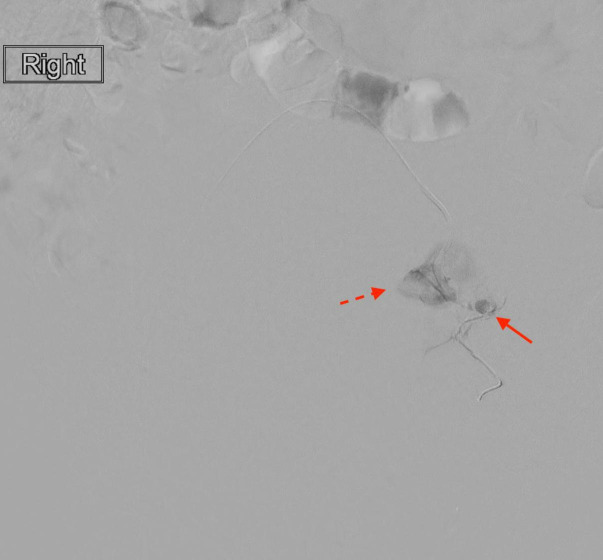
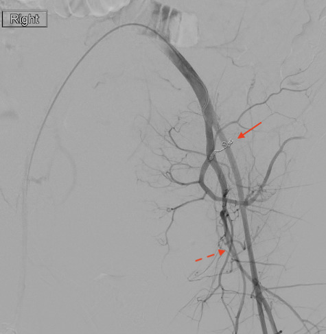
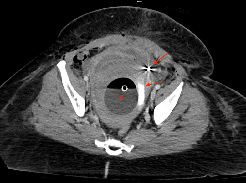
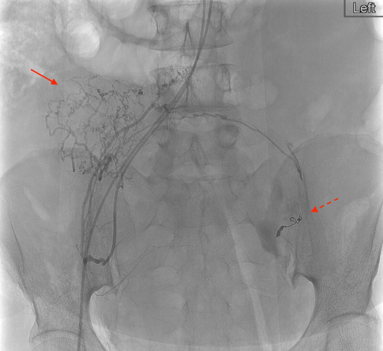
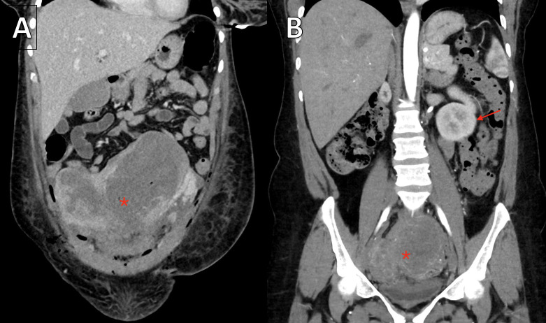
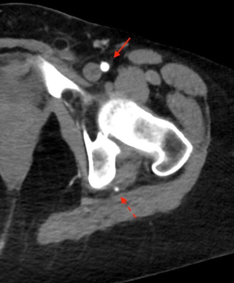
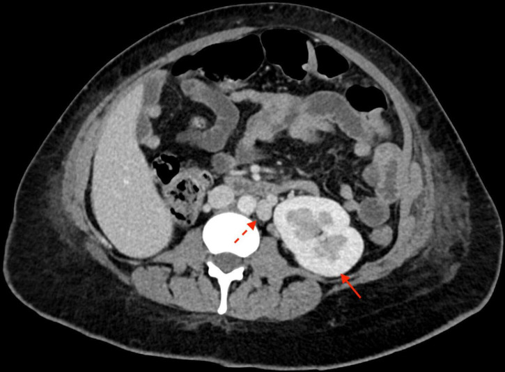
A 43-year-old woman presented with postpartum haemorrhage necessitating uterine artery embolisation. Prior to embolisation, angiography demonstrated the presence of a persistent sciatic artery (PSA). Due to the possibility of embolic particles inadvertently traveling to the lower extremity via this variant arterial pathway, care was taken to only embolise the uterine artery. PSAs are uncommon but important vascular pathways to screen for during pelvic intervention and are associated with other genitourinary anomalies.

Keywords: interventional radiology; obstetrics and gynaecology; radiology.


Conflict of interest statement

Competing interests: None declared.

Figures

Figure 1
Figure 1
Digital subtraction angiogram of the left uterine artery pseudoaneurysm (solid arrow) with blush of active extravasation (dotted arrow).
Figure 2
Figure 2
Digital subtraction angiogram with catheter tip in the left common iliac artery demonstrating the coiled left uterine artery (solid arrow) and patent persistent sciatic artery (dotted arrow).
Figure 3
Figure 3
Venous phase CT through the level of the pelvis performed immediately post partum and after embolisation. Bakri balloon is still in place (asterisk). Contrast staining can be seen within the uterine cavity (dotted arrow) consistent with recent extravasation. Left uterine artery embolic coils are also visualised (solid arrow).
Figure 4
Figure 4
Selective arteriogram of the right uterine artery branch vessels prior to gel foam embolisation (solid arrow). Left uterine artery pseudoaneurysm coils are in place (dotted arrow).
Figure 5
Figure 5
(A) Venous phase coronal CT performed in the immediate postpartum phase demonstrates a didelphys uterus markedly distended with blood products (asterisk). (B) Arterial phase coronal CT performed 2 weeks after discharge demonstrates the didelphys uterus significantly reduced in size (asterisk). Partially visualised single left kidney (arrow).
Figure 6
Figure 6
Arterial phase CT demonstrates the left persistent sciatic artery (dotted arrow) in addition to the normal left common femoral artery (solid arrow).
Figure 7
Figure 7
Venous phase CT through the level of the mid abdomen demonstrates a single left kidney (solid arrow) as well as a duplicated inferior vena cava (dotted arrow).

Similar articles

References

    1. van Hooft IM, Zeebregts CJ, van Sterkenburg SMM, et al. . The persistent sciatic artery. Eur J Vasc Endovasc Surg 2009;37:585–91. 10.1016/j.ejvs.2009.01.014 - DOI - PubMed
    1. Shaffer W, Maher M, Maristany M, et al. . Persistent sciatic artery: a favorable anatomic variant in a setting of trauma. Ochsner J 2017;17:189–94. - PMC - PubMed
    1. Gauffre S, Lasjaunias P, Zerah M. Sciatic artery: a case, review of literature and attempt of systemization. Surg Radiol Anat 1994;16:105–9. 10.1007/BF01627932 - DOI - PubMed
    1. Morisaki K, Yamaoka T, Iwasa K, et al. . Persistent sciatic artery aneurysm with limb ischemia: a report of two cases. Ann Vasc Dis 2017;10:44–7. 10.3400/avd.cr.16-00119 - DOI - PMC - PubMed
    1. Huang C-Y, Hsu H-L, Shen S-H, et al. . Persistent sciatic artery associated with solitary kidney and maldevelopment of mesonephric duct and urogenital sinus. Vasc Med 2016;21:471–2. 10.1177/1358863X15624531 - DOI - PubMed

Publication types