. 2021 Mar 23;14(675):eabd5605.
doi: 10.1126/scisignal.abd5605.

Essential requirement for JPT2 in NAADP-evoked Ca2+ signaling

Affiliations

Essential requirement for JPT2 in NAADP-evoked Ca2+ signaling

Gihan S Gunaratne et al. Sci Signal. .

Abstract

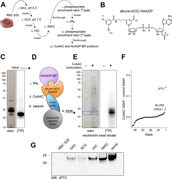
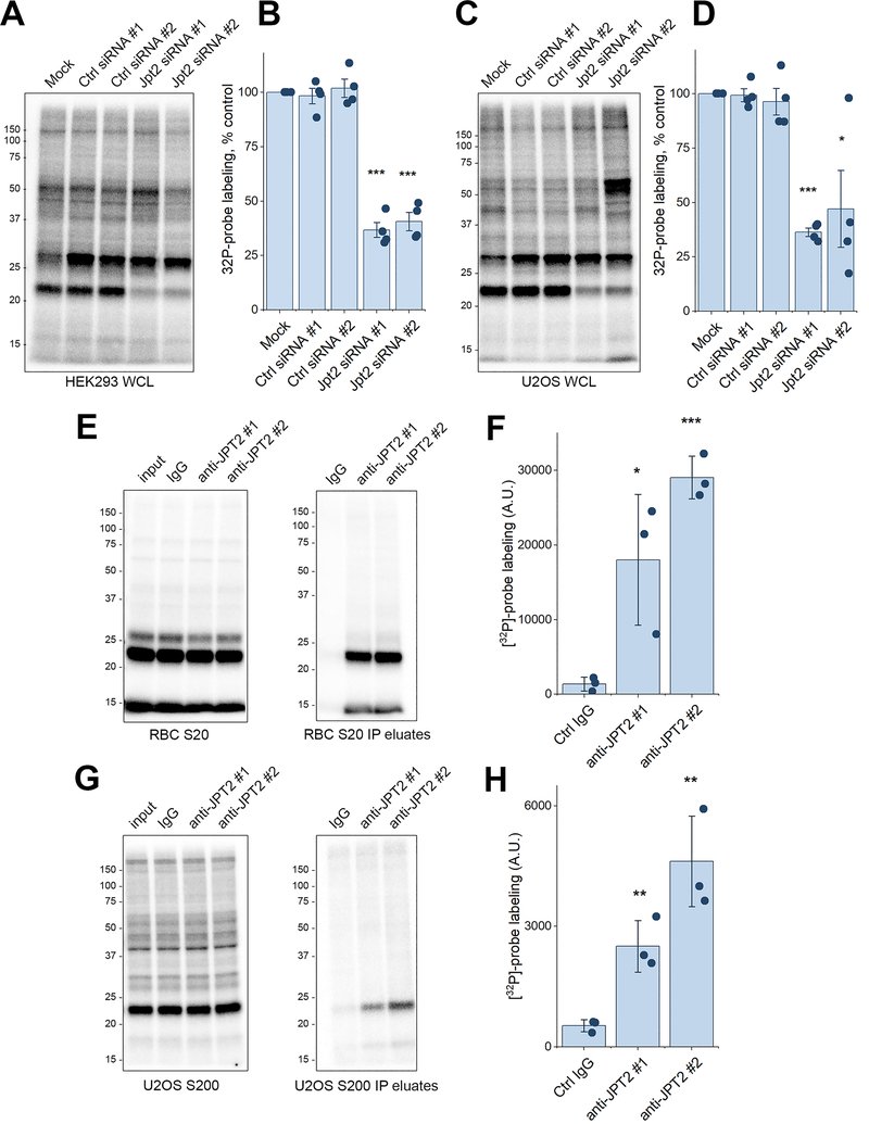
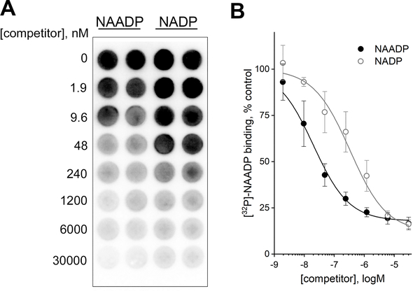
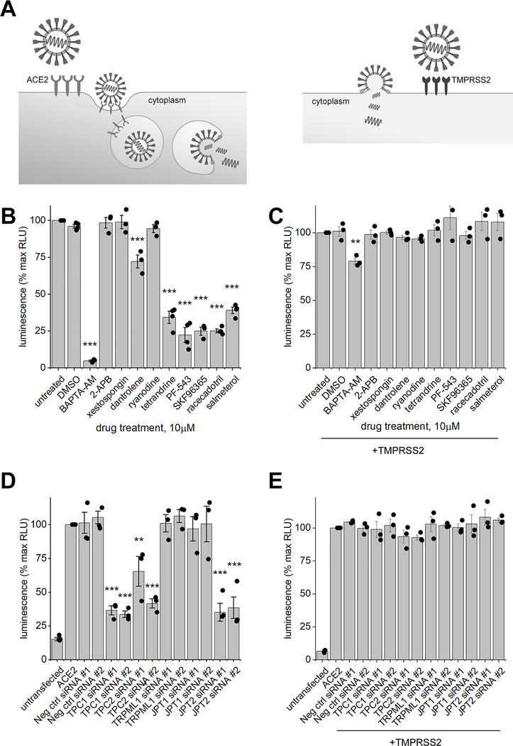
Nicotinic acid adenine dinucleotide phosphate (NAADP) is a second messenger that releases Ca2+ from acidic organelles through the activation of two-pore channels (TPCs) to regulate endolysosomal trafficking events. NAADP action is mediated by NAADP-binding protein(s) of unknown identity that confer NAADP sensitivity to TPCs. Here, we used a "clickable" NAADP-based photoprobe to isolate human NAADP-binding proteins and identified Jupiter microtubule-associated homolog 2 (JPT2) as a TPC accessory protein required for endogenous NAADP-evoked Ca2+ signaling. JPT2 was also required for the translocation of a severe acute respiratory syndrome coronavirus 2 (SARS-CoV-2) pseudovirus through the endolysosomal system. Thus, JPT2 is a component of the NAADP receptor complex that is essential for TPC-dependent Ca2+ signaling and control of coronaviral entry.


Conflict of interest statement

Competing interests: The authors declare that they have no competing interests.

Figures

Fig. 1.
Fig. 1.. Characterization of NAADP-binding proteins in human erythrocytes.
(A and B), Red blood cell (RBC), Jurkat, and U2OS whole cell lysates (WCL) were photolabeled with [32P]-alkyne-AIOC-NAADP (3nM) in the absence or presence of competing unlabeled NAADP (10μM). (C and D), RBC WCL, S20 cytosol fraction, and “ghost” membrane preparations were photolabeled with [32P]-alkyne-AIOC-NAADP (3nM) in the absence or presence of competing unlabeled NAADP (10μM). (E and F) RBC S20 was photolabeled with [32P]-alkyne-AIOC-NAADP (3nM) in the presence of the indicated compounds (10μM). (G and H) RBC S20 was photolabeled with [32P]-alkyne-AIOC-NAADP (3nM) in the presence of either NAADP or NADP at the indicated concentration. Data in (B, D, F, H) represent mean ± SD from densitometry analyses of n = 3 independent experiments, where representative gels for each specific assay (A, C, E and G) are shown. 30μg of protein was used for all labeling reactions. Data are shown as mean ± SEM. Statistical significance was assessed using a univariate regression model (* P<0.05, ** P<0.01, *** P<0.005). A.U., arbitrary units.
Fig. 2.
Fig. 2.. Biochemical enrichment and isolation of NAADP-binding proteins.
(A) Schematic representation of NAADP-binding protein enrichment. Red blood cell S20 proteins were sequentially fractionated in a series of chromatography steps: i. strong anion exchange (SAX) at pH 8.5, ii. strong cation exchange (SCX) at pH 7.0, iii. hydrophobic interaction chromatography (HIC), iv. immobilized metal affinity chromatography (IMAC). Eluates from a phosphoprotein enrichment resin were photoaffinity labeled, and fall-through was collected after passing protein through phosphoprotein enrichment resin a 2nd time (v-vii). t. (B) Structure of alkyne-AIOC-NAADP. Photolabile aryl azide (*) and ‘clickable’ alkyne (**) moieties are shown. (C) Silver stain (left) and phosphorimage (right) of 10μg of sample enriched with NAADP-binding protein. (D) Depiction of approach. NAADP-binding protein is crosslinked to the “clickable” alkyne-AIOC-NAADP photoprobe, which is biotinylated by copper-catalyzed azide-alkyne cycloaddition (CuAAC). Biotinylated protein is isolated using neutravidin agarose beads. (E) Probe-bound NAADP-binding protein was used in “click” chemistry reaction in absence or presence of biotin-azide-Plus. Biotinylated protein was captured with neutravidin agarose beads, stringently washed, and eluted. Silver stain (left) and phosphorimage (right) of eluates. Bands of interest are highlighted (blue box). (F) Gel bands of isolated NAADP-binding protein and corresponding bands from input samples and unbiotinylated controls were analyzed by mass spectrometry. Normalized spectral abundance factor (NSAF) after click and capture minus NSAF of unbiotinylated control samples is shown. (G) Western blot detection of JPT2 in erythrocyte S20 supernatant after fractionation by SAX, SCX, HIC, IMAC, or sequential fractionation through each of these steps (serial).
Fig. 3.
Fig. 3.. JPT2 expression profile in cells and organisms.
(A) mRNA expression profiling in 64 human cell lines ordered from high to low abundance for JPT2 (black), TPC1 (orange) and TPC2 (blue). SKBR3 and U2OS cells are highlighted (yellow). Data and Image credit to the Human protein atlas (17): ENSG00000206053-JPT2/cell, available from v19.proteinatlas.org. a.u., arbitrary units. Inset, schematic of JPT2 structure to highlight basic residues (circle), four repeat motifs (blue) and the ‘MASNIF’ motif. (B) Phylogenomic profile of JPT genes. Schematic depicting the number of JPT and TPC homologues in major animal phyla. Organisms, from left to right (accession numbers for JPT): Homo sapiens (NP_653171.1 and NP_001002032.1), Ciona intestinalis (XP_009861954.1), Branchiostoma floridae (XP_002611678.1), Strongylocentrotus purpuratus (XP_011684190.1), Asterias rubens (XP_033631490.1), Centruroides sculpturatus (XP_023222178.1), Drosophila melanogaster (Q9I7K0), Caenorhabditis elegans, Aplysia californica (XP_005102887.1), Capitella teleta (ELU08900.1), Schistosoma haematobium.
Fig. 4.
Fig. 4.. Photolabeling of NAADP-binding proteins following immunoprecipitation or knockdown of JPT2.
(A to D) Knockdown of JPT2 using two non-targeting control siRNAs and two discrete JPT2-specific siRNAs in HEK293 cells (A) and U2OS cells (C). Lysates were photolabeled with [32P]-alkyne-AIOC-NAADP (7nM). Densitometry analysis (B and D) of samples from n = 3 independent knockdown experiments. (E to H) JPT2 was immunoprecipitated from RBC S20 (E) and U2OS S200 (G) using control IgG or two discrete JPT2-specific antibodies. Protein-G beads were used to isolate antibody-protein complexes. Left, 10μg of input samples and cleared supernatants after immunoprecipitation were photolabeled with [32P]-alkyne-AIOC-NAADP (7nM). Right, immunoprecipitated protein was eluted and photolabeled with [32P]-alkyne-AIOC-NAADP (7nM). Densitometry analysis (F and H) of photolabeling of immunoprecipitated protein from three independent experiments. Data in (B, D, F, H) are collected from n = 3 independent experiments and shown as mean ± SEM. Statistical significance was assessed using a univariate regression model (* P<0.05, ** P<0.01, *** P<0.005).
Fig. 5.
Fig. 5.. Recombinant JPT2 binds to NAADP.
(A) Recombinant JPT2 was incubated with [32P]-NAADP in the presence of increasing concentrations of unlabeled NAADP or NADP. Representative phosphorimage of multi-well plates used in binding experiments. (B) Quantification of bound [32P]-NAADP in the presence of various concentrations of unlabeled NAADP (closed circle) or NADP (open circle). Data represents mean densitometry [32P] values ± SD from n = 3 independent experiments.
Fig. 6.
Fig. 6.. JPT2 interacts with TPC1 to regulate NAADP-evoked Ca2+ release.
(A) Co-immunoprecipitation of JPT2 with TPC1. Left, input controls of lysates from HEK293 cells overexpressing TPC1-GFP or TPC2-GFP. Middle, endogenous JPT2 immunoprecipitates immunoblotted for GFP to detect TPC1 or TPC2 and JPT2. Right, GFP immunoprecipitates immunoblotted for GFP to detect TPC1 or TPC2 and endogenous JPT2. Data show representative gels from n = 3 independent transfections. (B) Traces of intracellular Ca2+ flux in response to microinjection of buffer (black) or NAADP (blue, 10μM pipette concentration) in individual U2OS cells transfected with two different non-targeting control siRNAs (ctrl siRNA #1 & #2, green) or JPT2-specific siRNAs (siRNA #1 & #2, red). Individual single cell responses are shown, with the averaged trace bolded. (C) Averaged peak amplitude ± SD (left) and average area under the curve (AUC, right) is shown from n ≥ 5 independent cellular injections. P-values compared to NAADP injections in untransfected control cells. Data are shown as mean ± SEM, * P<0.05, ** P<0.01, *** P<0.005.
Fig. 7.
Fig. 7.. JPT2 regulates SARS-CoV-2 translocation through the endolysosomal system.
(A) Schematic to show different routes of SARS-CoV-2 cell entry mediated by ACE2 internalization and translocation through acidic Ca2+ stores (left) or fusion at the plasma membrane following spike protein activation by TMPRSS2 (right). (B) Luciferase activity was measured in HEK293 cells transduced with a luciferase-encoding SARS-CoV-2 pseudovirus and expressing ACE2 in the presence of the indicated compound (10μM), which inhibit IP3Rs (2-APB, xestospongin), RyRs (dantrolene, ryanodine), or NAADP-evoked Ca2+ release (tetrandrine, PF-543, SKF96365, racecadotril, and salmeterol). (C) Luciferase activity was measured in HEK293 cells transduced with a luciferase-encoding SARS-CoV-2 pseudovirus and expressing TMPRSS2. Cells were treated as in (B). (D and E) Luciferase activity in HEK293 cells transfected with the indicated siRNAs and ACE2 (D) or TMPRSS2 (E), except where indicated (untransfected). Data represent results from n = 3 independent assays, with values shown as mean ± SEM. Statistical significance was assessed using a univariate regression model (* P<0.05, ** P<0.01, *** P<0.005).

Similar articles

Cited by

References

    1. Patel S, Ramakrishnan L, Rahman T, Hamdoun A, Marchant JS, Taylor CW, Brailoiu E, The endo-lysosomal system as an NAADP-sensitive acidic Ca2+ store: Role for the two-pore channels. Cell Calcium 50, 157–167 (2011). - PMC - PubMed
    1. Lee HC, Cyclic ADP-ribose and nicotinic acid adenine dinucleotide phosphate (NAADP) as messengers for calcium mobilization. J. Biol. Chem. 287, 31633–31640 (2012). - PMC - PubMed
    1. Galione A, A primer of NAADP-mediated Ca2+ signalling: from sea urchin eggs to mammalian cells. Cell Calcium 58, 27–47 (2015). - PubMed
    1. Sakurai Y, Kolokoltsov AA, Chen CC, Tidwell MW, Bauta WE, Klugbauer N, Grimm C, Wahl-Schott C, Biel M, Davey RA, Ebola virus. Two-pore channels control Ebola virus host cell entry and are drug targets for disease treatment. Science 347, 995–998 (2015). - PMC - PubMed
    1. Gunaratne GS, Yang Y, Li F, Walseth TF, Marchant JS, NAADP-dependent Ca(2+) signaling regulates Middle East respiratory syndrome-coronavirus pseudovirus translocation through the endolysosomal system. Cell Calcium 75, 30–41 (2018). - PMC - PubMed

Publication types

MeSH terms