. 2021 Mar 23;12(2):e00007-21.
doi: 10.1128/mBio.00007-21.

Compartmentalization of Melanin Biosynthetic Enzymes Contributes to Self-Defense against Intermediate Compound Scytalone in Botrytis cinerea

Affiliations

Compartmentalization of Melanin Biosynthetic Enzymes Contributes to Self-Defense against Intermediate Compound Scytalone in Botrytis cinerea

Xue Chen et al. mBio. .

Abstract

In filamentous fungi, 1,8-dihydroxynaphthalene (DHN) melanin is a major component of the extracellular matrix, endowing fungi with environmental tolerance and some pathogenic species with pathogenicity. However, the subcellular location of the melanin biosynthesis pathway components remains obscure. Using the gray mold pathogen Botrytis cinerea, the DHN melanin intermediate scytalone was characterized via phenotypic and chemical analysis of mutants, and the key enzymes participating in melanin synthesis were fused with fluorescent proteins to observe their subcellular localizations. The Δbcscd1 mutant accumulated scytalone in the culture filtrate rather than in mycelium. Excessive scytalone appears to be self-inhibitory to the fungus, leading to repressed sclerotial germination and sporulation in the Δbcscd1 mutant. The BcBRN1/2 enzymes responsible for synthesizing scytalone were localized in endosomes and found to be trafficked to the cell surface, accompanied by the accumulation of BcSCD1 proteins in the cell wall. In contrast, the early-stage melanin synthesis enzymes BcPKS12/13 and BcYGH1 were localized in peroxisomes. Taken together, the results of this study revealed the subcellular distribution of melanin biosynthetic enzymes in B. cinerea, indicating that the encapsulation and externalization of the melanin synthetic enzymes need to be delicately orchestrated to ensure enzymatic efficiency and protect itself from the adverse effect of the toxic intermediate metabolite.IMPORTANCE The devastating gray mold pathogen Botrytis cinerea propagates via melanized conidia and sclerotia. This study reveals that the sclerotial germination of B. cinerea is differentially affected by different enzymes in the melanin synthesis pathway. Using gene knockout mutants and chemical analysis, we found that excessive accumulation of the melanin intermediate scytalone is inhibitory to B. cinerea. Subcellular localization analysis of the melanin synthesis enzymes of B. cinerea suggested two-stage partitioning of the melanogenesis pathway: the intracellular stage involves the steps until the intermediate scytalone was translocated to the cell surface, whereas the extracellular stage comprises all the steps occurring in the wall from scytalone to final melanin formation. These strategies make the fungus avert self-poisoning during melanin production. This study opens avenues for better understanding the mechanisms of secondary metabolite production in filamentous fungi.

Keywords: Botrytis cinerea; DHN melanin; endosome; peroxisome; scytalone; subcellular trafficking.


Figures

FIG 1
FIG 1
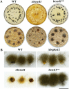
(A) Sclerotia formed by the WT, Δbcscd1, and bcscd1com strains (top row) and their germination in wetted sand soil after incubation for 1 month (bottom row). (B) Sclerotial germination is attenuated in the Δbcscd1 strain but not in the Δbcpks12 strain. Bar, 1 cm.
FIG 2
FIG 2
A melanin intermediate is deposited by the Δbcscd1 mutant extracellularly. (A) Thin-layer chromatography (TLC) analysis of crude extracts of hyphal homogenates and culture filtrates of the wild type (WT) and three mutant strains of B. cinerea. The arrow indicates the strong signal detected in the culture filtrate of the Δbcscd1 mutant. (B) The Δbcpks12 sclerotia gradually regained melanization when they were cocultured with the Δbcscd1 mutant, suggesting that the compound secreted by the Δbcscd1 mutant was utilized by the Δbcpks12 mutant to remedy its defect in melanin synthesis in sclerotia. Bar, 1 cm.
FIG 3
FIG 3
The melanin intermediate scytalone deposited by the Δbcscd1 mutant is inhibitory for sclerotial and spore germination. (A) The sclerotial color of WT and Δbcscd1 strains was altered when treated with the chemical inhibitor tricyclazole (50 μg/ml), which is supposed to inactivate the activity of the reductase BcBRN1/2 (bars, 1 mm). (B) Treatment with tricyclazole derepressed sclerotial germination of the Δbcscd1 mutant compared with the control group (CK) but caused no obvious effect on sclerotial germination of the WT (bar, 1 mm). (C) Spores produced by germinated sclerotia of the Δbcscd1 mutant were significantly reduced compared with the WT, Δbcpks12, and complementation strains, and tricyclazole treatment increased the spore quantity produced by Δbcscd1 sclerotia but caused no effect on the other strains. (D and E) Feeding assay by the addition of scytalone (400 μg/ml) reduces spore germination and germ tube growth rates. CK, control check.
FIG 4
FIG 4
The melanin intermediate scytalone is accumulated in old but not young cultures of the Δbcscd1 mutant. (A) Pigmentation patterns of culture filtrates of the wild type (WT) and the Δbcscd1 mutant were compared at a series of growth stages. (B) TLC analysis of the crude extracts from culture filtrates of the wild type and the Δbcscd1 mutant at a series of growth stages. “S” represents the loading of standard scytalone.
FIG 5
FIG 5
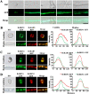
Localization of the reductases BcBRN1/2. (A) The fluorescence signal of GFP-labeled BcBRN1 in the WT+BcBRN1-GFP strain appeared at various subcellular sites in conidia and hyphae at different growth stages. DIC, differential interference contrast. (B to D) Simultaneous detection of GFP and RFP fluorescence in conidia and hyphae of the WT+BcBRN1-GFP+BcRAB5-RFP (B), WT+BcBRN1-GFP+BcRAB7-RFP (C), and WT+BcBRN1-GFP+BcBRN2-RFP (D) strains. Line scan graphs were generated at the indicated positions to show the relative localization of GFP (green) and RFP (red) signals. Conidia and hyphae were photographed 0 and 2 days after incubation, respectively. Bars, 10 μm (A) and 5 μm (B to D).
FIG 6
FIG 6
Relative localization analysis with BcBRN1-GFP and FM4-64 or neutral red staining signals. (A) The endocytic pathway was traced using FM4-64 in the WT+BcBRN1-GFP strain. Staining periods in minutes are given at the top. The dye entered the plasma membranes (5 min), and subsequently, plasma membrane-conglutinated endocytic vesicles were observed (60 min). Close views of the dotted rectangle areas indicated by arrows are shown on the right side. In the hyphae, BcBRN1-GFP is localized within the spherical vesicles, which can be partially colocalized with the endocytic vesicles. (B) Visualization of BcBRN1-GFP fluorescence and neutral red staining signals in the WT+BcBRN1-GFP strain. Vacuoles stained with neutral red and observed by DIC microscopy (black) are distinguishable from the vesicles localized with BcBRN1-GFP. Bars, 10 μm.
FIG 7
FIG 7
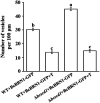
Effect of scytalone accumulation on the density of BcBRN1-GFP-labeled vesicles. Shown are the changes of BcBRN1-GFP-labeled vesicles in mycelia of the WT+BcBRN1-GFP and Δbcscd1+BcBRN1-GFP strains in the presence or absence of tricyclazole (T) (10 μg/ml). Different letters represent significant differences between columns (n = 150; P < 0.05).
FIG 8
FIG 8
Subcellular localization of enzymes at the early steps of melanin synthesis. (A) Subcellular localization of BcPKS12, BcPKS13, and BcYGH1 in the hyphae of the WT+BcPKS12-GFP, WT+BcPKS13-GFP, and WT+BcYGH1-GFP strains, respectively. (B) Relative localization of BcPKS13 and BcRAB5 in the WT+BcPKS13-GFP+BcRAB5-RFP strain. (C) The peroxisome membrane protein BcPEX3 colocalizes with BcPKS12, BcPKS13, and BcYGH1 in the hyphae of the WT+BcPKS12-GFP+BcPEX3-RFP, WT+BcPKS13-GFP+BcPEX3-RFP, and WT+BcYGH1-GFP+BcPEX3-RFP strains, respectively. Bars, 10 μm.
FIG 9
FIG 9
(A) Light and fluorescence microscopic views of spores, hyphae, and infection cushions of the bcscd1combcscd1+BcSCD1-GFP) strain. (B) Spores of the bcscd1com strain were subjected to cell wall digestion and regeneration assays. The samples for analysis included spores, protoplasts, and recovered spores obtained from the regenerated culture of protoplasts. Bars, 10 μm (A) and 20 μm (B).
FIG 10
FIG 10
Subcellular localization of BcSCD1-GFP and BRN2-RFP. Simultaneous detection of GFP-labeled BcSCD1 and RFP-labeled BcBRN2 was conducted in growing hyphae of the Δbcscd1+BcSCD1-GFP+BRN2-RFP strain. (A and B) BRN2-RFP signals are present in the cytoplasm and subcellular vesicles, being distinguished from the location of the BcSCD1-GFP signal on the cell wall. (C) BRN2-RFP and BcSCD1-GFP are colocalized on the cell wall. Bar, 10 μm.
FIG 11
FIG 11
Cytological regulation model for melanin synthesis in B. cinerea. N, nucleus; ER, endoplasmic reticulum; Ps, peroxisome; EEs, early endosomes; LEs, late endosomes; AT4HN, 2-acetyl-1,3,6,8-tetrahydroxynaphthalene.

Similar articles

Cited by

References

    1. Dean R, Van Kan JA, Pretorius ZA, Hammond-Kosack KE, Di Pietro A, Spanu PD, Rudd JJ, Dickman M, Kahmann R, Ellis J, Foster GD. 2012. The top 10 fungal pathogens in molecular plant pathology. Mol Plant Pathol 13:414–430. doi:10.1111/j.1364-3703.2011.00783.x. - DOI - PMC - PubMed
    1. Fillinger S, Walker A. 2016. Chemical control and resistance management of Botrytis diseases, p 189–216. In Fillinger S, Elad Y (ed), Botrytis—the fungus, the pathogen and its management in agricultural systems. Springer International Publishing, Cham, Switzerland.
    1. Leroux P. 2007. Chemical control of Botrytis and its resistance to chemical fungicides, p 195–222. In Elad Y, Williamson B, Tudzynski P, Delen N (ed), Botrytis: biology, pathology and control. Springer Netherlands, Dordrecht, Netherlands.
    1. Petrasch S, Silva CJ, Mesquida-Pesci SD, Gallegos K, van den Abeele C, Papin V, Fernandez-Acero FJ, Knapp SJ, Blanco-Ulate B. 2019. Infection strategies deployed by Botrytis cinerea, Fusarium acuminatum, and Rhizopus stolonifer as a function of tomato fruit ripening stage. Front Plant Sci 10:223. doi:10.3389/fpls.2019.00223. - DOI - PMC - PubMed
    1. Nakajima M, Akutsu K. 2014. Virulence factors of Botrytis cinerea. J Gen Plant Pathol 80:15–23. doi:10.1007/s10327-013-0492-0. - DOI

Publication types

Supplementary concepts

LinkOut - more resources