. 2021 Mar 23;12(2):e00288-21.
doi: 10.1128/mBio.00288-21.

Noncanonical Transmission of a Measles Virus Vaccine Strain from Neurons to Astrocytes

Affiliations

Noncanonical Transmission of a Measles Virus Vaccine Strain from Neurons to Astrocytes

Katrien C K Poelaert et al. mBio. .

Abstract

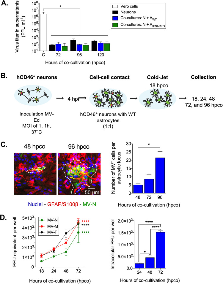
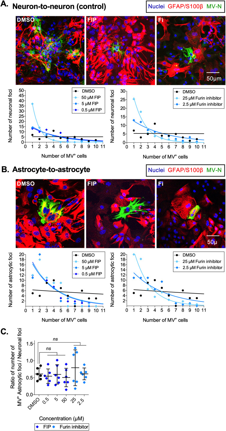
Viruses, including members of the herpes-, entero-, and morbillivirus families, are the most common cause of infectious encephalitis in mammals worldwide. During most instances of acute viral encephalitis, neurons are typically the initial cell type that is infected. However, as replication and spread ensue, other parenchymal cells can become viral targets, especially in chronic infections. Consequently, to ascertain how neurotropic viruses trigger neuropathology, it is crucial to identify which central nervous system (CNS) cell populations are susceptible and permissive throughout the course of infection, and to define how viruses spread between distinct cell types. Using a measles virus (MV) transgenic mouse model that expresses human CD46 (hCD46), the MV vaccine strain receptor, under the control of a neuron-specific enolase promoter (NSE-hCD46+ mice), a novel mode of viral spread between neurons and astrocytes was identified. Although hCD46 is required for initial neuronal infection, it is dispensable for heterotypic spread to astrocytes, which instead depends on glutamate transporters and direct neuron-astrocyte contact. Moreover, in the presence of RNase A, astrocyte infection is reduced, suggesting that nonenveloped ribonucleoproteins (RNP) may cross the neuron-astrocyte synaptic cleft. The characterization of this novel mode of intercellular transport offers insights into the unique interaction of neurons and glia and may reveal therapeutic targets to mitigate the life-threatening consequences of measles encephalitis.IMPORTANCE Viruses are the most important cause of infectious encephalitis in mammals worldwide; several thousand people, primarily the very young and the elderly, are impacted annually, and few therapies are reliably successful once neuroinvasion has occurred. To understand how viruses contribute to neuropathology, and to develop tools to prevent or ameliorate such infections, it is crucial to define if and how viruses disseminate among the different cell populations within the highly complex central nervous system. This study defines a noncanonical mode of viral transmission between neurons and astrocytes within the brain.

Keywords: astrocyte; measles virus; neuron; neurotransmitter transporters; ribonucleoprotein; synapse.


Figures

FIG 1
FIG 1
Astrocytes are the predominant infected cell type during MV reactivation in NSE-hCD46 mice in vivo. (A) NSE-hCD46+/RAG2 KO mice (n = 3) were inoculated with 1 × 104 PFU MV-Ed and monitored daily. At 30 dpi, mice were euthanized and perfused with paraformaldehyde, and brains were removed for cryosectioning and immunofluorescence. The number of infected cells within the cortex or hippocampus was then quantified. Results are shown as the number of infected neurons or astrocytes as a percentage of the total number of infected cells seen in 10 brain sections/mouse, with data collected at multiple axial levels. (B) Bone marrow chimeras were generated using NSE-hCD46+ recipients (n = 2) that had been infected with 1 × 104 PFU MV-Ed at least 90 days previously. Infected mice were reconstituted with RAG2 KO donor bone marrow and monitored daily. At 14 days after transplantation, mice were sacrificed, and brains were treated as described above. The number of infected cells within the cortex was quantified and is shown as the percentage of infected neurons or astrocytes as a proportion of the total number of infected cells, with data derived from 10 brain sections/mouse (n = 5). An unpaired t test for significance was used. ****, P < 0.0001.
FIG 2
FIG 2
Primary astrocytes lacking hCD46 are not permissive for cell-free MV infection in vitro. Primary astrocytes, isolated from NSE-hCD46+ mice, were inoculated with MV-Ed (MOI = 1) and collected at 3, 24, 48, and 72 hpi. (A) hCD46 RNA is detected only in neurons from NSE-hCD46+ mice; astrocytes do not synthesize hCD46 RNA. Unpaired t test for significance was used; **, P < 0.01. (B) RNA was collected from inoculated Vero cells, neurons, and astrocytes at the indicated time points. Random hexamers were used as primers for cDNA synthesis, followed by qPCR using primers specific for the MV nucleoprotein (MV-N). Data are represented as PFU equivalent per well (±400,000 cells) based on a standard curve. Data represent means plus SD for three independent experiments. A multiple-way analysis of variance (ANOVA), followed by a Tukey post hoc test, was used for significance; ****, P < 0.0001.
FIG 3
FIG 3
Primary astrocytes are permissive for MV infection when in direct contact with infected hCD46+ neurons. Primary neurons were inoculated with MV-Ed (MOI = 1) and cocultured 4 h thereafter either directly (cell-cell contact) or indirectly (shared medium) with uninfected primary astrocytes. (A) Schematic illustration of the experimental setup. (B) At 76 hpi (72 h postcoculture), neuron cultures or neuron-astrocyte cocultures were collected; MV-N and GFAP/S100β astrocyte cell markers were detected by immunostaining with Alexa Fluor 488 (green) and Alexa Fluor 555 (red), respectively; nuclei were counterstained with Hoechst 33342 (blue). (C) The number of astrocytic foci, in both the direct and indirect neuron-astrocyte cocultures, was quantified at the indicated time points. Data represent means plus SD for three independent experiments. An ANOVA, followed by a Tukey post hoc test, was used for significance; **, P < 0.01; ****, P < 0.0001.
FIG 4
FIG 4
Receptor-independent MV spread from neurons to astrocytes is neuron and astrocyte specific. (A) Representative confocal images of Vero-astrocyte cocultures at 48 hpco. MV-N and the GFAP/S100β astrocyte cell markers were identified by staining with Alexa Fluor 488 (green) and 555 (red), respectively. The dotted line marks the area of the x-z image. (B) The number of infected L929 cells, upon inoculation with cell-free virus or upon coculture with infected neurons, was counted at 48 hpco. Data represent means with SD for three independent experiments. An unpaired t test for significance was used. ns, not significant.
FIG 5
FIG 5
Infected astrocytes are permissive for MV reproduction and spread but do not release infectious viral progeny. (A) Extracellular virus titers from supernatants of Vero cells, neurons, or neuron-astrocyte cocultures were analyzed by plaque assay. Data shown are means with SD for three independent experiments. An ANOVA, followed by a Dunnett post hoc test, was used for significance; *, P < 0.05. (B) Schematic experimental setup. Neurons were removed selectively from the neuron-astrocyte cocultures at 18 hpco (cold jet). At the indicated time points postcocultivation, astrocytes were fixed. (C) MV-N and GFAP/S100β astrocyte cell markers were detected by immunostaining as before, with Alexa Fluor 488 (green) and Alexa Fluor 555 (red), respectively; nuclei were counterstained with Hoechst 33342 (blue). The regions marked with a white dashed line define one astrocytic focus at the indicated time points. Data represent means plus SD for three independent experiments. The number of infected cells within 30 astrocytic foci was counted. The nonparametric Kruskal-Wallis test, followed by a Dunnett post hoc test, was used for significance; *, P < 0.05. (D) RNA was collected from the remaining astrocytes at the indicated time points. (Left panel) cDNA was synthesized, followed by qRT-PCR using primers specific for MV-N, MV-M, and MV-F coding regions. Data are represented as PFU equivalent per well based on a standard curve. (Right panel) At the indicated time points, astrocytes were scraped into fresh medium, followed by a freeze-thaw cycle, to determine the intracellular virus titer using a standard plaque assay. An ANOVA, followed by a Tukey post hoc test, was used for significance, in which * indicates P < 0.05 and **** indicates P < 0.0001.
FIG 6
FIG 6
MV spread between heterotypic cell types of the brain is fusion independent. Neuronal-astrocyte cocultures were treated with two fusion-inhibitory drugs, fusion-inhibitory peptide (FIP; 0.5, 5, and 50 μM) and furin inhibitor (Fi; 2.5 and 25 μM), or equivalent volumes of DMSO. At 72 hpco, MV-N and GFAP/S100β astrocyte cell markers were identified with Alexa Fluor 488 (green) and 555 (red), respectively. (A) Representative confocal images of neuronal foci for each condition are shown (upper panel), as well as a graphical representation of the number of infected neurons within a neuronal focus for each reagent (lower panel). (B) Similarly organized data are shown for astrocytic foci for each condition. Data points for both panel A and panel B represent at least 60 foci from three independent replicates (20 per replicate). Each data point shows the cumulative total of foci containing a specific number of infected cells. The lines illustrate the trends for each reagent, plotted using GraphPad Prism. (C) The ratio of the number of astrocytic foci (numerator) over neuronal foci (denominator) within a scanning field (375 × 375 μm) was calculated. Each dot represents one replicate (n = 6). An ANOVA followed by a Dunnett post hoc test was used to gauge significance. ns, not significant.
FIG 7
FIG 7
MV spread between heterotypic cell types of the CNS depends on glutamate transporters. Neuron-astrocyte cocultures were treated with dl-threo-β-benzyloxyaspartic acid (dl-TBOA; 0.5 to 50 μM), dihydrokainic acid (DHK; 0.5 to 50 μM), sarcosine (50 μM), nipecotic acid (50 μM), or equivalent volumes of DMSO or distilled water (dH2O) (control). At 72 hpco, neuron-astrocyte cultures were fixed, followed by IF staining and confocal analysis. The ratio of the number of astrocytic foci (numerator) over the number of neuronal foci (denominator) within a scanning field (375 × 375 μm) was calculated. (A) dl-TBOA. (B) DHK. (C) The glycine transporter inhibitor sarcosine and the GABA uptake inhibitor nipecotic acid were included as controls. Each dot represents a replicate (n = 6). An ANOVA, followed by a Dunnett post hoc test, was used for significance. *, P < 0.05; ns, not significant.
FIG 8
FIG 8
RNase A treatment reduces viral spread between heterotypic cell types. (A) Neuron-astrocyte cocultures were treated with DNase (100 μg/ml), inactivated RNase, or RNase A (1 to 100 μg/ml). At 72 hpco, neuron-astrocyte cocultures were fixed, followed by IF staining and confocal analysis. The ratio of the number of astrocytic foci over the neuronal foci within a scanning field (375 × 375 μm) was calculated. (B) Virus stock was treated with inactivated RNase or 100 μg/ml RNase A for 45 min at RT and added on top of a monolayer of Vero cells. This plaque assay was fixed at 5 dpi, and the number of viral foci was counted per well. Each dot represents a replicate (n = 6). An ANOVA, followed by a Dunnett post hoc test, was used for significance. ****, P < 0.0001; ns, not significant.

Similar articles

Cited by

References

    1. Daneman R, Prat A. 2015. The blood-brain barrier. Cold Spring Harb Perspect Biol 7:a020412. doi:10.1101/cshperspect.a020412. - DOI - PMC - PubMed
    1. Reinhold AK, Rittner HL. 2017. Barrier function in the peripheral and central nervous system—a review. Pflugers Arch 469:123–134. doi:10.1007/s00424-016-1920-8. - DOI - PubMed
    1. Duprex WP, McQuaid S, Hangartner L, Billeter MA, Rima BK. 1999. Observation of measles virus cell-to-cell spread in astrocytoma cells by using a green fluorescent protein-expressing recombinant virus. J Virol 73:9568–9575. doi:10.1128/JVI.73.11.9568-9575.1999. - DOI - PMC - PubMed
    1. Dubois-Dalcq M, Coblentz JM, Pleet AB. 1974. Subacute sclerosing panencephalitis. Unusual nuclear inclusions and lengthy clinical course. Arch Neurol 31:355–363. doi:10.1001/archneur.1974.00490420021001. - DOI - PubMed
    1. Rima BK, Duprex WP. 2005. Molecular mechanisms of measles virus persistence. Virus Res 111:132–147. doi:10.1016/j.virusres.2005.04.005. - DOI - PubMed

Publication types

LinkOut - more resources