. 2021 Mar 30;118(13):e2008772118.
doi: 10.1073/pnas.2008772118.

Cancer-specific loss of TERT activation sensitizes glioblastoma to DNA damage

Affiliations

Cancer-specific loss of TERT activation sensitizes glioblastoma to DNA damage

Alexandra M Amen et al. Proc Natl Acad Sci U S A. .

Abstract

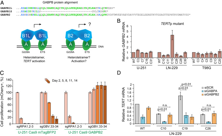
Most glioblastomas (GBMs) achieve cellular immortality by acquiring a mutation in the telomerase reverse transcriptase (TERT) promoter. TERT promoter mutations create a binding site for a GA binding protein (GABP) transcription factor complex, whose assembly at the promoter is associated with TERT reactivation and telomere maintenance. Here, we demonstrate increased binding of a specific GABPB1L-isoform-containing complex to the mutant TERT promoter. Furthermore, we find that TERT promoter mutant GBM cells, unlike wild-type cells, exhibit a critical near-term dependence on GABPB1L for proliferation, notably also posttumor establishment in vivo. Up-regulation of the protein paralogue GABPB2, which is normally expressed at very low levels, can rescue this dependence. More importantly, when combined with frontline temozolomide (TMZ) chemotherapy, inducible GABPB1L knockdown and the associated TERT reduction led to an impaired DNA damage response that resulted in profoundly reduced growth of intracranial GBM tumors. Together, these findings provide insights into the mechanism of cancer-specific TERT regulation, uncover rapid effects of GABPB1L-mediated TERT suppression in GBM maintenance, and establish GABPB1L inhibition in combination with chemotherapy as a therapeutic strategy for TERT promoter mutant GBM.

Keywords: CRISPR; TERT; cancer; glioblastoma; temozolomide.


Conflict of interest statement

Competing interest statement: The Regents of the University of California have patents issued and pending for CRISPR technologies on which J.A.D. is an inventor. C.F. is a co-founder of Mirimus, Inc. J.A.D. is a co-founder of Caribou Biosciences, Editas Medicine, Intellia Therapeutics, Scribe Therapeutics, and Mammoth Biosciences. J.A.D. is a scientific advisory board member of Caribou Biosciences, Intellia Therapeutics, eFFECTOR Therapeutics, Scribe Therapeutics, Synthego, Metagenomi, Mammoth Biosciences, and Inari. J.A.D. is a Director at Johnson & Johnson and has sponsored research projects by Pfizer, Roche Biopharma, and Biogen. J.F.C. is a co-founder of Telo Therapeutics, Inc. and has ownership interests. The other authors declare no competing interests.

Figures

Fig. 1.
Fig. 1.
GABPB1L-containing complexes bind and regulate the mutant TERT promoter. (A) Representative immunoblots of GABPB1 in U-251 and LN-18 cells expressing doxycycline-induced shRNAs targeting GABPB1S (shGB1S.82, shGB1S.77, shGB1S.32, and shGB1S.110) and GABPB1L (shGB1L.377, shGB1L.699, shGB1L.968, and shGB1L.1202) compared with negative control (olfactory receptor OR2B6, shOR2B6.83) and nontargeting (renilla luciferase, shRen.713) shRNAs. Cells were incubated with doxycycline for 6 d prior to harvest. The lower bands represent GABPB1S, and upper bands represent GABPB1L. (B) TERT mRNA expression measured via qRT-PCR in cell lines from A compared with a control cell line (U2OS) lacking TERT expression. (C) Representative gels of telomerase activity measured via TRAP assay in cell lines from A compared with a control cell line (U2OS) lacking TERT expression. The control condition reflects no lysate. (D and E) Representative gels (D) and quantification (E) of electrophoretic mobility shift assays (EMSAs) comparing binding affinity of GABPA-B1L heterodimers to the mutant (G228A) TERT promoter (additional ETS binding site), WT TERT promoter (native ETS binding sites), and control WT TERT promoter sequences lacking native ETS binding sites (G201T: native ETS single mutant; G201T and A197T: native ETS double mutant).
Fig. 2.
Fig. 2.
Inhibition of GABPB1L rapidly prevents growth of TERT promoter mutant cells. (A) GABPB1L exon 9 excision strategy. A diagram of the major human GABPB1 transcript variants, encoding either a short (GABPB1S) or long (GABPB1L) isoform. Red rectangles represent the coding sequence, and green rectangles represent the 3′ UTR. For clarity, the first exon containing the 5′ UTR is not shown. GABPB1L sgRNA targeting sites are highlighted in purple. (B) The editing strategy for scarless knockout of the long isoform of GABPB1 (GABPB1L). In each case, a pair of sgRNAs were used targeting GABPB1L intron number 8 and the 3′ UTR of GABPB1L, respectively. (C and D) Genotyping analysis of GABPB1L edited monoclonal TERT promoter mutant U-251, LN-229, and T98G GBM cell lines (C) and TERT promoter WT HEK293T, HAP1, NHA-S2, and LN-18 cell lines (D). The superscript characters indicate the sgRNA pair used for editing. WT: parental WT cell line. C, PCR no template control. Unedited, full-length genotyping band; Δ2-12, band after excision with sgGB2 and sgGB12; Δ3-14, band after excision with sgGB3 and sgGB14. (E) A comparison of GABPB1L editing outcomes between TERTp WT (blue) and TERTp mutant (yellow) lines. The bars represent the percentage of each editing outcome averaged over three TERTp mutant and four TERTp WT lines. Retained refers to clones where the GABPB1L exon 9 was overall retained, even though they may contain indels at either or both of the sgRNA target sites flanking exon 9. p, P value (unpaired, two-tailed Student’s t test). n.s., not significant (alpha level = 0.05). (F and G) Representative bioluminescence images (F) and Kaplan–Meier survival curve (G) of mice injected with U-251 cells expressing a control shRNA (shRen.713) or GABPB1L-targeting shRNAs (shGB1L.968, shGB1L.1202). Mice were fed doxycycline chow to induce shRNA expression starting at day 7 postinjection. n = 10 to 12 mice per condition. **P < 0.01 compared with shRen.713 (Kaplan–Meyer log-rank test). M, median survival.
Fig. 3.
Fig. 3.
GABPB2 up-regulation can compensate for loss of GABPB1L. (A) Similarity between GABP proteins. Partial protein alignment (MUSCLE) of GABPB1L, GABPB1S, and GABPB2. GABPB1L is the long isoform and GABPB1S the short isoform of the GABPB1 gene. GABPB2 is a distinct gene. Amino acids matching across all three proteins are highlighted in blue, and those matching across two proteins are highlighted in green. GABPA (alpha) subunits of the GABP complex bind to both the native ETS site (ETS) and the mutation-derived ETS sites (G228A or G250A) at the TERT promoter locus. The GABPB1 short (B1S) or long (B1L) isoform subunits bind to the alpha subunits to form either heterodimers (GABPA1B1S1) or heterotetramers (GABPBA2B1L2). Heterotetramer formation is presumably mediated through the leucine zipper-like domain of GABPB1L, which is also present in GABPB2. (B) GABPB2 mRNA expression measured via qRT-PCR in GABPB1L knockout clones, plotted relative to control (WT) cells, in TERTp mutant cell lines. The data represent mean ± SEM. (C) Competitive proliferation assay in U-251 cells using pairs of sgRNAs targeting a positive control locus (sgRPA1.2 and 3) and total GABPB1 (targeting exon 3, sgGBV.33 and 34) in the presence of a lentiviral vector expressing either GABPB2 or mTagBFP2 (control). The data represent mean ± SD of triplicates. (D) TERT mRNA expression measured via qRT-PCR following siRNA-mediated knockdown of GABPA or GABPB2 in LN-229 WT, full knockout, or heterozygous knockout cells. p, P value (unpaired, two-tailed Student’s t test). n.s., not significant (alpha level = 0.05). The data represent mean ± SEM.
Fig. 4.
Fig. 4.
Reduction of GABPB1L potentiates anti-tumor response of TERTp mutant GBM to chemotherapy. (A) A schematic for possible sensitization of TERT promoter mutant GBM cells to TMZ through GABPB1L inhibition. Inhibiting GABPB1L leads to reduced TERT expression, resulting in a blunted DDR and lack of normal cell cycle arrest, ultimately reducing cancer cell viability following DNA damage. SSB, single-strand break; DSB, double-strand break; DDR, DNA-damage response. (B) Representative immunoblots of yH2AX in U-251 cells expressing doxycycline-induced shRNAs targeting TERT (shTERT.3795 and shTERT.3592) or GABPB1L (shGB1L.968 and shGB1L.1202) compared with a nontargeting (renilla luciferase, shRen.713) shRNA. (C) Representative immunoblots (Bottom) and quantification (Top, from triplicate blots) of yH2AX in U-251 cells engineered to stably express either BFP (control) or TERT (rescue) in the presence of shRNAs targeting TERT (shTERT.3795 and shTERT.3592) or GABPB1L (shGB1L.968 and shGB1L.1202) compared with a nontargeting shRNA (renilla luciferase, shRen.713). (B and C) Cells were incubated with doxycycline for 6 d prior to harvest and treated with specified dose(s) of TMZ 20 h prior to harvest. (D) Representative images (Left) and G2/G1 ratio quantification (Right) from flow cytometry–based cell cycle analysis of U-251 WT and GABPB1L heterozygous knockout cells treated with a dose titration of TMZ 72 h prior to harvest. p, P value (unpaired two-tailed Student’s t test). (E) A schematic of in vivo TMZ treatment. Mice were xenografted with U-251 cells expressing doxycycline-inducible shRNAs and placed on doxycycline chow 7 d postorthotopic xenograft. Mice were then treated with TMZ or a vehicle control by oral gavage starting 12 d postxenograft for 5 d. IHC, immunohistochemistry. (F) Bioluminescence imaging of mice injected with U-251 cells engineered to express shRNAs targeting TERT (shTERT.3592), GABPB1L (shGB1L.968), or a nontargeting control shRNA (shRen.713). Mice were placed on doxycline chow and per os (p.o.) dosed with either TMZ or a vehicle control as described in E. (G) Representative images of immunofluorescence staining in tumors from mice described in E, cohort 1. Mice were euthanized 30 d postorthotopic xenograft for analysis. Doxycycline-induced tumor cells are GFP positive. n = 5 images per mouse, three mice per condition. (Scale bar, 50 μm.) (H) Kaplan–Meier survival curves for mice described in E, cohort 2. For statistics, survival of mice with no tumor burden was set at the experimental endpoint (130 d). n = 5 to 7 mice per condition. **P < 0.01, relative to all vehicle conditions (Kaplan–Meier log-rank test). M, median survival. (I) A comparison of the relative fold increase in median survival based on overall survival in H. The dashed line represents a simple addition of effects of TMZ plus GABPB1L or TERT knockdown. The survival of mice xenografted with WT cells and treated with vehicle control was used for normalization.

Similar articles

Cited by

References

    1. Reardon D. A., Rich J. N., Friedman H. S., Bigner D. D., Recent advances in the treatment of malignant astrocytoma. J. Clin. Oncol. 24, 1253–1265 (2006). - PubMed
    1. Stupp R.et al. .; European Organisation for Research and Treatment of Cancer Brain Tumor and Radiotherapy Groups; National Cancer Institute of Canada Clinical Trials Group , Radiotherapy plus concomitant and adjuvant temozolomide for glioblastoma. N. Engl. J. Med. 352, 987–996 (2005). - PubMed
    1. Louis D. N., et al. ., The 2016 World Health Organization classification of tumors of the central nervous system: A summary. Acta Neuropathol. 131, 803–820 (2016). - PubMed
    1. Hanahan D., Weinberg R. A., Hallmarks of cancer: The next generation. Cell 144, 646–674 (2011). - PubMed
    1. Kim N. W., et al. ., Specific association of human telomerase activity with immortal cells and cancer. Science 266, 2011–2015 (1994). - PubMed

Publication types

MeSH terms