

doi: 10.1038/s41421-021-00249-2.
SARS-CoV-2 cell tropism and multiorgan infection
Affiliations
- PMID: 33758165
- PMCID: PMC7987126
- DOI: 10.1038/s41421-021-00249-2
Item in Clipboard
SARS-CoV-2 cell tropism and multiorgan infection
Cell Discov.
.
No abstract available
Conflict of interest statement
The authors declare that they have no conflict of interest.
Figures
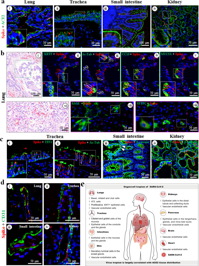
a Co-localization analysis of SARS-CoV-2 (spike proteins, red) and ACE2 (green) in the lung (i), trachea (ii), small intestine (iii), and kidney (iv) via dual-immunofluorescent staining. b Characterization of SARS-CoV-2-positive cell tropism in the lung. Co-localization of SARS-CoV-2 (spike proteins, red) with cell type-specific markers (green) was determined via multiplex-immunofluorescence staining. KRT5 (ii), Ac-Tub (iii), CC10 (iv), MUC5A (v), RAGE (vii), and SFTPC (viii) were used for basal, ciliated, club, goblet, AT1, and AT2 cells, respectively. The corresponding hematoxylin and eosin staining figures were shown in (i) and (vi), respectively. c Characterization of SARS-CoV-2 positive cell types in the trachea (i and ii), small intestine (iii), and kidney (iv). TFF1 was used for goblet cells (i), and Ac-Tub for ciliated cells (ii) in the trachea; β-catenin was used for epithelial cells in small intestine (iii) and kidney (iv). White arrows (iii) indicate the mucosa of the small intestine. d Co-localization analysis of SARS-CoV-2 (spike proteins, red) with CD31+ vascular endothelial cells (green) in the lung (i), trachea (ii), small intestine (iii), and kidney (iv). e Schematic of SARS-CoV-2 multiorgan infection and cell tropism in the human body.
References
Publication types
Grants and funding
- 31621061/National Natural Science Foundation of China (National Science Foundation of China)
- 2020FYC0844700/Chinese Ministry of Science and Technology | Department of S and T for Social Development (Department of S&T for Social Development)
- 2020FYC0841700/Chinese Ministry of Science and Technology | Department of S and T for Social Development (Department of S&T for Social Development)
- 2020FCA045/Department of Science and Technology, Hubei Provincial People's Government (Hubei Provincial Department of Science and Technology)
- 2020FCA003/Department of Science and Technology, Hubei Provincial People's Government (Hubei Provincial Department of Science and Technology)
LinkOut - more resources
Full Text Sources
Other Literature Sources
Miscellaneous
