. 2021 Mar 23;7(1):59.
doi: 10.1038/s41420-021-00443-x.

SIRT1 attenuates renal fibrosis by repressing HIF-2α

Affiliations

SIRT1 attenuates renal fibrosis by repressing HIF-2α

Peipei Li et al. Cell Death Discov. .

Abstract

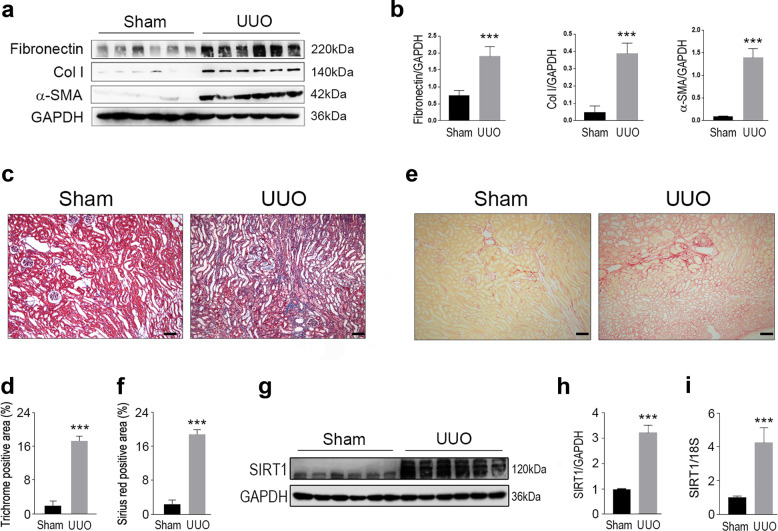
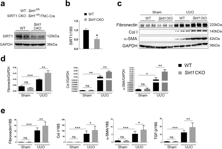
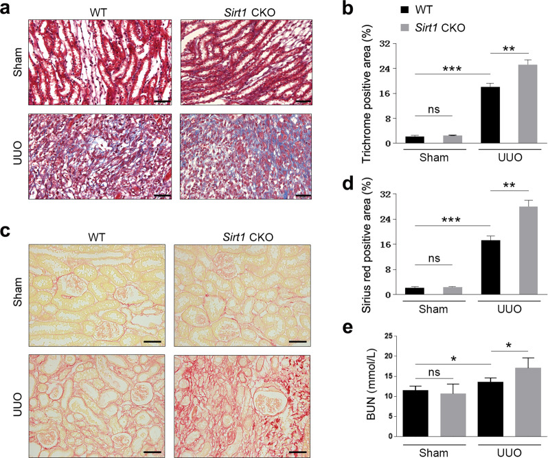
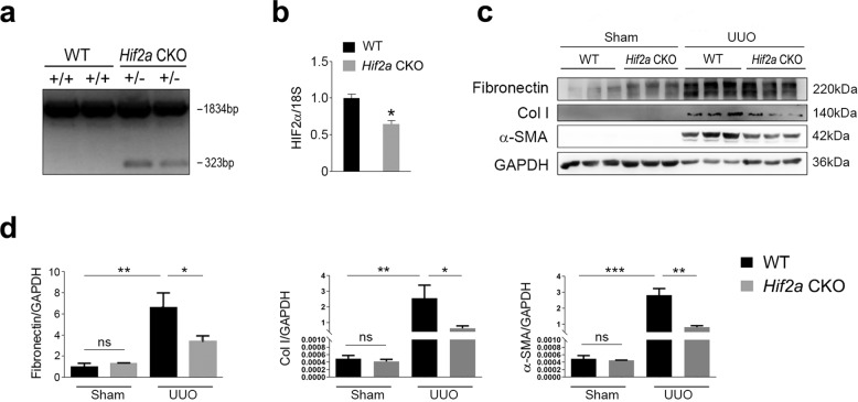
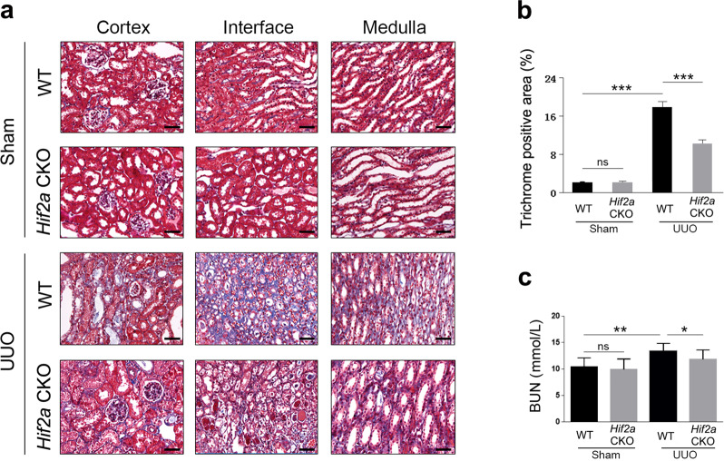
Sirtuin 1 (SIRT1) is a nicotinamide adenine dinucleotide (NAD+)-dependent deacetylase belonging to class III histone deacetylases. Previous studies have shown that SIRT1 is involved in kidney physiology regulation and protects the kidney from various pathological factors. However, the underlying mechanisms behind its function have yet to be fully elucidated. In our study, we found that ablation of Sirt1 in renal interstitial cells resulted in more severe renal damage and fibrosis in unilateral ureteral obstruction (UUO) model mice. We also observed that hypoxia-inducible factor (HIF)-2α expression was increased in Sirt1 conditional knockout mice, suggesting that HIF-2α might be a substrate of SIRT1, mediating its renoprotective roles. Therefore, we bred Hif2a deficient mice and subjected them to renal trauma through UUO surgery, ultimately finding that Hif2a ablation attenuated renal fibrogenesis induced by UUO injury. Moreover, in cultured NRK-49F cells, activation of SIRT1 decreased HIF-2α and fibrotic gene expressions, and inhibition of SIRT1 stimulated HIF-2α and fibrotic gene expressions. Co-immunoprecipitation analysis revealed that SIRT1 directly interacted with and deacetylated HIF-2α. Together, our data indicate that SIRT1 plays a protective role in renal damage and fibrosis, which is likely due to inhibition of HIF-2α.


Conflict of interest statement

The authors declare no competing interests.

Figures

Fig. 1
Fig. 1. SIRT1 is upregulated in the kidney from UUO modeled mice.
Male wild-type C57BL/6 mice, at 8–10 weeks of age, were subjected to UUO operation. Mice were euthanized 10 days after sham or UUO surgery. a Western blot analysis showed that fibronectin, collagen I (Col I), and a-SMA in the kidney were increased upon UUO surgery. GAPDH was used as a loading control. b Quantification of western blot data as shown in a. c Masson-trichrome staining. Scale bar = 50 μm. d Quantitative analysis for c. e Sirius red staining. Scale bar = 50 μm. f Quantitative analysis for e. g SIRT1 in the kidney was enhanced in UUO-operated mice. Protein levels were analyzed by western blot analysis and GAPDH has used a loading control. h Quantification of western blot data as shown in e. i SIRT1 mRNA levels were increased in the kidney from UUO mice. n = 6. Data are means ± SEM. ***P < 0.001.
Fig. 2
Fig. 2. SIRT1 deficiency aggravates UUO injury in the kidney.
a SIRT1 was decreased in the kidney in kidney conditional knockout mice (Sirt1 CKO). Protein levels were analyzed by western blot analysis and GAPDH was used as a loading control. b SIRT1 mRNA levels in the kidney were also reduced in Sirt1 CKO mice. c SIRT1 deficiency further exacerbates kidney fibrosis induced by UUO surgery. Protein levels were analyzed by western blot analysis and GAPDH was used as a loading control. d Quantification of western blot data as shown in c. e Sirt1 deficiency in the kidney stimulates kidney fibrosis-related gene expressions in UUO mice. Gene expression was analyzed by qRT-PCR and 18S has used a house-keeping gene. n = 6. Data are means ± SEM. *P < 0.05, **P < 0.01, and ***P < 0.001. ns means no significance.
Fig. 3
Fig. 3. Renal interstitial cell-specific ablation of Sirt1 enhances fibrogenesis during UUO injury.
Male WT (Sirt1flox/flox) and Sirt1 CKO (Sirt1flox/flox/TNCCreER), at 8–10 weeks of age, were subjected to UUO operation. Mice were sacrificed on the 10th day after sham or UUO surgery. a Paraffin section from kidneys of sham or UUO surgery mice was analyzed by Masson-trichrome staining. Scale bar = 20 μm. b Quantitative analysis of Masson-trichrome staining. c Sirius red staining. Scale bar = 20 μm. d Quantitative analysis of Sirius red staining. e BUN levels. n = 6. Data are means ± SEM; ns: no significance. *P > 0.05, **P < 0.01, ***P < 0.001.
Fig. 4
Fig. 4. HIF-2α in the kidney is stimulated by UUO injury.
a UUO injury increases HIF-2α protein levels in kidneys of WT and Sirt1 CKO mice. Protein levels were analyzed by western blot analysis and GAPDH was used as a loading control. b Quantitative analysis of western blot data as shown in a. c HIF-2α mRNA levels were not altered by UUO surgery. Gene expression was analyzed by qRT-PCR and 18S was used as a house-keeping gene. de HIF-2α in the renal tubule (d) and glomerulus (e) were analyzed by immunofluorescence. Scale bar = 20 μm. n = 6. Data are means ± SEM. ns: no significance. *P > 0.05, **P < 0.01.
Fig. 5
Fig. 5. Hif2a deficiency in renal interstitial cells attenuates fibrogenesis induced by UUO injury.
a Genotyping for Hif2a CKO mice. The length of PCR products for the wild-type Hif2a allele is 1834 bp, and the length for the knockout Hif2a allele is 323 bp. b HIF-2α expression was reduced in kidneys from Hif2a CKO mice. Gene expression was analyzed by qRT-PCR and 18S has used a house-keeping gene. c Renal fibrosis was attenuated by Hif2a deficiency. Protein levels were analyzed by western blot and GAPDH was used as a loading control. d Quantitative analysis of western blot data as shown in c. n = 6. Data are means ± SEM. ns: no significance. *P > 0.05, **P < 0.01, ***P < 0.001.
Fig. 6
Fig. 6. Hif2a deficiency ameliorates renal fibrosis induced by UUO surgery.
a Masson-trichrome staining showing fibrosis was reduced. Scale bar = 20 μm. b Quantitative analysis of the data shown in a. c BUN levels was decreased in Hif2a CKO mice. n = 6. Data are means ± SEM. ns means no significance. *P > 0.05, **P < 0.05, ***P < 0.001.
Fig. 7
Fig. 7. Modulation of SIRT1 activity affects HIF-2α and fibrosis gene expression.
a NRK-49F cells were treated with CoCl2, NMN, Sirtinol as indicated. Cell lysates were prepared for western blot analysis. HIF-1α, HIF-2α, Fibronectin, and Col I were increased by inhibition of SIRT1. GAPDH was used as a loading control. b Quantitative analysis of western blot data as shown in a. c SIRT1 interacts with and deacetylates HIF-2α. NRK-49F cells were treated with NMN, Sirtinol as indicated. Total cell lysates were prepared and subjected to co-immunoprecipitation analysis using an anti-HIF-2α antibody. The immunocomplex was analyzed by western blot. IP: immunoprecipitation. IB: immunoblot. Data are means ± SEM. ns means no significance. *P > 0.05, **P < 0.01, ***P < 0.001.

References

    1. Webster AC, Nagler EV, Morton RL, Masson P. Chronic kidney disease. Lancet. 2017;389:1238–1252. doi: 10.1016/S0140-6736(16)32064-5. - DOI - PubMed
    1. Tonelli M, Riella M. Chronic kidney disease and the ageing population. Lancet. 2014;383:1278–1279. doi: 10.1016/S0140-6736(14)60155-0. - DOI - PubMed
    1. Astor BC, et al. Lower estimated glomerular filtration rate and higher albuminuria are associated with mortality and end-stage renal disease. A collaborative meta-analysis of kidney disease population cohorts. Kidney Int. 2011;79:1331–1340. doi: 10.1038/ki.2010.550. - DOI - PMC - PubMed
    1. Humphreys BD. Mechanisms of renal fibrosis. Annu. Rev. Physiol. 2018;80:309–326. doi: 10.1146/annurev-physiol-022516-034227. - DOI - PubMed
    1. Hao CM, Haase VH. Sirtuins and their relevance to the kidney. J. Am. Soc. Nephrology. 2010;21:1620–1627. doi: 10.1681/ASN.2010010046. - DOI - PMC - PubMed

LinkOut - more resources