. 2021 Mar 23;12(1):1849.
doi: 10.1038/s41467-021-22054-x.

Axonal chemokine-like Orion induces astrocyte infiltration and engulfment during mushroom body neuronal remodeling

Affiliations

Axonal chemokine-like Orion induces astrocyte infiltration and engulfment during mushroom body neuronal remodeling

Ana Boulanger et al. Nat Commun. .

Abstract

The remodeling of neurons is a conserved fundamental mechanism underlying nervous system maturation and function. Astrocytes can clear neuronal debris and they have an active role in neuronal remodeling. Developmental axon pruning of Drosophila memory center neurons occurs via a degenerative process mediated by infiltrating astrocytes. However, how astrocytes are recruited to the axons during brain development is unclear. Using an unbiased screen, we identify the gene requirement of orion, encoding for a chemokine-like protein, in the developing mushroom bodies. Functional analysis shows that Orion is necessary for both axonal pruning and removal of axonal debris. Orion performs its functions extracellularly and bears some features common to chemokines, a family of chemoattractant cytokines. We propose that Orion is a neuronal signal that elicits astrocyte infiltration and astrocyte-driven axonal engulfment required during neuronal remodeling in the Drosophila developing brain.


Conflict of interest statement

The authors declare no competing interests.

Figures
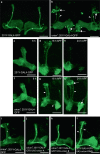
Fig. 1
Fig. 1. The orion gene is necessary for MB remodeling.
al γ neurons are visualized by the expression of 201Y-GAL4-driven UAS-mCD8-GFP (green). In adults, this GAL4 line also labels the αβ-core axons shown here by asterisks. a, b Adult γ axons in control (a) and orion1 (b). Note the presence of unpruned γ axon bundles (arrowhead) and the high amount of uncleared axonal debris (arrows) in orion1 compared to wild-type. (n ≥ 100 MBs for control and orion1. See quantitation in Table 1 and Supplementary Fig. 2.) ch γ axon development in wild-type (ce) and orion1 (fh) at 6, 18, and 24 h APF as indicated. Unpruned axons (arrowhead) in orion1 are already apparent at 18 h APF (compare g with d) although no differences are detected at 6 h APF (c, f). Note the presence of unpruned γ axons (arrowhead) and debris (arrow) in orion1 at 24 h APF (n = 40 MBs for each developmental stage). ik The adult orion1 phenotype (i) is completely rescued by expression in MBs of UAS-orion-A (n = 89 MBs) (j) or UAS-orion-B (n = 387 MBs) (k). l UAS-orion-RNAi expression in MBs results in unpruned γ axon phenotypes (arrowheads) (n = 20 MBs). Scale bars represent 40 µm. All the images are composite confocal images. Genotypes are listed in the Supplementary list of fly strains.
Fig. 2
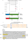
Fig. 2. The orion gene encodes for a CX3C motif-containing protein.
a Complementation map of orion with the tested duplications and deficiencies in the 7B–7D region. Duplications are drawn with a heavy line and deficiencies with a light line. If orion+ is present on the chromosome carrying a duplication or deficiency, it is indicated in the status column with a “+”; and if it is not present, it is marked “−.” The red line indicates the location of the 72 kb to which orion is mapped based on the complementation results. b Linear representation of the polypeptide chain of the two Orion isoforms. Green represents the common region of the two Orion proteins, blue is the specific N-terminal region of Orion-A, and red the specific N-terminal region of Orion-B. The signal peptide of Orion-A and Orion-B (SP) are colored in light blue or light red, respectively. The CX3C chemokine motif as well as the location of the orion1 mutation present in the common region of Orion-A and Orion-B is indicated. c Amino acid sequence lineups of human, mouse, and chicken fractalkines with the common CX3C-bearing motif of the Drosophila Orion proteins is shown. The number within parentheses after the species’ names indicates the total length of the protein. The underlined sequences in the fractalkine sequences indicate the junctions at which their signal peptides are cleaved. The numbers at the beginning and end of the sequence indicate the protein regions in the lineup. The CX3C (CXXXC) and conserved downstream cysteines in the fractalkine species are indicated in blue. Fractalkine intramolecular disulfide bonds between conserved cysteines are specified with brackets. The CX3C motif in the Orions and the downstream cysteines are indicated in green. The Orion downstream cysteines are offset by one and two amino acids, respectively, from those in fractalkine relative to the CX3C motif cysteines. The Orions differ from fractalkine by the inclusion of considerable extensions upstream to the CX3C motif, while the fractalkine CX3C motifs lie within ten amino acids of the mature signal peptide-cleaved proteins. d Orion-B amino acid sequence where the signal peptide is in bold, the three putative GAG binding sites (GAG1, GAG2, GAG3) are highlighted in yellow, the basic residues involved in GAG binding (R = Arg and K = Lys) are in red, and the CX3C site is in brown. An asterisk is located at the end of the Orion-B-specific amino acid sequence/beginning of the common region. The glycine (GGC) that is mutated to an aspartic acid (GAC) in orion1 is indicated by an arrow.
Fig. 3
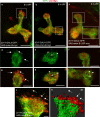
Fig. 3. Orion is extracellularly present on MB γ axons.
ak Six-hour APF γ axons are visualized by the expression of 201Y-GAL4-driven UAS-mCD8-GFP (green). a, b, j, k γ axons expressing the wild-type Orion-B-Myc protein (red) (n = 10 MBs). c γ axons expressing the Orion-B-Myc protein lacking the signal peptide (∆SP) (n = 9 MBs). ac are confocal Z-projections and j is a unique confocal plane. d, g Higher magnification images of the region indicated by a rectangle shows a representative unique confocal plane. Note the presence of Myc-labeled Orion-B outside the γ axon bundle (arrows). e, h Higher magnification images of the region indicated by a rectangle in b showing a representative unique confocal plane. Note the presence of Myc-labeled Orion-B inside the hole-like structures present in the γ axon bundle (arrowheads). f, i Higher magnification images of the vertical and medial γ lobes, respectively (rectangles in c). Orion-B-∆SP-Myc is observed neither outside the γ axons (f) nor in the hole-like structures (arrowheads in i). j, k Presence of Myc-labeled Orion-B extracellular proteins not associated with GFP-labeled axon membranes can be observed outside the γ axon bundle (arrows). k Three-dimensional surface-rendering (3D) of the confocal image. j Reveals close apposition of GFP-labeled axons and Myc-labeled Orion and reveals Orion is present as small extracellular globules. Scale bars represent 40 µm in ac, 20 µm in di and 5 µm in j, k. Full genotypes are listed in the Supplementary list of fly strains.
Fig. 4
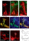
Fig. 4. Orion is required for the infiltration of astrocytes into the MB γ bundle and engulfment of the larval axons at 6 h APF.
ac Single confocal planes of 6 h APF brains expressing repo-GAL4-driven UAS-mCD8-GFP (green) in controls (a, b) and orion1 (c) focused on the MB dorsal lobe (n = 12 MBs controls and n = 20 MBs orion1). Anti-Fas2 staining (red) reveals spherical hole-like structures occupied by glial processes infiltrating into the γ bundle (green, arrows) in wild-type (a, b), but not in orion1 individuals (c). Scale bars are 20 µm. df A single confocal plane showing the expression of 201Y-GAL4-driven UAS-mCD8-GFP (green, df) and Orion-B-Myc (red, d, f) in 6 h APF MB. Anti-Drpr antibody (blue) was used to visualize the glial cells (blue, e, f). df display the same MB. d displays Orion-B-Myc expression outside the axons at the top of the vertical γ bundle (arrows) as well as in hole-like structures (arrowheads). e displays an astrocyte positioned at the top of the γ bundle (asterisk in its nuclei) as well as several astrocyte processes occupying hole-like structures (arrowheads). Note the colocalization of Orion-B-Myc and glia processes in the hole-like structures (arrowheads in f). The astrocyte cell membrane (continuous line) and the membrane contacting the tip of the γ bundle (dotted line), where phagocytosis is taking place, based on our interpretation of the astrocyte limits according to the green and the blue channels for GFP and Drpr, respectively, are indicated in f. n = 10 MBs. Scale bar is 40 µm. g, h Representative images to illustrate how the quantitation of Orion-B-Myc expression (red) (g) and Orion-B-∆SP-Myc (h) driven by 201Y-GAL4 in a traced 60 µm line contained in a γ axon vertical bundle was performed. The position of an astrocyte (dotted line), labeled by anti-Drpr staining and its nucleus (solid circle) are indicated. i, j Representative plotted intensity profiles of Orion expression (gray value) in Orion-B-Myc- (i) or Orion-B-∆SP-Myc-expressing MBs (j), according to the distance from the tips (0 µm) to the bottoms (60 µm) of vertical γ bundles. The highest peaks are always located at <7 µm distance to the tip of the vertical bundle (red bar) when Orion-B-Myc is expressed (n = 10), although this was never the case (n = 9) when secretion-deficient Orion-B-∆SP-Myc expression was quantified. Scale bar in g, h are 30 µm. Source data are provided as a Source Data file. Full genotypes are listed in the Supplementary list of fly strains.

References

    1. Luo L, O’Leary DD. Axon retraction and degeneration in development and disease. Annu. Rev. Neurosci. 2005;28:127–156. doi: 10.1146/annurev.neuro.28.061604.135632. - DOI - PubMed
    1. Neukomm LJ, Freeman MR. Diverse cellular and molecular modes of axon degeneration. Trends Cell Biol. 2014;24:515–523. doi: 10.1016/j.tcb.2014.04.003. - DOI - PMC - PubMed
    1. Schuldiner O, Yaron A. Mechanisms of developmental neurite pruning. Cell. Mol. Life Sci. 2015;72:101–119. doi: 10.1007/s00018-014-1729-6. - DOI - PMC - PubMed
    1. Neniskyte U, Gross CT. Errant gardeners: glial-cell-dependent synaptic pruning and neurodevelopmental disorders. Nat. Rev. Neurosci. 2017;18:658–670. doi: 10.1038/nrn.2017.110. - DOI - PubMed
    1. Watts RJ, Hoopfer ED, Luo L. Axon pruning during Drosophila metamorphosis: evidence for local degeneration and requirement of the ubiquitin-proteasome system. Neuron. 2003;38:871–885. doi: 10.1016/S0896-6273(03)00295-2. - DOI - PubMed

Publication types

LinkOut - more resources