. 2021 Mar 23;11(1):6620.
doi: 10.1038/s41598-021-86005-8.

A new trifocal corneal inlay for presbyopia

Affiliations

A new trifocal corneal inlay for presbyopia

Walter D Furlan et al. Sci Rep. .

Abstract

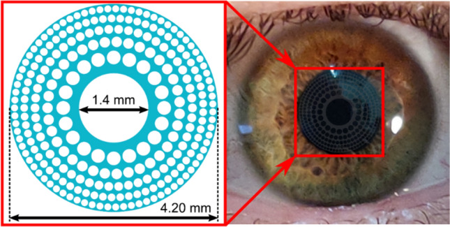
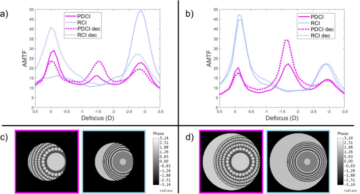
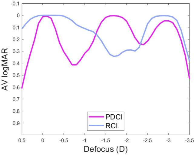
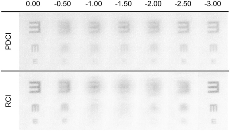
Corneal inlays (CIs) are the most recent surgical procedure for the treatment of presbyopia in patients who want complete independence from the use of glasses or contact lenses. Although refractive surgery in presbyopic patients is mostly performed in combination with cataract surgery, when the implantation of an intraocular lens is not necessary, the option of CIs has the advantage of being minimally invasive. Current designs of CIs are, either: small aperture devices, or refractive devices, however, both methods do not have good performance simultaneously at intermediate and near distances in eyes that are unable to accommodate. In the present study, we propose the first design of a trifocal CI, allowing good vision, at the same time, at far, intermediate and near vision in presbyopic eyes. We first demonstrate the good performance of the new inlay in comparison with a commercially available CI by using optical design software. We next confirm experimentally the image forming capabilities of our proposal employing an adaptive optics based optical simulator. This new design also has a number of parameters that can be varied to make personalized trifocal CI, opening up a new avenue for the treatment of presbyopia.


Conflict of interest statement

The authors declare no competing interests.

Figures

Figure 1
Figure 1
PDCI design. The blue areas in the left figure represent a biocompatible (transparent), hydrogel-based, material of refractive index of 1.458 (see “Methods” section for details). The image on the right is a simulation of the appearance of the PDCI (in stark contrast) on a real eye.
Figure 2
Figure 2
AMTFs of PDCI (magenta) and RCI (blue) for 3.0 mm (a), and 4.5 mm pupil (b). Dotted lines correspond to the AMTFs with the optical inlays decentered 1.0 mm with respect to the pupil center as shown in the phase maps shown with the corresponding color frames in (c) for 3.0 mm and (d) for 4.5 mm pupil diameters.
Figure 3
Figure 3
Through-focus VA curves for the PDCI (magenta), and RCI (blue) obtained from the AMTFs using Eq. (1). The pupil size is 4.5 mm and the abscissa axis has the origin (0.0 D defocus) at the distance focus of each lens.
Figure 4
Figure 4
Images of a tumbling E optotype corresponding to 0.4, 0.2 and 0 logMAR VA obtained the VAO system simulating the PDCI and RCI with the object at different vergences from 0.0 D to − 3.0 D.

References

    1. Fricke TR, et al. Global prevalence of presbyopia and vision impairment from uncorrected presbyopia: Systematic review, meta-analysis, and modelling. Ophthalmology. 2018;125:1492–1499. doi: 10.1016/j.ophtha.2018.04.013. - DOI - PubMed
    1. Charman WN. Developments in the correction of presbyopia II: Surgical approaches. Ophthal. Physiol. Opt. 2014;34:397–426. doi: 10.1111/opo.12129. - DOI - PubMed
    1. Moarefi MA, Bafna S, Wiley W. A review of presbyopia treatment with corneal inlays. Opthamol. Ther. 2017;6:55–65. doi: 10.1007/s40123-017-0085-7. - DOI - PMC - PubMed
    1. Beer SMC, et al. A 3-year follow-up study of a new corneal inlay: Clinical results and outcomes. Brit. J. Ophthalmol. 2020;104:723–728. doi: 10.1136/bjophthalmol-2019-314314. - DOI - PubMed
    1. Limnopoulou AN, et al. Visual outcomes and safety of a refractive corneal inlay for presbyopia using femtosecond laser. J. Refract. Surg. 2013;29:12–18. doi: 10.3928/1081597X-20121210-01. - DOI - PubMed

Publication types