. 2021 Mar 23;11(1):6628.
doi: 10.1038/s41598-021-85019-6.

Test conditions can significantly affect the results of in vitro cytotoxicity testing of degradable metallic biomaterials

Affiliations

Test conditions can significantly affect the results of in vitro cytotoxicity testing of degradable metallic biomaterials

Eva Jablonská et al. Sci Rep. .

Abstract

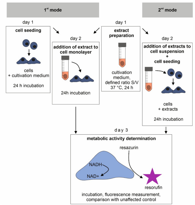
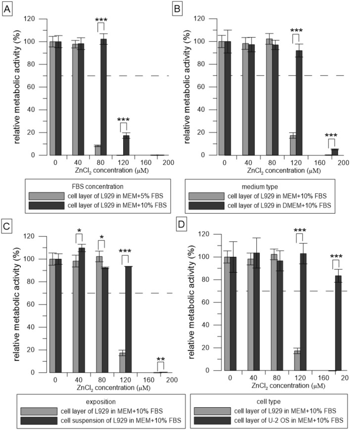
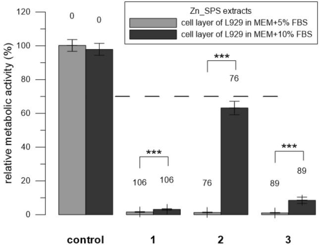
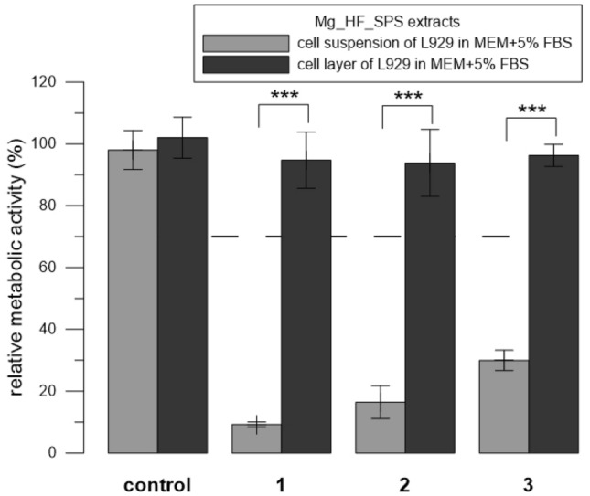
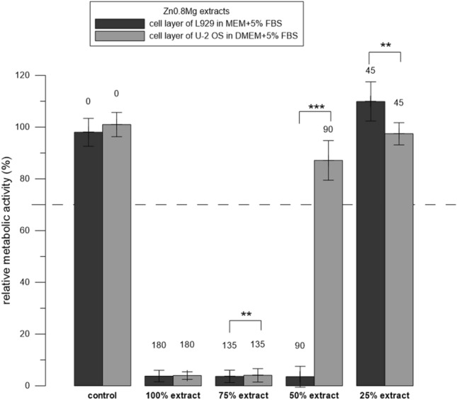
In vitro cytotoxicity testing is an indispensable part of the development of new biomaterials. However, the standard ISO 10993-5 enables variability in the testing conditions, which makes the results of the test incomparable. We studied the influence of media composition on the results of the cytotoxicity test. Solutions of ZnCl2 served as simulated extracts and we also used extracts of three types of Zn-based and Mg-based degradable metals. We incubated the cells with the solutions prepared in two types of media with two concentrations of serum (5 and 10%). We compared the toxic effect of the extracts on L929 murine fibroblast-derived cell line, which is recommended by ISO standard and on "osteoblast-like cells" U-2 OS. We also compared two methods of exposition: solutions were added either to a sub-confluent layer or to the cell suspension. We evaluated the metabolic activity of the cells using the resazurin test. We found out that in vitro cytotoxicity is dramatically influenced by the concentration of serum and by the type of the medium as well as by the type of exposition and type of cells. Therefore, when performing in vitro cytotoxicity testing of biomaterials, the authors should carefully specify the conditions of the test and comparison of different studies should be carried out with caution.


Conflict of interest statement

The authors declare no competing interests.

Figures

Figure 1
Figure 1
Two modes of exposition. Extracts were either added to a sub-confluent cell monolayer 24 h post-seeding (1st mode) or were added to freshly suspended cells (2nd mode). The figure was created using CorelDRAW 2019, version 21.3.0.755.
Figure 2
Figure 2
Relative metabolic activity of the cells (resazurin assay) after 24 h incubation with solutions of ZnCl2. Metabolic activity of the medium without ZnCl2 was taken as 100%. (AD) represent the different conditions used: (A) compares two concentrations of FBS, (B) compares two types of media, (C) compares two ways of exposition, and (D) compares two cell types. The dashed line stands for the normative limit of 70% metabolic activity of the control. Error bars stand for the standard error deviation of six replicates. Significance codes according to p value: 0 ‘***’0.001 ‘**’0.01 ‘*’0.05 (two-sample t-test).
Figure 3
Figure 3
Relative metabolic activity of L929 cells (resazurin assay) after 24 h incubation with extracts of Zn_SPS. Three replicates 1–3 were used. Extracts were prepared in medium either with 5% or with 10% FBS. Sole extraction medium served as a control (unaffected cells which metabolic activity was taken as 100%). Numbers above the columns stand for the concentration of measured Zn in µM. The dashed line stands for the normative limit of 70% metabolic activity of the control. Error bars stand for the standard error deviation of six measurements. Significance codes according to p value: 0 ‘***’0.001 ‘**’0.01 ‘*’0.05 (two-sample t-test).
Figure 4
Figure 4
Relative metabolic activity of L929 cells (resazurin assay) after 24 h incubation with extracts of Mg_HF_SPS. Three replicates 1–3 were used. The extract was added either to the cell layer or to the cell suspension. Sole extraction medium served as a control (unaffected cells which metabolic activity was taken as 100%). The dashed line stands for the normative limit of 70% metabolic activity of the control. Error bars stand for the standard error deviation of six measurements. Significance codes according to p value: 0 ‘***’0.001 ‘**’0.01 ‘*’0.05 (two-sample t-test).
Figure 5
Figure 5
Relative metabolic activity of the cells (resazurin assay) after 24 h incubation with extracts of Zn0.8 Mg. Three replicates 1–3 were used. The extract was added either to L929 or to U-2 OS cells. Sole extraction medium served as a control (unaffected cells which metabolic activity was taken as 100%). Numbers above the columns stand for the concentration of measured Zn in µM. The dashed line stands for the normative limit of 70% metabolic activity of the control. Error bars stand for the standard error deviation of six measurements. Significance codes according to p value: 0 ‘***’0.001 ‘**’0.01 ‘*’0.05 (two-sample t-test).

References

    1. Wolf MF, Coleman KP, Lewerenz GM. Chapter II. 3.3—In vitro assessment of cell and tissue compatibility A2—Ratner, Buddy D. In: Hoffman AS, Schoen FJ, Lemons JE, editors. Biomaterials Science. 3. Cambridge: Academic Press; 2013. pp. 593–608.
    1. Jablonská E, Horkavcová D, Rohanová D, Brauer DS. A review of in vitro cell culture testing methods for bioactive glasses and other biomaterials for hard tissue regeneration. J. Mater. Chem. B. 2020;8(48):10941–10953. doi: 10.1039/D0TB01493A. - DOI - PubMed
    1. J.M. Anderson, R.W. Bianco, J. Grehan, F., B.C. Grubbs, S.R. Hanson, K.D. Hauch, M. Lahi, J.P. Mrachek, S. Northup, J., Biological Testing of biomaterials, in: B.D. Ratner, A.S. Hoffman, F.J. Schoen, J.E. Lemons (Eds.) Biomaterials science: an introduction to materials in medicine, Elsevier Amsterdam, 2004.
    1. Zheng YF, Gu XN, Witte F. Biodegradable metals. Mater. Sci. Eng. R. Rep. 2014;77:1–34. doi: 10.1016/j.mser.2014.01.001. - DOI
    1. Fischer J, Profrock D, Hort N, Willumeit R, Feyerabend F. Reprint of: Improved cytotoxicity testing of magnesium materials. Mater. Sci. Eng. B-Adv. 2011;176(20):1773–1777. doi: 10.1016/j.mseb.2011.06.002. - DOI

Publication types

LinkOut - more resources