

Bio-multifunctional noncovalent porphyrin functionalized carbon-based nanocomposite
- PMID: 33758300
- PMCID: PMC7988124
- DOI: 10.1038/s41598-021-86119-z
Bio-multifunctional noncovalent porphyrin functionalized carbon-based nanocomposite
Erratum in
-
Author Correction: Bio-multifunctional noncovalent porphyrin functionalized carbon-based nanocomposite.Sci Rep. 2023 May 3;13(1):7186. doi: 10.1038/s41598-023-33771-2. Sci Rep. 2023. PMID: 37137929 Free PMC article. No abstract available.
Abstract
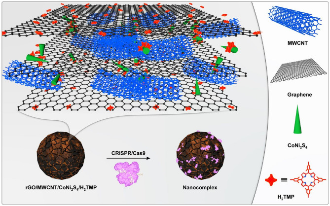
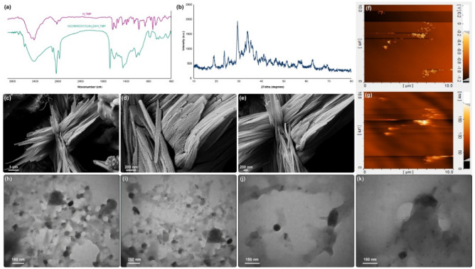
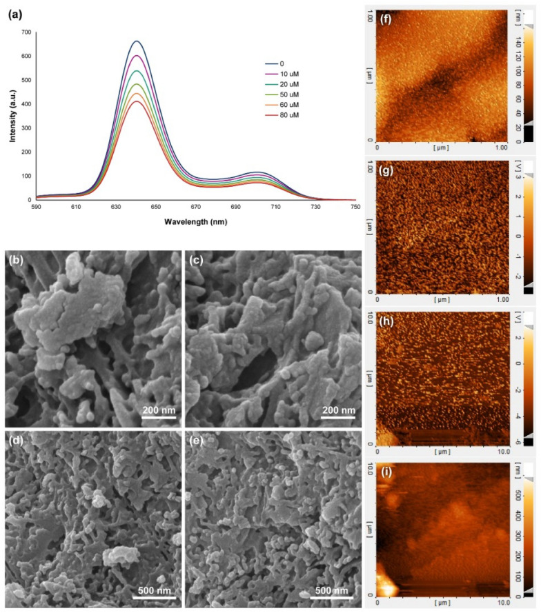
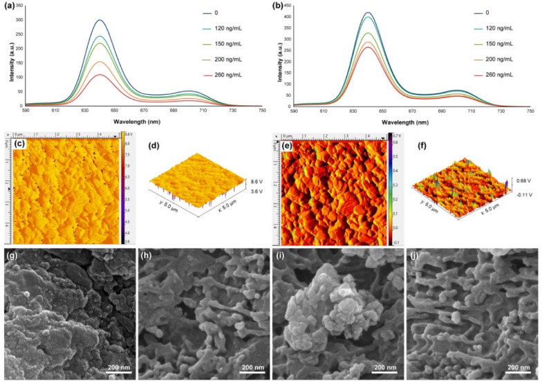
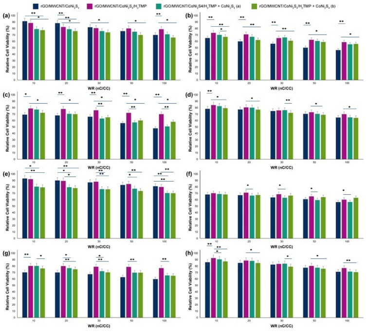
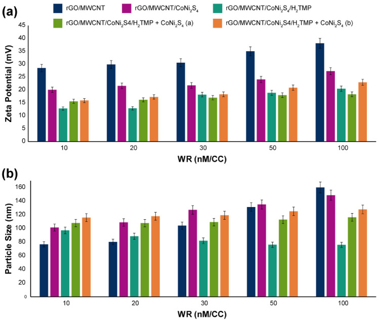
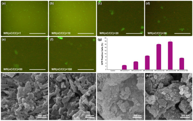
Herein, in a one-pot method, the reduced graphene oxide layers with the assistance of multiwalled carbon nanotubes were decorated to provide a suitable space for the in situ growth of CoNi2S4, and the porphyrins were incorporated into the layers as well to increase the sensitivity of the prepared nanostructure. The prepared nanocomposite can establish π-π interactions between the genetic material and on the surface of porphyrin rings. Also, hydrogen bonds between genetic domains and the porphyrin' nitrogen and the surface hydroxyl groups are probable. Furthermore, the potential donor-acceptor relationship between the d7 transition metal, cobalt, and the genetic material provides a suitable way to increase the interaction and gene loading , and transfections. The reason for this phenomenon was optimized to increase the EGFP by up to 17.9%. Furthermore, the sensing ability of the nanocomposite towards H2O2 was investigated. In this regard, the limit of detection of the H2O2 obtained 10 µM. Also, the in situ biosensing ability in the HEK-293 and PC12 cell lines was evaluated by the addition of PMA. The nanocomposite showed the ability to detect the released H2O2 after adding the minimum amount of 120 ng/mL of the PMA.
Conflict of interest statement
The authors declare no competing interests.
Figures
Similar articles
-
Porphyrin Molecules Decorated on Metal-Organic Frameworks for Multi-Functional Biomedical Applications.Biomolecules. 2021 Nov 17;11(11):1714. doi: 10.3390/biom11111714. Biomolecules. 2021. PMID: 34827712 Free PMC article.
-
Turning Toxic Nanomaterials into a Safe and Bioactive Nanocarrier for Co-delivery of DOX/pCRISPR.ACS Appl Bio Mater. 2021 Jun 21;4(6):5336-5351. doi: 10.1021/acsabm.1c00447. Epub 2021 Jun 10. ACS Appl Bio Mater. 2021. PMID: 35007014
-
Sonochemical synthesis and anchoring of zinc oxide on hemin-mediated multiwalled carbon nanotubes-cellulose nanocomposite for ultra-sensitive biosensing of H2O2.Ultrason Sonochem. 2020 May;63:104917. doi: 10.1016/j.ultsonch.2019.104917. Epub 2019 Dec 4. Ultrason Sonochem. 2020. PMID: 31945552
-
A novel hierarchical 3D N-Co-CNT@NG nanocomposite electrode for non-enzymatic glucose and hydrogen peroxide sensing applications.Biosens Bioelectron. 2017 Mar 15;89(Pt 2):970-977. doi: 10.1016/j.bios.2016.09.077. Epub 2016 Sep 23. Biosens Bioelectron. 2017. PMID: 27816584
-
Direct electrochemistry of myoglobin at reduced graphene oxide-multiwalled carbon nanotubes-platinum nanoparticles nanocomposite and biosensing towards hydrogen peroxide and nitrite.Biosens Bioelectron. 2014 Mar 15;53:420-7. doi: 10.1016/j.bios.2013.09.075. Epub 2013 Oct 18. Biosens Bioelectron. 2014. PMID: 24211453
Cited by
-
Bioactive hybrid metal-organic framework (MOF)-based nanosensors for optical detection of recombinant SARS-CoV-2 spike antigen.Sci Total Environ. 2022 Jun 15;825:153902. doi: 10.1016/j.scitotenv.2022.153902. Epub 2022 Feb 17. Sci Total Environ. 2022. PMID: 35182622 Free PMC article.
-
Metal-Organic Frameworks (MOFs) for Cancer Therapy.Materials (Basel). 2021 Nov 28;14(23):7277. doi: 10.3390/ma14237277. Materials (Basel). 2021. PMID: 34885431 Free PMC article.
-
Synthesis of Carbon-Encapsulated Magnetic Iron Oxide Nanocomposites for Bioapplication.Int J Biomater. 2022 Sep 20;2022:3302082. doi: 10.1155/2022/3302082. eCollection 2022. Int J Biomater. 2022. PMID: 36176284 Free PMC article.
-
Gelatin-Graphene Oxide Nanocomposite Hydrogels for Kluyveromyces lactis Encapsulation: Potential Applications in Probiotics and Bioreactor Packings.Biomolecules. 2021 Jun 22;11(7):922. doi: 10.3390/biom11070922. Biomolecules. 2021. PMID: 34206397 Free PMC article.
-
Porphyrin Molecules Decorated on Metal-Organic Frameworks for Multi-Functional Biomedical Applications.Biomolecules. 2021 Nov 17;11(11):1714. doi: 10.3390/biom11111714. Biomolecules. 2021. PMID: 34827712 Free PMC article.
References
-
- Patel KD, Singh RK, Kim H-W. Carbon-based nanomaterials as an emerging platform for theranostics. Mater. Horizon. 2019;6:434–469. doi: 10.1039/C8MH00966J. - DOI
-
- Liu Y, et al. A novel non-enzymatic electrochemical biosensor based on the nanohybrid of bimetallic PdCu nanoparticles/carbon black for highly sensitive detection of H2O2 released from living cells. Sensors Actuators B: Chem. 2019;290:249–257. doi: 10.1016/j.snb.2019.03.129. - DOI
-
- Zhao Y, et al. Biosensor based on 3D graphene-supported Fe3O4 quantum dots as biomimetic enzyme for in situ detection of H2O2 released from living cells. Sensors Actuators B Chem. 2017;244:1037–1044. doi: 10.1016/j.snb.2017.01.029. - DOI
Publication types
LinkOut - more resources
Full Text Sources
Other Literature Sources
Research Materials
Miscellaneous
