Review
. 2021 May;21(5):281-297.
doi: 10.1038/s41568-021-00344-2. Epub 2021 Mar 23.

Autophagy in tumour immunity and therapy

Affiliations
Review

Autophagy in tumour immunity and therapy

Houjun Xia et al. Nat Rev Cancer. 2021 May.

Abstract

Autophagy is a regulated mechanism that removes unnecessary or dysfunctional cellular components and recycles metabolic substrates. In response to stress signals in the tumour microenvironment, the autophagy pathway is altered in tumour cells and immune cells - thereby differentially affecting tumour progression, immunity and therapy. In this Review, we summarize our current understanding of the immunologically associated roles and modes of action of the autophagy pathway in cancer progression and therapy, and discuss potential approaches targeting autophagy to enhance antitumour immunity and improve the efficacy of current cancer therapy.


Conflict of interest statement

Competing interests

The authors declare no competing interests.

Figures

Fig. 1 |
Fig. 1 |. The machinery of canonical and non-canonical autophagy.
Canonical autophagy is normally induced in response to the cell undergoing an energy crisis. Autophagy-inducing signals, such as nutrient deprivation, trigger the activation of 5′-AMP-activated protein kinase (AMPK), whose kinase activity simultaneously inhibits mTOR and activates the pre-initiation complex (unc-51-like kinase 1 (ULK1), autophagy-related protein 13 (ATG13), ATG101 and FAK family interacting protein of 200 kDa (FIP200)). This complex, in turn, phosphorylates components in the autophagy initiation complex composed of vacuolar protein sorting 34 (VPS34), VPS15 and Beclin 1, along with ATG14 and AMBRA1. The initiation complex converts phosphoinositides in the endoplasmic reticulum to phosphatidylinositol-3-phosphate (PI3P), which recruits ligation machinery composed of the E1 ligase ATG7, the E2 ligase ATG3 and a complex of ATG16L1 and an ATG5–ATG12 conjugate that together act as an E3 ligase. The ligase machinery functions to ligate lipidate microtubule-associated protein 1A/1B light chain 3 (LC3) family proteins to phosphatidylethanolamine (PE) to form LC3–PE. The activity and coordination of the ATG5–ATG12 system and the LC3–PE system facilitate the curvature and sealing of the autophagosome, as well as the lipidation and embedding of LC3–PE into the autophagosomal membrane. The resulting autophagosomes then fuse to lysosomes (forming autolysosomes), resulting in digestion of the autophagosome contents loaded by the LC3 binding proteins. The non-canonical autophagy pathway also functions to lipidate LC3 family proteins on a single membrane. LC3-associated phagocytosis (LAP) and LC3-associated endocytosis (LANDO) process independently of the autophagy pre-initiation (ULK1) complex, and utilize a different initiation complex composed of VPS34, VPS15, Beclin 1, UVRAG and Rubicon without requirements for ATG14 or AMBRA1. mTORC1, mTOR complex 1; ROS, reactive oxygen species; TLR, Toll-like receptor; WIPI2, WD repeat domain phosphoinositide-interacting protein 2.
Fig. 2 |
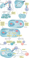
Fig. 2 |. Impact of tumour autonomous autophagy on immunity.
a | Autophagy in dead tumour cells promotes dendritic cell (DC)-mediated cross-presentation via increasing production of autophagosome with tumour antigen. In addition, neighbour of BRCA1 gene 1 protein (NBR1) mediates major histocompatibility complex (MHC) class I degradation via the autophagy pathway, resulting in reduced MHC class I surface expression in pancreatic cancer cells and abolished cytotoxic T lymphocyte (CTL) recognition. b | Autophagy blockade or deficiency results in accumulated reactive oxygen species (ROS) in tumour cells and subsequent mitochondria and genome damage, thereby releasing DNA into cytoplasm. Absent in melanoma 2 (AIM2) senses cytosolic DNA, activates inflammasome signalling and promotes tumorigenesis via recruiting immunosuppressive immune cells. c-GAS–stimulator of interferon genes (STING) can also sense cytosolic DNA, stimulate type I interferon responses and enhance antitumour immunity via recruiting CD8+ T and natural killer (NK) cells. c | Hypoxia induces autophagy in tumour cells through hypoxia-induced factor 1α (HIF1α). Elevated autophagy mediates degradation of granzyme B in tumour cells, which is secreted by activated CD8+ T cells and NK cells, and blocks CTL-mediated and NK cell-mediated tumour killing. Meanwhile, autophagy inhibits chemokine CCL5 expression, which recruits the NK cell migration to the tumour microenvironment (TME). MDSC, myeloid-derived suppressor cell; TAA, tumour-associated antigen; Treg cell, regulatory T cell.
Fig. 3 |
Fig. 3 |. autophagy in immune cells in the tumour microenvironment.
a | In the tumour microenvironment (TME), T cells rely on autophagy to support their survival and differentiation. Basal levels of autophagy in naive T cells maintain their quiescence and protect them from mitochondria-derived reactive oxygen species (ROS)-induced apoptosis. High levels of lactate in tumours disrupt autophagy of naive T cells and impair the antitumour response in mouse models. High levels of potassium or low levels of nutrients in the TME constrain the nutrient uptake in effector T cells and cause functional caloric restriction, which induces autophagy through mTOR complex 1 (mTORC1) inhibition. Autophagy is critical for mitochondrial integrity, which determines effector T cell differentiation via metabolic reprogramming. High levels of arginase 2 (ARG2) expression in tumour-infiltrating regulatory T cells (Treg cells) may maintain their high level of autophagy. The loss of autophagy in Treg cells switches their metabolism from oxidative phosphorylation (OXPHOS) to glycolysis with active mTORC1 and MYC, which leads to FOXP3 instability. Increased apoptosis and defective function in autophagy-deficient Treg cells contribute to greater tumour resistance. b | Tumour-associated macrophages (TAMs) take advantage of lipidate microtubule-associated protein 1A/1B light chain 3 (LC3)-associated phagocytosis (LAP) to degrade apoptotic cancer cells. LAP deficiency in TAMs leads to the release of mitochondrial DNA from apoptotic cancer cells, which induces type I interferon response through the cGAS–stimulator of interferon genes (STING) pathway and antitumour activity. c | In ovarian cancer metastasis, TIM4+ TAMs are embryonically originated and locally sustained, whereas TIM4 TAMs are replenished from circulating monocytes. Relative to TIM4 TAMs, TIM4+ TAMs manifest high levels of oxidative phosphorylation and adapt mitophagy to alleviate oxidative stress. High levels of ARG1 in TIM4+ TAMs contribute to potent mitophagy activities via weakened mTORC1 activation due to low arginine levels resulting from ARG1-mediated metabolism. Furthermore, genetic deficiency of autophagy element FAK family interacting protein of 200 kDa (FIP200) results in TIM4+ TAM loss via ROS-mediated apoptosis, and elevated T cell immunity and tumour inhibition in vivo. ATG13, autophagy-related protein 13; BCL2, B cell lymphoma 2; MCT1, monocarboxylate transporter 1; TIM4, T cell immunoglobulin and mucin domain-containing molecule 4; ULK1, unc-51-like kinase 1.
Fig. 4 |
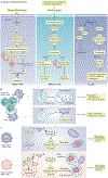
Fig. 4 |. Targeting autophagy for tumour immunotherapy.
a | Targeting autophagy remodels the tumour microenvironment (TME). Tumour autophagy inhibition can upregulate production of the T helper 1 (TH1)-type chemokines, promoting effector immune cell tumour infiltration. Targeting tumour autophagy may be combined with adoptive transfer of engineered chimeric antigen receptor T cells (CAR T cells) and immune checkpoint blockade (ICB). b | Autophagy can mediate degradation of potential tumour antigens and major histocompatibility complex (MHC) components in antigen-presenting cells (APCs). Autophagy inhibition can enhance the antitumour T cell response through elevated antigen procession and presentation. c | Autophagy can mediate degradation of MHC class I and granzyme B to avoid cytotoxic T lymphocyte (CTL)-mediated and natural killer (NK) cell-mediated killing. Autophagy inhibition can result in increased levels of MHC class I on the cell surface and prevents the autophagic degradation of granzyme B, thereby allowing cell lysis. Autophagy inhibition can enhance ICB therapeutic efficacy. d | High-dose interleukin-2 (IL-2) treatment causes systemic autophagic syndrome. Autophagy inhibition protects patients with cancer from systemic damage and improves IL-2 therapy. DAMP, damage-associated molecular pattern; DC, dendritic cell; TAM, tumour-associated macrophage; Treg cell, regulatory T cell.

Similar articles

Cited by

References

    1. Morishita H & Mizushima N Diverse cellular roles of autophagy. Annu. Rev. Cell Dev. Biol 35, 453–475 (2019). - PubMed
    1. Amaravadi RK, Kimmelman AC & Debnath J Targeting autophagy in cancer: recent advances and future directions. Cancer Discov 9, 1167–1181 (2019). - PMC - PubMed
    1. Mizushima N, Yoshimori T & Ohsumi Y The role of Atg proteins in autophagosome formation. Annu. Rev. Cell Dev. Biol 27, 107–132 (2011). - PubMed
    2. This detailed review discusses the protein and membrane interactions required for autophagosome formation.

    1. Marino G, Niso-Santano M, Baehrecke EH & Kroemer G Self-consumption: the interplay of autophagy and apoptosis. Nat. Rev. Mol. Cell Biol 15, 81–94 (2014). - PMC - PubMed
    1. Dikic I & Elazar Z Mechanism and medical implications of mammalian autophagy. Nat. Rev. Mol. Cell Biol 19, 349–364 (2018). - PubMed

Publication types

Substances