

Coronil, a Tri-Herbal Formulation, Attenuates Spike-Protein-Mediated SARS-CoV-2 Viral Entry into Human Alveolar Epithelial Cells and Pro-Inflammatory Cytokines Production by Inhibiting Spike Protein-ACE-2 Interaction
- PMID: 33758527
- PMCID: PMC7981146
- DOI: 10.2147/JIR.S298242
Coronil, a Tri-Herbal Formulation, Attenuates Spike-Protein-Mediated SARS-CoV-2 Viral Entry into Human Alveolar Epithelial Cells and Pro-Inflammatory Cytokines Production by Inhibiting Spike Protein-ACE-2 Interaction
Abstract
Purpose: Coronil is a tri-herbal formulation containing extracts from Withania somnifera, Tinospora cordifolia, and Ocimum sanctum. Recently, it was shown that Coronil rescued humanized zebrafish from SARS-CoV-2 induced pathologies. Based on reported computational studies on the phytochemicals present in Coronil, it could be a potential inhibitor of SARS-CoV-2 entry into the host cell and associated cytokines' production.
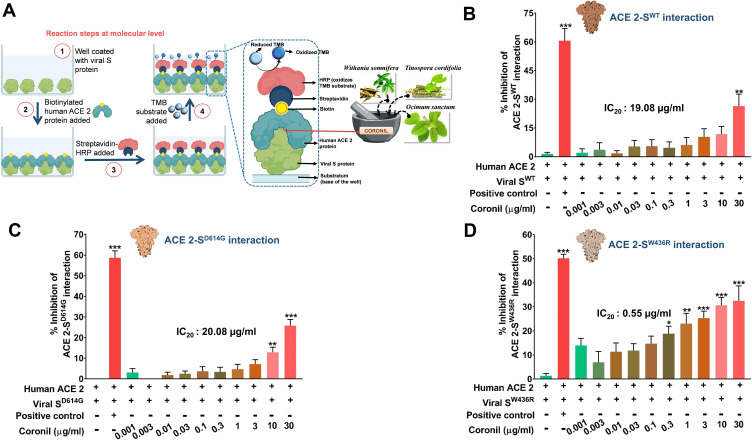
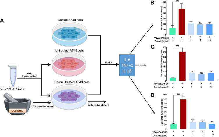
Methods: Through an ELISA-based biochemical assay, effects of Coronil on interaction between ACE-2 and different mutants of viral spike (S) protein, crucial for viral invasion of host cell, were evaluated. Additionally, using recombinant pseudoviruses having SARS-CoV-2 spike (S) protein in their envelopes and firefly luciferase reporter in their genomes, effects of Coronil on virus entry into human alveolar epithelial cells were evaluated through luciferase assay. UHPLC profiled Coronil also modulated S-protein mediated production of pro-inflammatory cytokines in A549 cells, like interleukin-6 (IL-6), interleukin-1β (IL-1β), and tumor necrosis factor-α (TNF-α), as evaluated through RT-qPCR and ELISA.
Results: Coronil effectively inhibited the interaction of ACE-2 not only with the wild-type S protein (SWT) but also with its currently prevalent and more infectious variant (SD614G) and another mutant (SW436R) with significantly higher affinity toward ACE-2. Treatment with Coronil significantly reduced the increased levels of IL-6, IL-1β, and TNF-α in A549 cells incubated with different S-protein variants in a dose-dependent manner. Likewise, it also prevented the SARS-CoV-2 S-protein pseudotyped vesicular stomatitis virus (VSVppSARS-2S) mediated cytokine response in these cells by reducing entry of pseudoviruses into host cells.
Conclusion: Coronil prevented SARS-CoV-2 S-protein mediated viral entry into A549 cells by inhibiting spike protein-ACE-2 interactions. SARS-CoV-2 S protein induced inflammatory cytokine response in these cells was also moderated by Coronil.
Keywords: ACE-2; Coronil; SARS-CoV-2; pro-inflammatory cytokines; pseudovirus; spike protein.
© 2021 Balkrishna et al.
Conflict of interest statement
The test article was provided by Divya Pharmacy, Haridwar, Uttarakhand, India. Acharya Balkrishna is an honorary trustee in Divya Yog Mandir Trust, in addition he holds an honorary managerial position in Patanjali Ayurved Ltd, Haridwar, India. Besides, providing the test article, Divya Pharmacy was not involved in any aspect of this study. All other authors have no conflicts of interest to declare.
Figures


Similar articles
-
Herbo-mineral formulation, Divya-Swasari-Vati averts SARS-CoV-2 pseudovirus entry into human alveolar epithelial cells by interfering with spike protein-ACE 2 interaction and IL-6/TNF-α /NF-κB signaling.Front Pharmacol. 2022 Oct 26;13:1024830. doi: 10.3389/fphar.2022.1024830. eCollection 2022. Front Pharmacol. 2022. PMID: 36386162 Free PMC article.
-
Application of Humanized Zebrafish Model in the Suppression of SARS-CoV-2 Spike Protein Induced Pathology by Tri-Herbal Medicine Coronil via Cytokine Modulation.Molecules. 2020 Nov 2;25(21):5091. doi: 10.3390/molecules25215091. Molecules. 2020. PMID: 33147850 Free PMC article.
-
Withanone from Withania somnifera Attenuates SARS-CoV-2 RBD and Host ACE2 Interactions to Rescue Spike Protein Induced Pathologies in Humanized Zebrafish Model.Drug Des Devel Ther. 2021 Mar 11;15:1111-1133. doi: 10.2147/DDDT.S292805. eCollection 2021. Drug Des Devel Ther. 2021. PMID: 33737804 Free PMC article.
-
Therapeutic potential of green tea catechin, (-)-epigallocatechin-3-O-gallate (EGCG) in SARS-CoV-2 infection: Major interactions with host/virus proteases.Phytomed Plus. 2023 Feb;3(1):100402. doi: 10.1016/j.phyplu.2022.100402. Epub 2022 Dec 30. Phytomed Plus. 2023. PMID: 36597465 Free PMC article. Review.
-
Mechanistic Aspects of Medicinal Plants and Secondary Metabolites against Severe Acute Respiratory Syndrome Coronavirus 2 (SARS-CoV-2).Curr Pharm Des. 2021;27(38):3996-4007. doi: 10.2174/1381612827666210705160130. Curr Pharm Des. 2021. PMID: 34225607 Review.
Cited by
-
Effects of growth hormone/estrogen/androgen on COVID-19-type proinflammatory responses in normal human lung epithelial BEAS-2B cells.BMC Mol Cell Biol. 2022 Sep 29;23(1):42. doi: 10.1186/s12860-022-00442-5. BMC Mol Cell Biol. 2022. PMID: 36175845 Free PMC article.
-
Sodium-calcium exchanger isoform-3 targeted Withania somnifera (L.) Dunal therapeutic intervention ameliorates cognition in the 5xFAD mouse model of Alzheimer's disease.Sci Rep. 2022 Jan 27;12(1):1537. doi: 10.1038/s41598-022-05568-2. Sci Rep. 2022. PMID: 35087161 Free PMC article.
-
A study on the effect of natural products against the transmission of B.1.1.529 Omicron.Virol J. 2023 Aug 25;20(1):191. doi: 10.1186/s12985-023-02160-6. Virol J. 2023. PMID: 37626376 Free PMC article. Review.
-
Comparative retrospective open-label study of ayurvedic medicines and their combination with allopathic drugs on asymptomatic and mildly-symptomatic COVID-19 patients.J Herb Med. 2021 Oct;29:100472. doi: 10.1016/j.hermed.2021.100472. Epub 2021 May 21. J Herb Med. 2021. PMID: 34055580 Free PMC article.
-
Network pharmacology and in-silico studies for molecular mechanisms of analgesic, anti-inflammatory and anti-arthritic effects of Withania somnifera (L.) Dunal phytoconstituents.J Ayurveda Integr Med. 2025 Jul-Aug;16(4):101088. doi: 10.1016/j.jaim.2024.101088. Epub 2025 Jun 26. J Ayurveda Integr Med. 2025. PMID: 40578054 Free PMC article.
References
LinkOut - more resources
Full Text Sources
Other Literature Sources
Miscellaneous
